这个网页非常非常大,需要等待一段时间才能完全加载。
Chapter 1 Introduction
1.1 The Scope of Human Physiology
Physiology is the study of how living organisms function.
1.2 Internal Environment and Homeostasis
1.2.1 Body Organization
1.2.1.1 Tissue
Muscle cells and tissue
- Skeletal, Cardiac, Smooth muscle cell
Neurons and Nervous tissue
- Neuron controls other cell through conducting electrical signals
Epithelial Cells and Epithelium
- Cuboidal, Columnar, Squamous, Ciliated cells
- Simple, stratified epithelium
Connective-Tissue Cells and Connective Tissue
- Bone, Cartilage, adipose tissue; loose, dense connective tissue;
- Blood
1.2.1.2 Organs and Organ Systems
- Organs are composed of two or more of the four kinds of tissues arranged in various proportions and patterns.
- Organ system is a collection of organs that together perform an overall function.
1.2.2 Body Fluid Compartments
Intracellular fluid 细胞内液
- the fluid contained within all the cells of the body (67%).
Extracellular fluid 细胞外液
- plasma (20-25%) 血浆
- interstitial fluid (75-80%) 组织液
1.2.3 Homeostasis
Homeostasis was defined as a state of reasonably stable balance between physiological variables such as those just described. 体内平衡被定义为刚才描述的生理变量之间合理稳定的平衡状态。
Homeostasis is a state of dynamic constancy.
1.3 Homeostatic Control Systems in the Body
Homeostatic control systems
- Feedback control systems: negative / positive feedback
- Adaptation and Acclimatization 适应和顺应: Resetting of set points
- Feedforward control systems
1.4 Forms of Functional Regulations in Human Body
Neural Regulation: Reflexes 神经调节:反射
- stimulus–response sequences
Humoral Regulation 体液调节
Autoregulation 自身调节
Chapter 2 Basic Function of Cell
2.1 Movement of Molecules Across the Cell Membranes
2.1.1 Functional Structure of the Cell Membranes
The plasma membrane forms the cell’s flexible outer surface, separating the cell’s internal environment from the external environment: selective barrier and communication 质膜形成细胞柔韧的外表面,将细胞内部环境与外部环境分隔开:选择性屏障和通讯
The cytoplasm consists of all the cellular contents between the plasma membrane and the nucleus: cytosol and organelles 细胞质由质膜和细胞核之间的所有细胞内容物组成:细胞溶胶和细胞器
The nucleus is a large organelle that houses most of a cell’s DNA; Chromosome, a single molecule of DNA associated with several proteins, contains genes. 细胞核是一个⼤型细胞器,容纳了细胞的⼤部分 DNA;染色体是与几种蛋⽩质相关的单个 DNA 分子,包含基因
Functions
- Regulate the passage of substances into and out of cells and between cell organelles and cytosol. 调节物质进出细胞
- Detect chemical messengers arriving at the cell surface. 检测到达细胞表面的化学信使
- Link adjacent cells together by membrane junctions. 通过膜连接将相邻的细胞连接在⼀起
- Anchor cells to the extracellular matrix. 将细胞锚定于细胞外基质
2.1.1.1 Membrane Lipids
2.1.1.1.1 Membrane Structure
- The plasma membrane, a flexible yet sturdy barrier that surrounds and contains the cytoplasm of a cell. 质膜是一种灵活而坚固的屏障,包围并包含细胞的细胞质
- Membranes are fluid structures because the lipids and many of the proteins are free to rotate and move sideways in their own half of the bilayer. 膜是流体结构,因为脂质和许多蛋⽩质可以在双层膜的各自一半中自由旋转和侧向移动。
- All membranes consist of a double layer of lipid molecules in which proteins are embedded. The major membrane lipids are phospholipids 所有膜均由双层脂质分子组成,蛋⽩质嵌⼊其中。主要的膜脂是磷脂。
2.1.1.1.2 Phospholipids 磷脂
- Phospholipids are amphipathic molecules 磷脂是两亲性分子
- Polar head and non-polar tail
- Lipid bilayer has the characteristics of a fluid 具有流体的特性
- Without chemical bonds, and fatty acids tail can bend ⽆化学键,脂肪酸尾部可弯曲
- Cholesterol is inserted into the lipid bilayer 胆固醇插⼊脂质双层
- limit the ordered packing of fatty acids 限制脂肪酸的有序堆积
2.1.1.1.3 Membrane Proteins
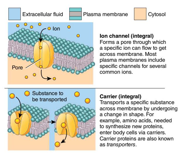
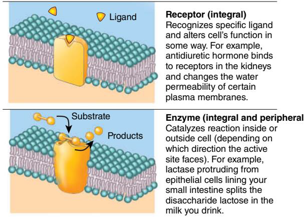
- Integral proteins extend into or through the lipid bilayer and are firmly embedded in it. Most integral proteins are transmembrane proteins, which means that they span the entire lipid bilayer and protrude into both the cytosol and extracellular fluid. 整合蛋白延伸至脂质双层内或穿过脂质双层,并牢固地嵌入其中。大多数整合蛋白都是跨膜蛋白,这意味着它们跨越整个脂质双层,并突出到细胞溶胶和细胞外液中。
- Peripheral proteins are attached to the polar heads of membrane lipids or to integral proteins at the inner or outer surface of the membrane. 外周蛋白附着在膜脂的极性头部或膜内表面或外表面的整合蛋白上。
2.1.1.1.4 The Fluid-Mosaic Model 流体镶嵌模型
- certain membrane proteins are anchored to cytoplasmic proteins 某些膜蛋⽩锚定在细胞质蛋⽩上
- Proteins are covalently linked with membrane lipids to form structures called “lipid rafts” 蛋白质与膜脂质共价连接,形成称为“脂筏”的结构
2.1.1.1.5 Membrane junctions
- Extracellular matrix-membrane 细胞外基质膜
- Integrins 整合素
- Desmosomes 桥粒
- Dense plaque: Cadherins 致密斑块:钙粘蛋⽩
- Tight junction 紧密连接
- Limited to a disk-shaped area of the membrane 仅限于膜的圆盘状区域
- Gap junction 间隙连接
- protein channels linking the cytosols of adjacent cells 连接相邻细胞胞质的蛋⽩质通道
2.1.1.2 Cell Organelles
Nucleus
- Nuclear envelope, nuclear pore, chromatin, nucleolus 核膜、核孔、染色质、核仁
Ribosome: protein factory 核糖体:蛋⽩质工厂
Endoplasmic reticulum 内质⽹
- protein folding, lipid, Calcium
Golgi Apparatus 高尔基体
Secretory vesicles 分泌囊泡
Endosome
Mitochondria: bioenergetic hub 线粒体:⽣物能量中⼼
Peroxisome 过氧化物酶体
Membrane-less organelles
- nuclear stress bodies, processing bodies (P bodies) and stress granules
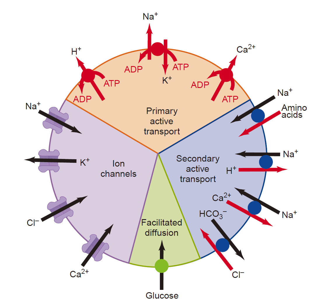
2.1.2 Transmembrane Transport of Molecules
Transport through the cell membrane, either directly through the lipid bilayer or through the proteins, occurs by one of two basic processes:
-
Passive transport, is a movement of ions or other substances across cell membranes from a region of their higher concentration-in the direction down the concentration gradient.
Simple diffusion; Facilitated diffusion; Osmosis 简单扩散;协助扩散;渗透
-
Active transport is a movement of ions or other substances across the membrane against a concentration gradient.
Primary active transport; Secondary active transport 初级主动运输;次级主动运输
2.1.2.1 Simple Diffusion
The movement of molecules from one location to another solely as a result of their random thermal motion is known as diffusion. (The energy is from heat. Random movement with no preferred direction of movement) 分子从一个位置移动到另一个位置,完全是由于其随机热运动而发⽣的,这种现象称为扩散。(能量来自热量。随机运动,没有首选运动⽅向)
The rate at which a substance is transported across a membrane refers to the number of molecules that cross the membrane in a unit of time, which is called the flux. 物质穿过膜的速率是指单位时间内穿过膜的分子数量,称为通量。
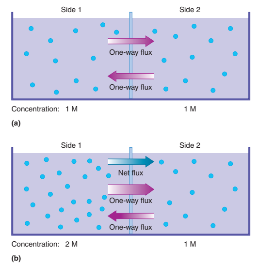
The magnitude of the net flux depends on several factors: the magnitude of the driving force, the membrane surface area, and the permeability of the membrane 净通量的⼤⼩取决于几个因素:驱动⼒的⼤⼩、膜表面积和膜的渗透性
concentration difference 浓度差: the greater the concentration difference, the greater the magnitude of the net flux; ↑
temperature 温度: the higher the temperature,the greater the speed of molecular movement and the greater the net flux; ↑
mass of the molecule 分子质量: large molecules (for example,proteins) have a greater mass and lower speed than smaller molecules (for example, glucose) and thus have a smaller net flux; ↓
surface area 表面积: the greater the surface area between two regions, the greater the space available for diffusion and thus the greater the net flux; ↑
the medium 分子运动的介质: through which the molecules are moving-most polar molecules diffuse very slowly or not at all,whereas nonpolar molecules diffuse much more rapidly.
2.1.2.2 Facilitated Diffusion
- Like simple diffusion, molecules move across a membrane from high concentration to low concentration, or “downhill” in facilitated diffusion.
- Neither simple diffusion nor facilitated diffusion is coupled to ATP derived from metabolism.
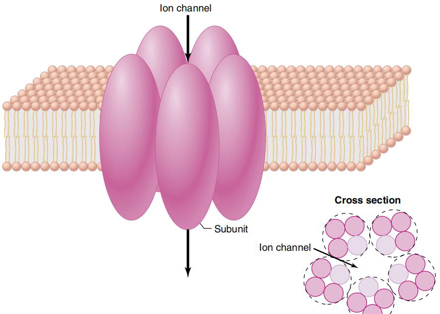
2.1.2.2.1 Diffusion Through Ion Channels
Ions such as Na^+^, K^+^, Cl^-^, Ca^2+^ diffuse across plasma membranes at rates that are much faster than would be predicted from their very low solubility in membrane lipids.
Different cells have quite different permeabilities to these ions, whereas nonpolar substances have similar permeabilities. 不同细胞对这些离子的通透性有相当⼤的差异,而⾮极性物质的通透性则相似。
Ion channels show a selectivity for the type of ion that can pass through them. This selectivity is based partially on the channel diameter and partially on the charged and polar surfaces of the protein subunits that form the channel walls and electrically attract or repel the ions. 离子通道对能通过的离子类型具有选择性。这种选择性部分取决于通道直径,部分取决于形成通道壁的蛋⽩质亚基的带电和极性表面,这些表面以电⽅式吸引或排斥离子。
Effect of electrical forces on ion movement:
-
Positive and negative charges are distributed unequally between the inside and outside the cell. Separation of electrical charge exist across the membrane, known as membrane potential. 跨膜存在电荷分离,称为膜电位。Membrane potential function as electrical force to influence the movement of ions across the membrane. 膜电位作为电⼒发挥作用,影响离子跨膜的运动。
-
Both the concentration difference and the electrical difference of ion determine the magnitude and direction of ion movement. 离子的浓度差和电性差共同决定离子运动的⼤⼩和⽅向。
-
These two driving forces are collectively known as electrochemical gradient across the membrane. 这两种驱动⼒统称为跨膜的电化学梯度。
An electrochemical gradient is a gradient of electrochemical potential, usually for an ion that can move across a membrane. The gradient consists of two parts:
- The chemical gradient, or difference in solute concentration across a membrane.
- The electrical gradient, or difference in charge across a membrane.
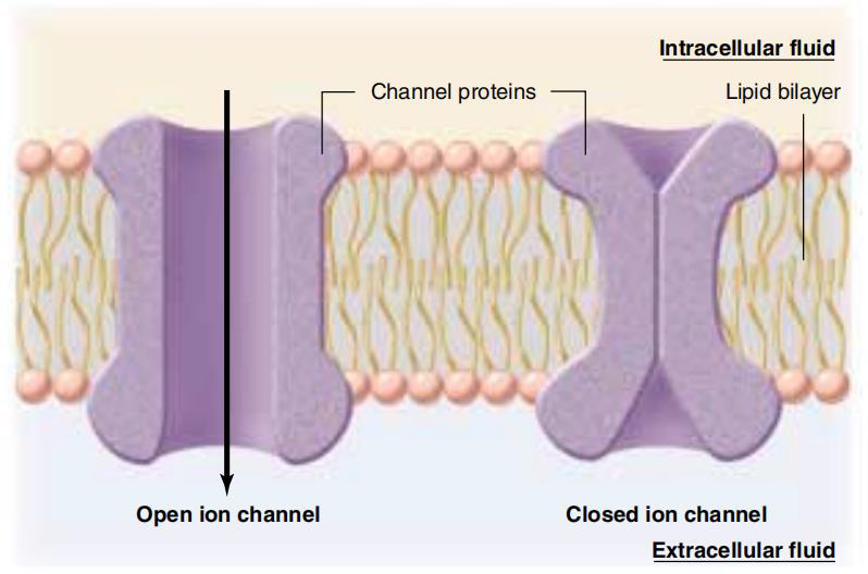
Regulation of diffusion through ion channels
Three factors can alter the channel protein conformations:
- the binding of specific molecules to channel proteins may directly or indirectly produce either an change in the shape of the channel protein;such channels are termed ligand-gated channels, and the ligands that influence them are often chemical messengers. 特定分子与通道蛋⽩的结合可能直接或间接地导致通道蛋⽩形状的改变;这种通道被称为配体门控通道,影响它们的配体通常是化学信使。
- Changes in the membrane potential can cause movement of the charged regions on a channel protein, altering its shape: voltage-gated channels. 膜电位的变化可能导致通道蛋⽩上带电区域的移动,从而改变其形状:电压⻔控通道。
- Stretching the membrane may affect the conformation of some channel proteins: mechanically gated channels. 拉伸膜可能会影响某些通道蛋⽩的构象:机械门控通道。
2.1.2.2.2 Facilitated Diffusion via Carrier
The movement of substances through a membrane by transporter (carrier) is called mediated transport.
- The solute must first binds to a specific site of transporter. 溶质必须首先与转运蛋⽩的特定位点结合
- The molecules can move in either direction.
- Transporter share similar characteristic with ion channel, but low efficiency. 转运体与离子通道具有相似的特性,但效率较低。
- Transporter can be saturated. (All binding site are occupied) 转运蛋⽩可能饱和。(所有结合位点均被占据)
Three factors determine the magnitude of solute flux through a mediated-transport system:
- The extent to which the transporter binding sites are saturated 转运蛋⽩结合位点的饱和程度
- the number of transporters in the membrane 膜中的转运蛋⽩数量
- the rate at which the conformational change in the transport protein occurs 运输蛋⽩发⽣构象变化的速率
2.1.2.3 Active transport
- Moves a substance “uphill” across a membrane (against substance’s concentration gradient).
- Consumes ATP
- Needs transporter, or usually called pump
- the direct use of ATP in primary active transport
- the use of an ion concentration difference across a membrane to drive the process in secondary active transport
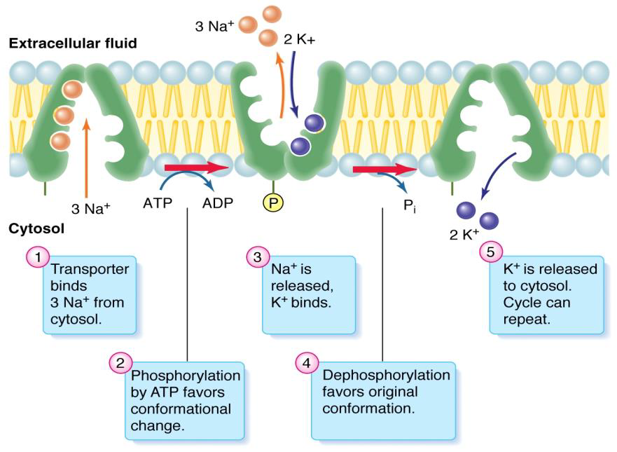
Primary Active Transport
- The transporter is an enzyme (ATPase) that catalyzes the breakdown of ATP and,in the process, phosphorylates itself. 转运蛋⽩是一种酶(ATPase),可催化 ATP 的分解,并在此过程中对其进行自身磷酸化。
- Phosphorylation of the transporter protein (covalent modulation) changes its conformation and affinity binding with solutes. 转运蛋⽩的磷酸化(共价调节)改变其构象和与溶质的亲和⼒结合。
- For example, $Na^+/K^+ATPase,Ca^{2+}-ATPase, H^±ATPase,H^+/K^+ATPase$
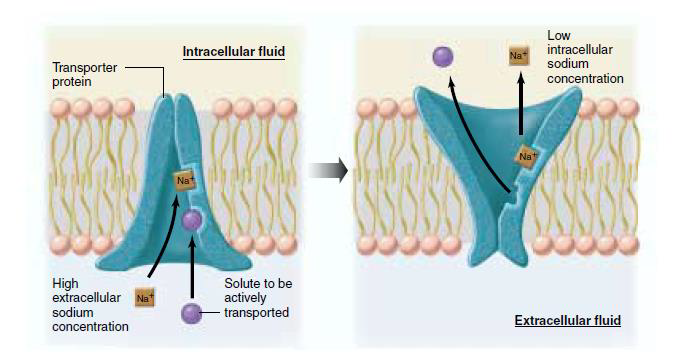
Secondary Active Transport
- Moves ion (normally but not always Na^+^) across a membrane through established electrochemical gradient
- Need transporter
- Transport of another molecule is also coupled to ion movement (piggyback).
- Both molecules bind to transporter.
- The creation and maintenance of electrochemical gradient relies on the ATPase.
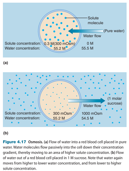
2.1.2.4 Osmosis
Water (polar) diffuses across the plasma membranes of most cells very rapidly mediated by proteins known as aquaporins. ⽔(极性)在⽔通道蛋⽩的介导下⾮常迅速地扩散到⼤多数细胞的质膜上。
The net diffusion of water across a membrane is called osmosis. ⽔穿过膜的净扩散称为渗透。
Water concentration depends on the number of solute particles, regardless of chemical composition. ⽔的浓度取决于溶质粒子的数量,与化学成分无关。
The total solute particle concentration of a solution is known as its osmolarity. 溶液的总溶质颗粒浓度称为其渗透压。
When a solution containing solutes is separated from pure water by a semipermeable membrane, the pressure that must be applied to the solution to prevent the net flow of water into it is known as the osmotic pressure of the solution.
Extracellular osmolarity and cell shape
- Unequal distribution of charged ion between inside and outside the cell contributes to the osmolarity of extracellular fluid.
- A solution of nonpenetrating solute with the same osmolarity as normal extracellular fluid (about 300 mOsm) is said to be isotonic.
- Isotonic solution would not cause the change in cell shape.
- Hypertonic solution causes cells to shrink is, whereas hypotonic solution causes cells to swell.
- Penetrating solutes do not contribute to the tonicity of a solution.
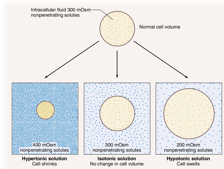
All hypoosmotic solutions are also hypotonic, whereas a hyperosmotic solution can be hypertonic, isotonic, or hypotonic. 所有低渗溶液也都是低张的,而高渗溶液可以是高张的、等张的或低张的。如果溶质分子不能透过细胞膜时,等渗即意味着等张。
2.1.2.5 Endocytosis and Exocytosis
When living cells are observed under a light microscope,regions of the plasma membrane can be seen to fold into the cell, forming small pockets that pinch off to produce intracellular, membrane-bound vesicles that enclose a small volume of extracellular fluid. This process is known as endocytosis. 在光学显微镜下观察活细胞时,可以看到质膜区域折叠进⼊细胞,形成⼩⼝袋,这些⼝袋会收缩形成细胞内膜结合囊泡,囊泡内会包裹少量细胞外液。这个过程称为内吞作用。
A similar process in the reverse direction,known as exocytosis,occurs when membrane-bound vesicles in the cytoplasm fuse with the plasma membrane and release their contents to the outside of the cell. 当细胞质中的膜结合囊泡与质膜融合并将其内容物释放到细胞外部时,就会发⽣一个相反⽅向的类似过程,称为胞吐作用。
2.1.2.5.1 Transport of Molecules into Cells by Endocytosis
In pinocytosis also known as fluid endocytosis an endocytotic vesicle encloses a small volume of extracellular fluid. (soluble molecules) 在胞饮作用(也称为液体内吞作用)中,内吞囊泡包裹少量细胞外液。(可溶性分子)
In phagocytosis cells engulf bacteria or large particles such as cell debris from damaged tissues.(pathogens or debris) In receptor-mediated endocytosis certain molecules in the extracellular fluid bind to specific proteins on the outer surface of the plasma membrane.(specific ligands) 在吞噬作用中,细胞吞噬细菌或⼤颗粒,如受损组织的细胞碎⽚。(病原体或碎⽚)在受体介导的内吞作用
中,细胞外液中的某些分子与质膜外表面的特定蛋⽩质结合。(特定配体)
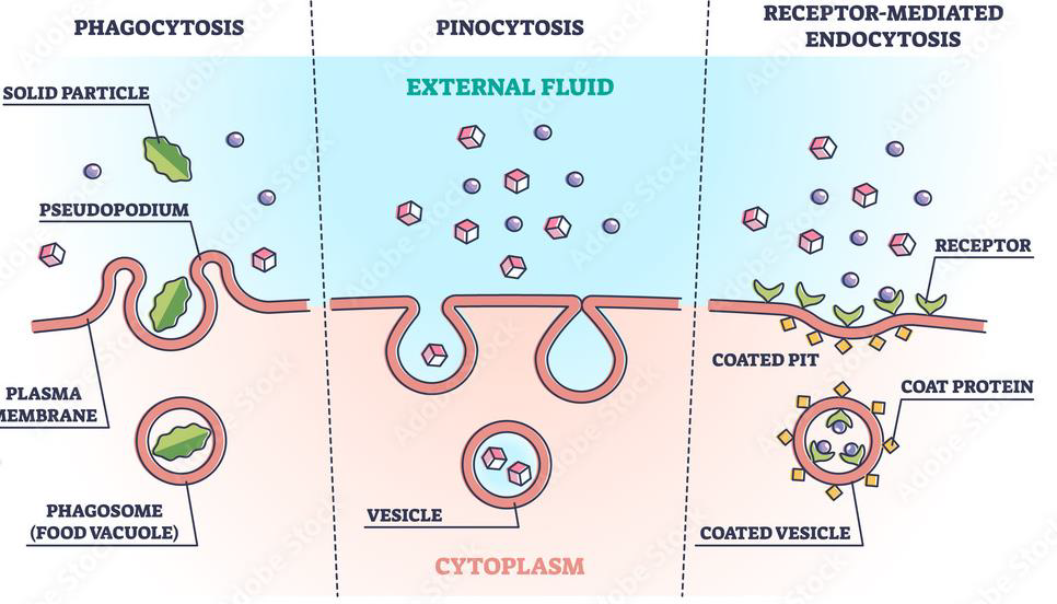
2.1.2.5.2 Transport of Molecules Out of Cells by Exocytosis
Exocytosis is basically endocytosis in reverse: vesicle inside the cell fuses with the plasma membrane and releases its contents into the extracellular fluid.
Exocytosis has three functions:
- to add components to the plasma membrane 向质膜添加成分
- to recycle receptors removed from the plasma membrane by endocytosis 回收通过内吞作用从质膜上去除的受体
- to secrete specific substances out of the cell and into the extracellular fluid 将特定物质分泌出细胞并进⼊细胞外液
2.2 Transmembrane Signal Transductions
2.2.1 Overview
The process by which intercellular chemical signals communicate with cells and thereby elicit a physiological response is called signal transduction. 细胞间化学信号与细胞通讯并从而引起生理反应的过程称为信号转导。
Signal transduction enables coordination of cell function:
- Cell division (proliferation)
- Cell development (differentiation)
- Secretion
- Contraction / relaxation
- Firing of nerves
Cellular Signalling
- Aberrant cellular signalling underlies disease processes 异常细胞信号是疾病过程的基础
- Many medications target to cellular signalling processes 许多药物针对的是细胞信号传导过程
- Improved knowledge of cellular signalling processes continues to identify novel targets for drug design and improved therapy 对细胞信号传导过程的了解不断提高,为药物设计和改善治疗提供了新的靶点
Intercellular Signals
- neurotransmitters and hormones 神经递质和激素
- proteins, e.g. insulin 蛋⽩质,例如胰岛素
- small peptides, e.g. bradykinin ⼩肽,例如缓激肽
- amino acids, e.g. glutamate 氨基酸,例如⾕氨酸
- steroids, e.g. oestrogen, testosterone 类固醇,例如雌激素、睾酮
- vitamins
- fatty acid derivatives, e.g. prostaglandins, leukotrienes 脂肪酸衍⽣物,例如前列腺素、⽩三烯
- gas, e.g. nitric oxide (NO), H2S
2.2.2 Receptors
- typically integral membrane protein at the plasma membrane 通常是质膜上的整合膜蛋⽩
- recognise and bind to specific chemicals (ligands) 识别并结合特定化学物质(配体)
- receptor numbers can be increased (up-regulation) and decreased (down-regulation) 受体数量可以增加(上调)或减少(下调)
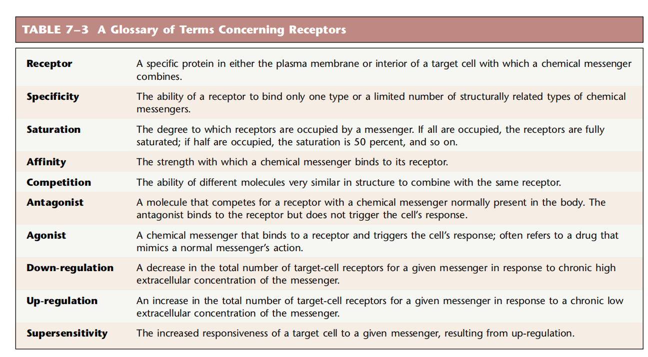
- Specificity: Receptors show specificity for the messenger; that is,they generally bind only one messenger or a class of messengers. 特异性:受体对信使表现出特异性;也就是说,它们通常只与一种信使或一类信使结合
- Affinity: The strength of the binding between a messenger and its receptor 亲和⼒:信使与其受体之间的结合强度
- Saturation 饱和
- Competition 竞争
Ligands 配体
Ligands are chemical messengers which bind to receptor proteins
Agonist – ligand with two important properties 激动剂
Antagonist – ligand which blocks the receptor has affinity but not efficacy 拮抗剂
Six steps of cellular signalling
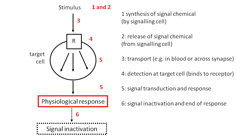
2.2.3 Signal Transduction pathway
Signal transduction pathways: The "signal"is the receptor activation, and “transduction” denotes the process by which a stimulus is transformed into a response.
Signal transduction pathways differ at the very outset for lipid-soluble and lipid-insoluble messengers, the receptors for these two broad chemical classes of messenger are in different locations-the former inside the cell and the latter in the plasma membrane of the cell.
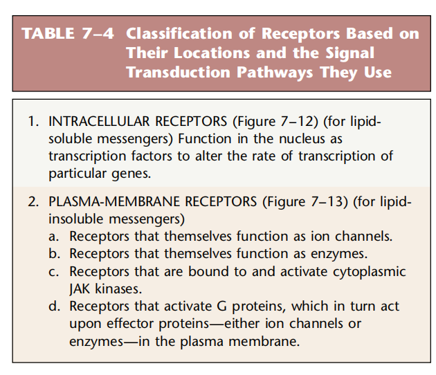
2.2.3.1 Pathways Initiated by Lipid-Soluble Messengers
(Mediated by Intracellular Receptors)
Most lipid-soluble messengers are hormones like steroid hormones, the thyroid hormones,and the steroid derivative, 1,25-dihydroxy vitamin D3. Structurally these hormones are all closely related, and their receptors constitute the steroid hormone receptor “superfamily”. ⼤多数脂溶性信使都是激素,如类固醇激素、甲状腺激素和类固醇衍⽣物 1,25‑二羟基维⽣素 D3。从结构上看,这些激素都密切相关,它们的受体构成类固醇激素受体“超家族”。
The receptors are intracellular and are inactive when no messenger is bound to them; the inactive receptors are mainly in the cell nucleus 受体位于细胞内,当没有信使与其结合时,受体处于⾮活性状态;⾮活性受体主要位于细胞核中
Step of signalling
- A receptor is located in the nucleus, the hormone diffuses into the nucleus and binds to it, forming a hormone-receptor complex. 受体位于细胞核内,激素扩散到细胞核内并与之结合,形成激素‑受体复合物
- Inside the nucleus,the hormone-receptor complex functions as a transcription factor by binding to a region of DNA called the hormone response element (HRE), which is located at the beginning of a specific gene. 在细胞核内,激素受体复合物通过与位于特定基因起始处的称为激素反应元件(HRE)的 DNA 区域结合发挥转录因子的作用
- Binding of the complex to the HRE activates or deactivates the gene, which affects transcription of mRNA and ultimately increases or decreases synthesis of the protein coded by the gene. 复合物与 HRE 的结合会激活或抑制基因,从而影响 mRNA 的转录,最终增加或减少基因编码的蛋⽩质的合成
- The mRNA moves into the cytosol. mRNA 进⼊细胞质
- The mRNA is translated by ribosomes to yield proteins. mRNA 由核糖体翻译产⽣蛋⽩质
Mechanism of Action for Steroid Hormones
- The receptors for steroid hormones are known as nuclear receptors. For the steroid hormone-receptor complex to activate (or deactivate) a particular gene, two complexes must bind to the HRE in a process called dimerization. 类固醇激素受体被称为核受体。类固醇激素受体复合物要激活(或停用)特定基因,必须有两个复合物与 HRE 结合,这个过程称为二聚化
2.2.3.2 Pathways Initiated by Water-Soluble Messengers
(Mediated by Membrane-Bound Receptors)
- Signal transduction mediated by G-protein-linked receptor G蛋⽩连接受体介导的信号转导
- Signal transduction mediated by ionotropic receptor 离子型受体介导的信号转导
- Signal transduction mediated by enzyme-linked receptor 酶联受体介导的信号转导
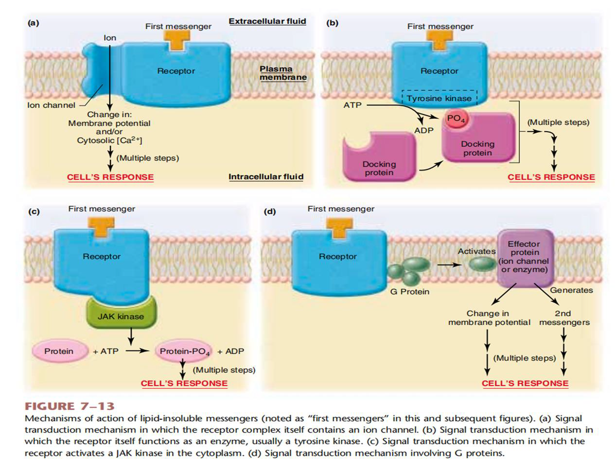
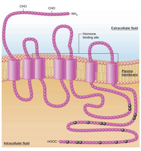
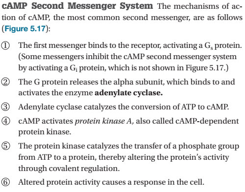
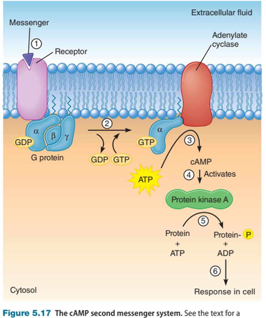
2.2.3.2.1 Signal transduction mediated by G-protein-linked receptor
- The signal transduction mediated by G-protein-linked receptor is achieved by the cascade activities of the membranous receptors,G protein,G protein effector, second messenger and other molecules in cell membrane and cytoplasm. G蛋⽩连接受体介导的信号转导是通过细胞膜和细胞质中的膜性受体、G蛋⽩、G蛋⽩效应物、第二信使等分子的级联作用来实现的
- G-Protein-linked receptor: The structure consisted by a peptide chain that traverses the membrane seven times,belong to the same family: 7- transmembrane receptors G蛋⽩连接受体:由一条穿过膜七次的肽链组成的结构,属于同一家族:7‑跨膜受体
- The conformation of the receptor changes after the ligand binding to them, and then activate G protein. 配体与其结合后,受体构象发⽣改变,从而激活G蛋⽩
G protein
- Bound to the receptor is a protein located on the inner (cytosolic) surface of the plasma membrane and belonging to the family of proteins known as G proteins. 与受体结合的蛋⽩质位于质膜内表面(胞质),属于 G 蛋⽩家族
- The binding of a first messenger to the receptor changes the conformation of the receptor. This change causes one of the three subunits of the G protein to link up with still another plasma-membrane protein, either an ion channel or an enzyme. 第一信使与受体的结合改变了受体的构象。这种变化导致 G 蛋⽩的三个亚基之一与另一个质膜蛋⽩(离子通道或酶)连接起来。
- These ion channels and enzymes are termed plasma membrane effector proteins since they mediate the next steps in the sequences of events leading to the cell’s response. 这些离子通道和酶被称为质膜效应蛋⽩,因为它们介导导致细胞反应的一系列事件的下一步。
G protein effector G蛋⽩效应器
- Enzymes can catalyse the generation of second messengers,such as adenylyl cyclase phospholipase c, guanylyl cyclase… 酶可以催化第二信使的产⽣,例如腺苷酸环化酶、磷脂酶c、⻦苷酸环化酶…
- Ion channels 离子通道
- They mediate the next steps in the sequence of events leading to the cell’s response.
Second messenger
- First messengers: the intercellular chemical messengers that reach the cell from the extracellular fluid and bind to their specific receptors. 第一信使:从细胞外液到达细胞并与特定受体结合的细胞间化学信使
- Second messengers: substances that enter or are generated in the cytoplasm as a result of receptor activation by the first messengers. 第二信使:由于第一信使的受体激活而进⼊细胞质或在细胞质中产⽣的物质
- Serve as chemical relays from the plasma membrane to the biochemical machinery inside the cell.
- CAMP, IP~3~, DG, cGMP, Ca^2+^
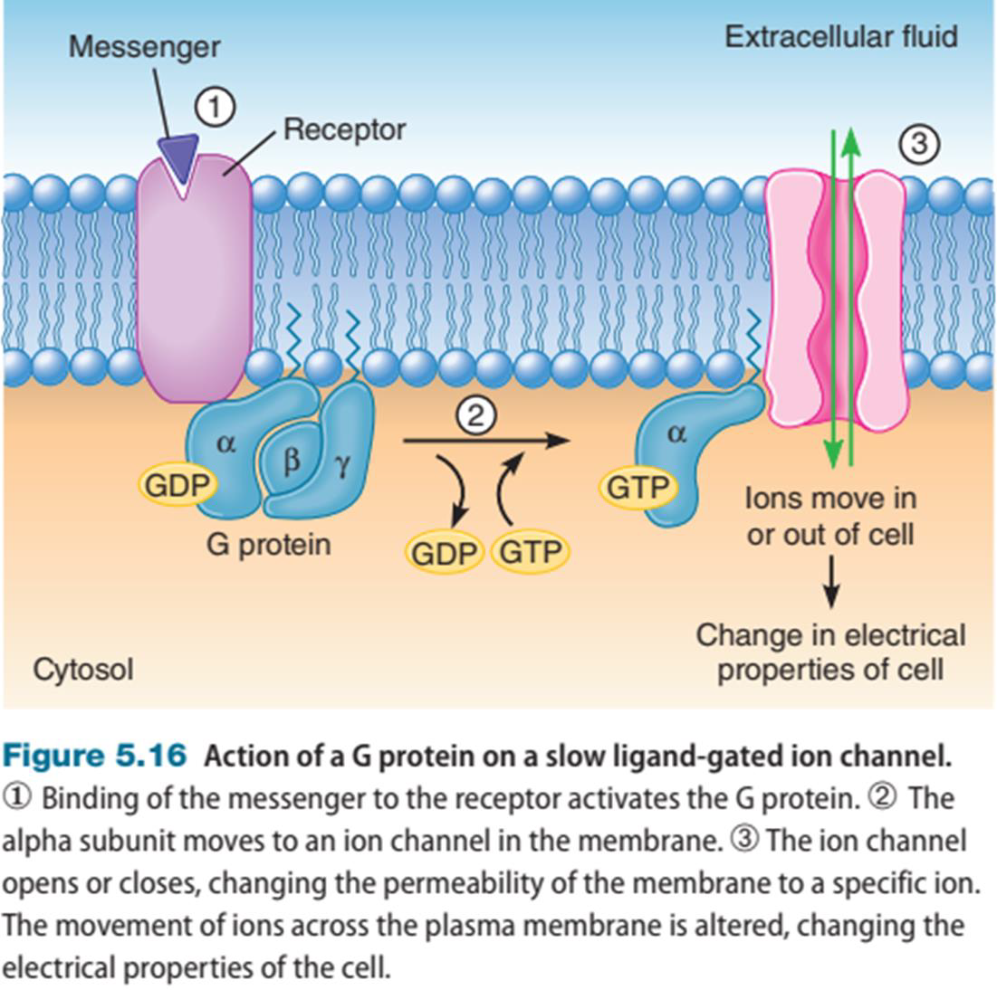
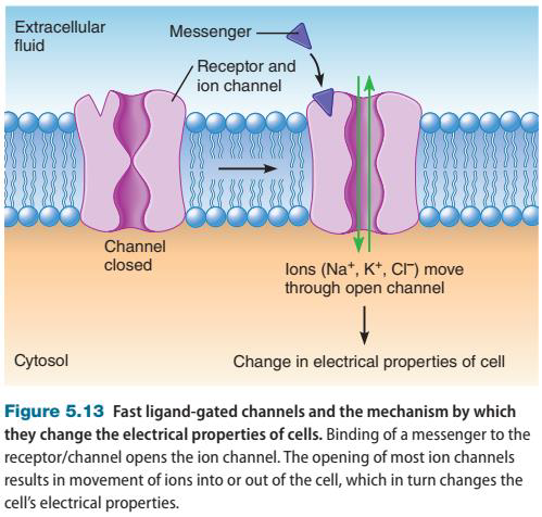
2.2.3.2.2 Signal Transduction Mediated by Ionotropic Receptor
- Ionotropic Receptors are a type of ligand-gated ion channel in which the ligand is a messenger that binds to a receptor.
- The binding of a messenger to the receptor / ion channel causes the channel to open, increasing the membrane’s permeability for that specific ion. 信使与受体/离子通道的结合导致通道打开,从而增加膜对该特定离子的通透性
- Open ion channels allow a specific ion or class of ions to move across the plasma membrane down its electrochemical gradient. 开放的离子通道允许特定离子或一类离子沿着电化学梯度穿过质膜
- Ion movement into or out of the cell can have two different effects on the target cell: (1) Ions entering and leaving can change the electrical properties of the cell and (2) entering ions can interact with proteins inside the cell to induce a response such as muscle contraction, secretion, change in metabolism, or altered transport of a substance. )离子的进⼊和离开可以改变细胞的电特性,进⼊的离子可以与细胞内的蛋⽩质相互作用,引起肌⾁收缩、分泌、代谢变化或物质运输改变等反应。
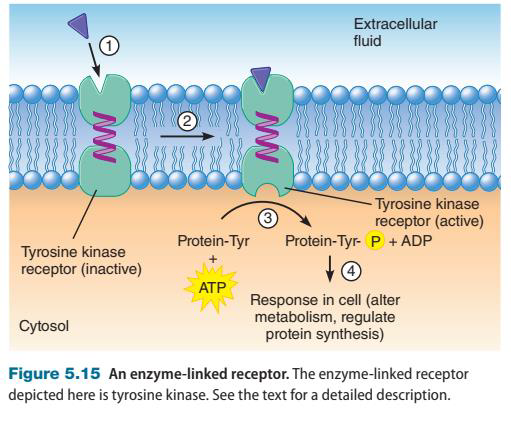
2.2.3.2.3 Signal Transduction Mediated by Enzyme-linked Receptor
- The receptor side faces the interstitial fluid and the enzyme side faces the cytosol 受体一侧朝向间质液,酶一侧朝向细胞溶胶
- A messenger binds to the receptor, changing its conformation.
- The conformation change activates the tyrosine kinase. 构象变化激活酪氨酸激酶
- The tyrosine kinase then catalyses phosphorylation of an intracellular protein. 酪氨酸激酶催化细胞内蛋⽩质的磷酸化
- Phosphorylation of the protein changes its activity by covalent regulation, bringing about a response in the target cell. 蛋⽩质的磷酸化通过共价调节改变其活性,从而引起靶细胞的反应
2.3 Electrical Activities of the Cell
2.3.1 Basic Physical Principles of Bioelectricity
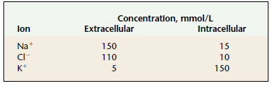
- The predominant solutes in the extracellular fluid are sodium and chloride ions. The intracellular fluid contains high concentrations of potassium ions and ionized nonpenetrating molecules, particularly proteins, with negatively charged side chains and phosphate compounds. 细胞外液中的主要溶质是钠离子和氯离子。细胞内液含有高浓度的钾离子和离子化的⾮渗透性分子,特别是侧链带负电荷的蛋⽩质和磷酸盐化合物
- According to the physical principle, the oppositely charged molecules will move toward each other.
- Plasma membrane separate the electrical charges with opposite sign.

Bioelectricity
- Separated electric charges of opposite sign have the potential to come together, which is called an electric potential (V), or a potential difference because it is determined by the difference in the amount of charge between two points.
- The movement of electric charge is called a current (I). The hindrance to electrical charge movement is known as resistance ®.
- The lipid layers of the plasma membrane (insulator) sperate two aqueous compartments (conductor): the intracellular fluid and the extracellular fluid.
2.3.2 Resting Potential
- All living cells have a potential difference across their plasma membrane.
- When the cell is at rest this potential difference is called the resting membrane potential of the cell A tiny excess of negative ions inside the cell and an excess of positive ions outside. 当细胞处于静⽌状态时,这种电位差称为细胞的静息膜电位。细胞内部有少量负离子过量,而细胞外部有⼤量的正离子过量
- Extracellular fluid is designated as the voltage reference point,and the polarity of the membrane potential is stated in terms of the sign of the excess charge on the inside of the cell by comparison. 以细胞外液为电压参考点,通过⽐较,以细胞内部过量电荷的符号来表⽰膜电位的极性
- The ion concentration difference between inside and outside of a cell is established due to Na^+^/K^+^-ATPase activity.
- One certain ion moves across the membrane creates the potential difference.
- The differences in membrane permeabilities to different ion determines their movement.
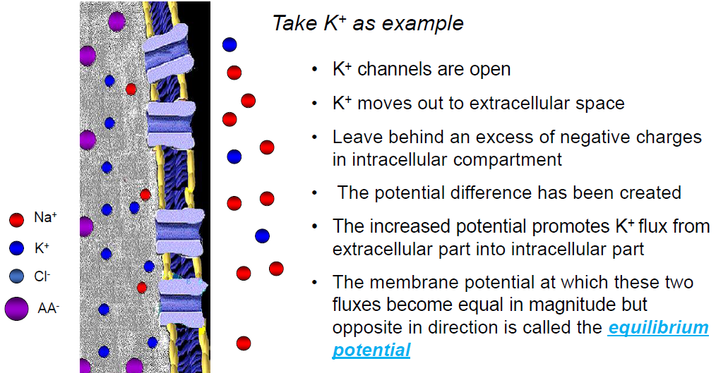
outflux: The concentration gradient (the chemical gradient)
influx: The electrical gradient
The equilibrium potential for K^+^ in cell
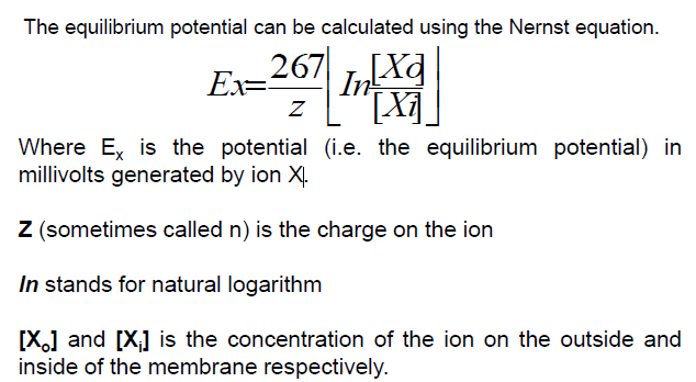
A simple cell has a concentration of 150 mM K^+^ inside and 5 mM K^+^ outside. At these concentrations, the equilibrium potential for K^+^ can be calculated out as -90mV.
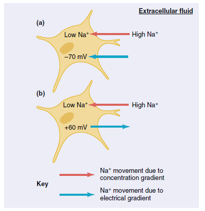
If only Na^+^ channels are open in plasma membrane, Na^+^ flux will move under its electrochemical gradient as similar as K^+^ does, but at reverse direction.
According to the Nernst equation, the Na^+^ flux through open channels will tend to bring the membrane potential toward +60 mV at its typical concentration.
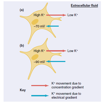
The greater the membrane permeability to an ion species, the greater the contribution that ion species will make to the membrane potential. 膜对离子种类的通透性越⼤,该离子种类对膜电位的贡献越⼤
The neuron compensates for the movement of Na^+^ and K^+^
-
the resting potential is generated across the plasma membrane largely because of the movement of K^+^out of the cell down its concentration gradient through open K^+^channels (called leak K^+^ channels)
-
a small number of open Na^+^ channels does pull the membrane potential slightly toward the Na^+^ equilibrium potential.
-
The Na^+^/K^+^-ATPase pump maintains the resting membrane potential
-
A membrane enzyme actively transports ions to compensate for Na^+^ and K^+^ leaks.
The pump uses the energy of ATP to move Na^+^ and K^+^ against their electrochemical gradients. Three Nat ions are pumped out of the neuron for every two K^+^ ions that are pumped in (electrogenic pump).
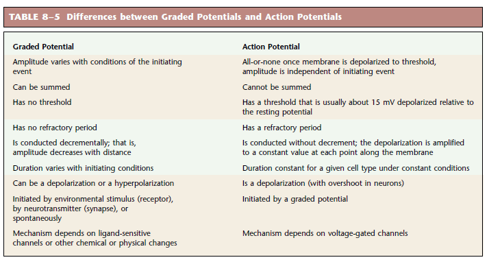
2.3.3 Graded Potentials and Action Potentials
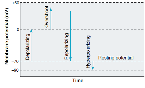
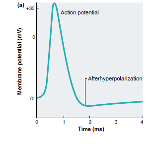
Transient changes in the membrane potential from its resting level produce electric signals. These signals occur in two forms: graded potentials and action potentials.
The membrane is said to be depolarized when its potential is less negative (closer to zero) than the resting level. Overshoot refers to a reversal of the membrane potential polarity-that is, when the inside of a cell becomes positive relative to the outside. When a membrane potential that has been depolarized returns toward the resting value, it is said to be repolarized. The membrane is hyperpolarized when the potential is more negative than the resting level. 当膜电位⽐静息⽔平更低(更接近于零)时,膜被称为去极化。过冲是指膜电位极性的逆转,即当细胞内部相对于外部变为正值时。当去极化的膜电位恢复到静息值时,它被称为复极化。当电位⽐静息⽔平更负时,膜是超极化的。
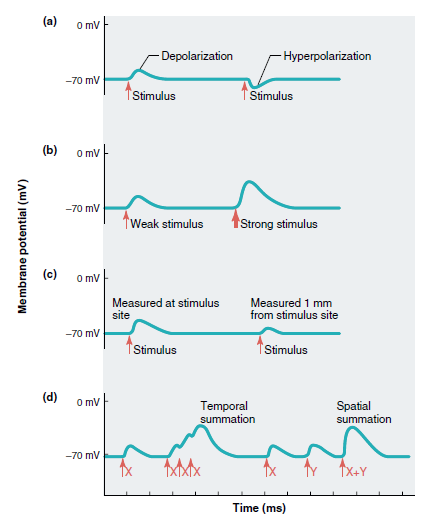
2.3.3.1 Graded Potentials
Result from a net gain of Na^+^ ions
- Small and confined to a localised small region of membrane ⼩且局限于膜的局部⼩区域
- The potential dissipates (in other words the charge leaks away from the membrane) 电位消散(换句话说,电荷从膜中泄漏)
- Ions are diluted down by the bulk of the intracellular fluid and K^+^ moves out to restore the resting membrane potential
- Its size is dependent upon the strength of the stimulus. 它的⼤⼩取决于刺激的强度
- Areas downstream of the initial depolarisation are affected by the movement of these ions (i.e. these areas become depolarized). 初始去极化下游的区域受到这些离子运动的影响(即这些区域变得去极化)
- The larger the potential the further it will tend to travel. 电位越⼤,它趋向于传播得越远
- Depending upon the initiating event, graded potentials can occur in either a depolarizing or hyperpolarizing direction and their magnitude is related to the magnitude of the initiating event. 根据起始事件,分级电位可以发⽣在去极化或超极化⽅向,并且其幅度与起始事件的幅度有关
- The magnitude of the current decreases with the distance from the initial site of the potential change. 电流的⼤⼩随着距电位变化的初始位置的距离而减⼩
- Local current is decremental; that is, its amplitude decreases with increasing distance from the site of origin of the potential. The resulting change in membrane potential from resting level therefore also decreases with the distance from the potential’s site of origin. 局部电流是递减的,也就是说,其幅度随着与电位起源点距离的增加而减⼩。因此,膜电位相对于静息⽔平的变化也随着与电位起源点距离的增加而减⼩
2.3.3.2 The Action Potential
2.3.3.2.1 Properties of Action Potentials
Nerve and muscle cells as well as some endocrine, immune, and reproductive cells have plasma membranes capable of producing action potentials. These membranes are called excitable membranes, and their ability to generate action potentials is known as excitability. Whereas all cells are capable of conducting graded potentials, only excitable membranes can conduct action potentials. The propagation of action potentials is the mechanism used by the nervous system to communicate over long distances. 神经细胞、肌⾁细胞以及一些内分泌细胞、免疫细胞和⽣殖细胞具有能够产⽣动作电位的质膜。这些膜称为可兴奋膜,其产⽣动作电位的能⼒称为兴奋性。虽然所有细胞都能够传导分级电位,但只有可兴奋膜才能传导动作电位。动作电位的传播是神经系统用于长距离通信的机制。
2.3.3.2.2 Voltage-Gated Ion Channels
Na^+^and K^+^channels (voltage-gated) are similar in having sequences of charged amino acid residues in their structure that make the channels reversibly change shape in response to changes in membrane potential. When the membrane is at a negative potential (for example, at the resting membrane potential), both types of channels tend to close, whereas membrane depolarization tends to open them. ,它们的结构中都有带电氨基酸残基序列,这使得通道能够响应膜电位的变化而可逆地改变形状。当膜处于负电位(例如,处于静息膜电位)时,这两种类型的通道都倾向于关闭,而膜去极化则倾向于打开它们。

2.3.3.2.3 Action Potential Mechanism
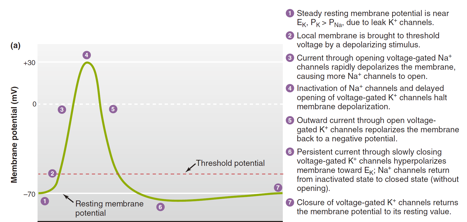
- Steady resting membrane potential is near E~K~, P~K~ > P~Na~, due to leak K^+^ channels. 由于 K^+^ 通道泄漏,稳定静息膜电位接近 E~K~、P~K~ > P~Na~。
- Local membrane is brought to threshold voltage by a depolarizing stimulus. 局部膜通过去极化刺激达到阈值电压。
- Current through opening voltage-gated Na^+^ channels rapidly depolarizes the membrane, causing more Na^+^ channels to open. 通过打开电压门控 Na^+^ 通道的电流使膜快速去极化,导致更多 Na^+^ 通道打开。
- Inactivation of Na^+^ channels and delayed opening of voltage-gated K^+^ channels halt membrane depolarization. Na^+^ 通道失活和电压门控 K^+^ 通道延迟打开会阻止膜去极化。
- Outward current through open voltage-gated K^+^ channels repolarizes the membrane back to a negative potential. 通过开放电压门控 K^+^ 通道的外向电流使膜重新极化回负电位。
- Persistent current through slowly closing voltage-gated K^+^ channels hyperpolarizes membrane toward E~K~; Na^+^ channels return from inactivated state to closed state (without opening). 通过缓慢关闭电压门控 K^+^ 通道的持续电流使膜向 E~K~ 超极化; Na^+^通道从失活状态返回到关闭状态(不打开)。
- Closure of voltage-gated K^+^ channels returns the membrane potential to its resting value. 电压门控 K^+^ 通道的关闭使膜电位恢复至其静息值。
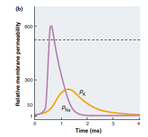
The depolarizing phase of the action potential is due to the opening of voltage-gated sodium channels, which increases the membrane permeability to sodium ions several hundredfold. (1) the sodium channels that opened during the depolarization phase undergo inactivation near the peak of the action potential, which causes them to close; and (2) voltage-gated potassium channels, which open more slowly than sodium channels,open in response to the depolarization. 动作电位的去极化相是由于电压⻔控钠通道的开放,这使膜对钠离子的通透性增加了几百倍。(1)在去极化阶段打开的钠通道在动作电位峰值附近失活,导致其关闭;(2)电压⻔控钾通道⽐钠通道打开得慢,在去极化反应中打开。
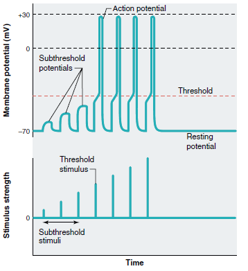
Threshold and the All-or-None Response
- Action potentials occur only when the net movement of positive charge through ion channels is inward. The membrane potential at which this occurs is called the threshold potential, and stimuli that are just strong enough to depolarize the membrane to this level are threshold stimuli. 动作电位仅当正电荷通过离子通道的净运动向内时才会发⽣。发⽣这种情况的膜电位称为阈值电位,强度刚好足以使膜去极化到这个⽔平的刺激是阈值刺激。
- The threshold of most excitable membranes is about 15 mV less negative than the resting membrane potential. Thus, if the resting potential of a neuron is 70 mV, the threshold potential may be 55 mV. ⼤多数可兴奋膜的阈值⽐静息膜电位低约 15 mV。因此,如果神经元的静息电位为 70 mV,则阈值电位可能为 55 mV。
- At depolarizations less than threshold, outward potassium movement still exceeds sodium entry, and the positive-feedback cycle cannot get started despite the increase in sodium entry. In such cases, the membrane will return to its resting level as soon as the stimulus is removed, and no action potential is generated. These weak depolarizations are subthreshold potentials, and the stimuli that cause them are subthreshold stimuli. 当去极化低于阈值时,钾离子向外运动仍超过钠离子进⼊,尽管钠离子进⼊增加,正反馈循环仍无法启动。在这种情况下,一旦刺激消失,膜就会恢复到静息⽔平,不会产⽣动作电位。这些弱去极化是阈下电位,引起它们的刺激是阈下刺激。
- Changes in the membrane potential with increasing strength of depolarizing stimulus. When the membrane potential reaches threshold, action potentials are generated. Increasing the stimulus strength above threshold level does not cause larger action potentials. 将刺激强度增加到阈值以上不会引起更⼤的动作电位。
- Action potentials are all-or-none. The actual shape and amplitude of the action potential depends on the membrane conditions existing at a given time. 动作电位是全有或全无的。动作电位的实际形状和幅度取决于给定时间存在的膜条件。
- A single action potential cannot convey information about the magnitude of the stimulus that initiated it. 单个动作电位无法传达有关引发该动作电位的刺激幅度的信息。
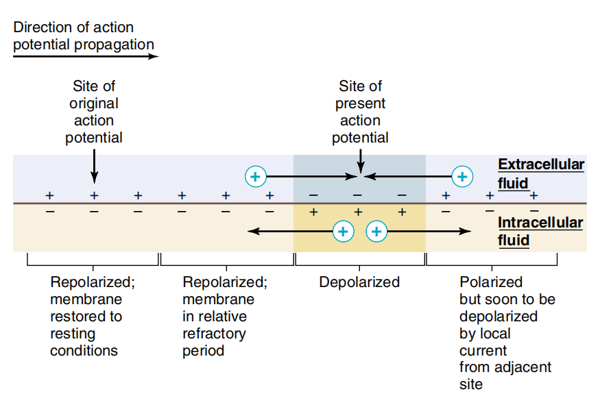
2.3.3.2.4 Refractory Periods
- During the action potential, a second stimulus, no matter how strong, will not produce a second action potential, and the membrane is said to be in its absolute refractory period. (the voltage-gated Na^+^ channels are inactivated) 在动作电位期间,第二个刺激无论多强都不会产⽣第二个动作电位,并且膜被称为处于绝对不应期(电压⻔控 Na^+^ 通道失活)
- Following the absolute refractory period, there is an interval during which a second action potential can be produced, but only if the stimulus strength is considerably greater than usual. This is the relative refractory period, which can last 10 to 15 ms or longer in neurons and coincides roughly with the period of afterhyperpolarization. (only partial Na^+^ channels are recovered and K^+^ permeability is still above resting period.) 绝对不应期之后,有一个间隔,在此期间可以产⽣第二个动作电位,但前提是刺激强度远高于平常。这是相对不应期,在神经元中可持续 10 到 15 毫秒或更长时间,⼤致与超极化后期相吻合(只有部分 Na^+^ 通道恢复,K^+^ 通透性仍高于静息期。)
- Supernormal period is most of after-depolarization potential when the threshold intensity is lower than normal value resulting from the membrane potential close to the threshold potential, and the action potential size is still less than the normal. 超正常期是指膜电位接近阈值电位时,其阈值强度低于正常值而引起的后去极化电位,而动作电位⼤⼩仍⼩于正常。
- Subnormal period is the last stage coincides roughly with the period of after-hyperpolarization. Large stimulus is needed to depolarize the membrane above the threshold potential due to the hyperpolarized membrane potential. 亚正常期是最后一个阶段,⼤致与超极化后期相吻合。由于膜电位超极化,需要很⼤的刺激才能使膜去极化到阈值电位以上。
2.3.3.2.5 Action Potential Propagation
How does the action potential move along the length of the axon? 动作电位如何沿着轴突的长度移动?
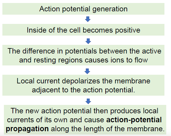
Propagation of Action Potentials in Unmyelinated Axons 无髓鞘轴突中动作电位的传播
- Action potential doesn’t move but “sets off” a new action potential in the region of the axon just ahead of it. 动作电位不会移动,但会在其前⽅的轴突区域“引发”新的动作电位。
- Excitable membranes are able to conduct action potentials in either direction, the direction of propagation being determined by the stimulus location. 可兴奋膜能够向任一⽅向传导动作电位,传播⽅向由刺激位置决定。
- The larger the fibre diameter, the faster the action potential propagates. 纤维直径越⼤,动作电位传播越快。
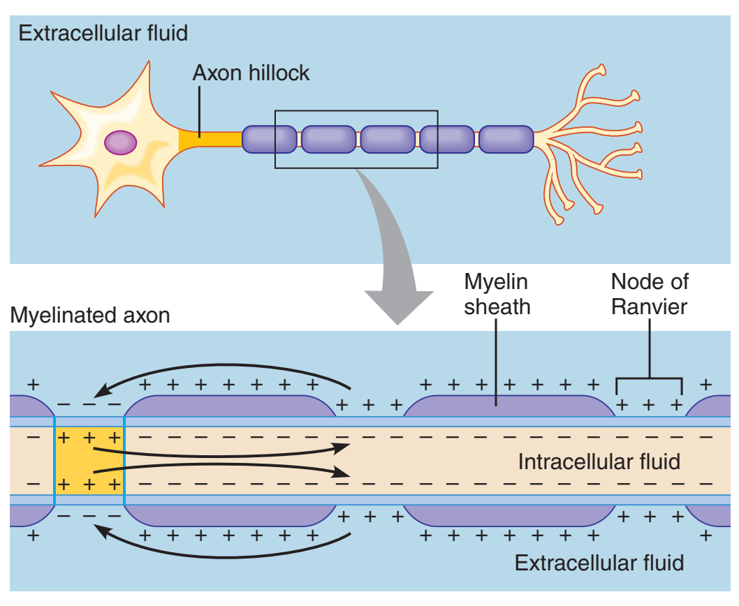
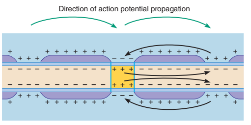
Why does myelination speed up conduction? (Saltatory Conduction) 为什么髓鞘形成会加速传导?(跳跃传导)
- In axons that are sheathed in myelin, action potentials are propagated by a specialized type of electrotonic conduction called saltatory conduction. 在被髓鞘包裹的轴突中,动作电位通过一种特殊类型的电紧张传导(称为跳跃传导)传播。
- The nodes of Ranvier are gaps in the myelin where the axon membrane lacks insulation, is exposed to the interstitial fluid, and has a high concentration of voltage gated sodium and potassium channels. 郎⻜⽒结是髓鞘中的空隙,其中轴突膜缺乏绝缘,暴露于间质液,并且具有高浓度的电压⻔控钠通道和钾通道。
- The separation of charge in the intracellular fluid causes current to flow from one node of Ranvier to the next. 细胞内液中电荷的分离导致电流从一个郎⻜⽒结流向另一个郎⻜⽒结。
2.3.4 Synapses
Transmitter 神经递质:
- diffusible molecule 扩散分子
- released from nerve terminal 从神经末梢释放
- triggers response in innervated cell 触发受支配细胞的反应
Receptor:
- chemical group on cell surface
- part of protein molecule
- binds transmitter
- mediates response
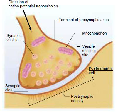
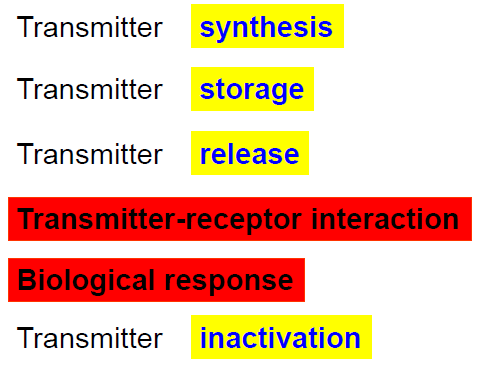
- Calcium channels open when the axon terminal is depolarized. 当轴突末端去极化时,钙通道开放
- More calcium ions flow into the axon terminal. 更多的钙离子流⼊轴突末端
- Calcium causes synaptic vesicles to release the neurotransmitters into the synaptic cleft. 钙导致突触⼩泡释放神经递质进⼊突触间隙
- Neurotransmitter molecules bind to receptors and 神经递质分子与受体结合
- induce next response.
- Some neurotransmitter molecules are degraded by enzymes. 一些神经递质分子被酶降解
- Neurotransmitter molecules can be reuptaked into the presynaptic neuron 神经递质分子可以被重新吸收到突触前神经元中
- others simply diffuse out of the cleft. 其余的则从裂缝中扩散出来
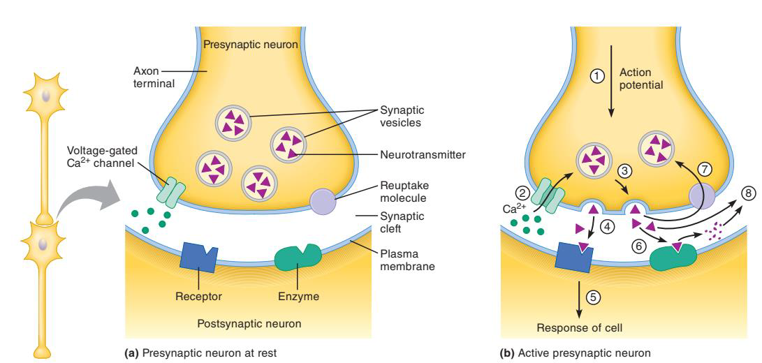
Signal Transduction Mechanisms at Chemical Synapses 化学突触的信号转导机制
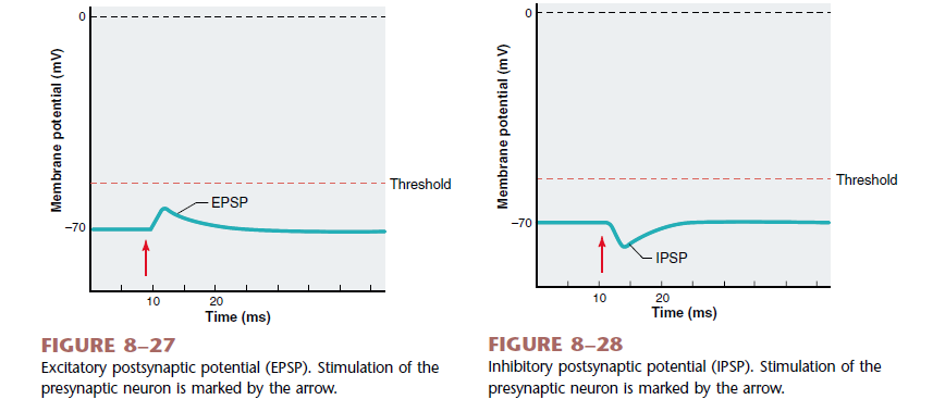
- The binding of the neurotransmitter opens the ion channel and changes the electrical properties of the postsynaptic neuron. The typical response is a change in the membrane potential, called a postsynaptic potential (PSP). 神经递质的结合会打开离⼦通道并改变突触后神经元的电特性。典型的反应是膜电位的变化,称为突触后电位 (PSP)
- An excitatory synapse is one that brings the membrane potential of the postsynaptic neuron closer to the threshold for generating an action potential. This depolarization, which is called an excitatory postsynaptic potential (EPSP). 兴奋性突触使突触后神经元的膜电位更接近产生动作电位的阈值。这种去极化被称为兴奋性突触后电位 (EPSP)
- An inhibitory synapse is one that decreases the likelihood that an action potential will be generated in the postsynaptic neuron. When a neurotransmitter causes potassium channels to open,potassium will move out of the cell, hyperpolarizing it. This hyperpolarization is called an
inhibitory postsynaptic potential (IPSP) . 抑制性突触会降低突触后神经元产生动作电位的可能性。当神经递质导致钾通道打开时,钾会移出细胞,使细胞超极化。这种超极化称为抑制性突触后电位 (IPSP)
2.4 Muscular Contraction
2.4.1 Skeletal Characteristics
The contractile cells of the body can be classified into three major groups based on their shape, number and position of nuclei, presence of striations and whether they are under voluntary or involuntary control. 根据收缩细胞的形状、细胞核的数量和位置、是否有条纹以及是否受自主或⾮自主控制,可将人体的收缩细胞分为三大类。
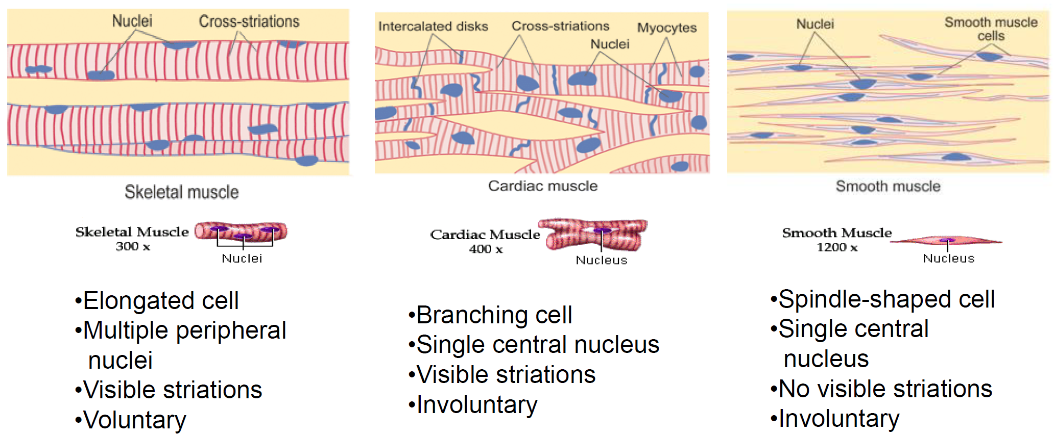
The term muscle refers to a number of muscle fibres bound together by connective tissue. Muscles are usually linked to bones by bundles of collagen fibres known as tendons, which are located at each end of the muscle. 肌⾁一词指的是通过结缔组织结合在一起的多条肌⾁纤维。肌⾁通常通过位于肌⾁两端的胶原纤维束(称为肌腱)与⻣骼连接。
2.4.1.1 Skeletal Muscle
- The most striking feature seen when observing skeletal-or cardiac-muscle fibres through a light microscope is a series of light and dark bands perpendicular to the long axis of the fibre. Because of this characteristic banding, both types are known as striated muscle. ⽤光学显微镜观察⻣骼肌或⼼肌纤维时,最显著的特征是一系列垂直于纤维长轴的明暗带。由于这种特征性带状结构,这两种类型的肌纤维都被称为横纹肌。
- The striated pattern in skeletal and cardiac fibres results from the arrangement of numerous thick and thin filaments in the cytoplasm into approximately cylindrical bundles (1 to 2 um in diameter) known as myofibrils. ⻣骼纤维和⼼脏纤维中的条纹图案是由细胞质中⽆数根粗细不同的细丝排列成近似圆柱形的束(直径 1 至 2 微米),称为肌原纤维。
- Most of the cytoplasm of a fibre is filled with myofibrils,each of which extends from one end of the fibre to the other and is linked to the tendons at the ends of the fibre. 纤维的细胞质中大部分充满着肌原纤维,每根肌原纤维从纤维的一端延伸到另一端,并与纤维末端的肌腱相连。
2.4.1.2 Muscle fibre (single muscle cell) 肌纤维(单个肌细胞)
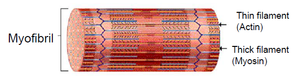
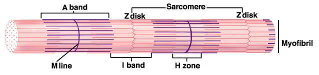
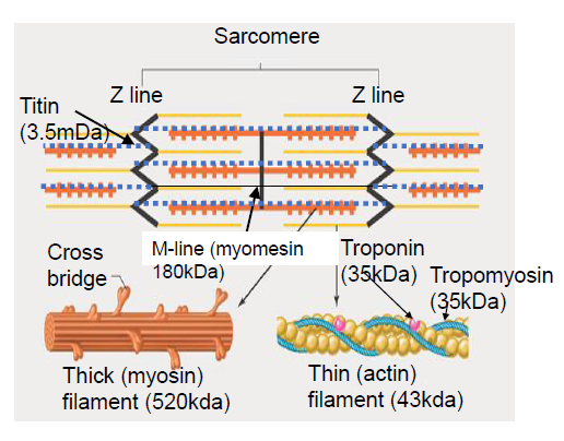
The thick and thin filaments in each myofibril are arranged in a repeating pattern along the length of the myofibril. One unit of this repeating pattern is known as a sarcomere. 每根肌原纤维中的粗肌丝和细肌丝沿着肌原纤维的长度以重复的方式排列。这种重复模式的一个单位称为肌节。
Structure of Myofibril 肌原纤维的结构
- The thick filaments are composed almost entirely of the contractile protein myosin. 粗丝几乎完全由收缩蛋白肌球蛋白组成。
- The thin filaments (which are about half the diameter of the thick filaments) contain the contractile protein actin, as well as to two other proteins - troponin and tropomyosin - that play important roles in regulating contraction. 细丝(其直径约为粗丝的一半)含有收缩蛋白肌动蛋白,以及其他两种蛋白质肌钙蛋白和原肌球蛋白,它们在调节收缩中发挥重要作⽤。
Arrangements of Myofilaments 肌丝的排列
- The arrangement of thick and thin myofilaments forms the light and dark bands (striations) along the myofibril. 粗肌丝和细肌丝的排列沿着肌原纤维形成明带和暗带(条纹)。
- The thick filaments are located in the middle of each sarcomere, where their orderly parallel arrangement produces a wide, dark band known as the A band. 粗肌丝位于每个肌节的中间,它们有序的平行排列形成一条宽而暗的带,称为 A 带。
- Each sarcomere contains two sets of thin filaments, one at each end. One end of each thin filament is anchored to a network of interconnecting proteins known as the Z line, whereas the other end overlaps a portion of the thick filaments. 每个肌节包含两组细丝,每组位于两端。每组细丝的一端锚定在称为 Z 线的互连蛋白质⽹络上,而另一端与粗丝的一部分重叠。
Structure of Myofibril 肌原纤维的结构
- A light band, known as the I band, lies between the ends of the A bands of two adjacent sarcomeres and contains those portions of the thin filaments that do not overlap the thick filaments. 一条亮带(称为 I 带)位于两个相邻肌节的 A 带末端之间,包含不与粗肌丝重叠的细肌丝部分。
- The H zone is a narrow light band in the centre of the A band. It corresponds to the space between the opposing ends of the two sets of thin filaments in each sarcomere. H 区是 A 带中⼼的一条窄光带。它对应于每个肌节中两组细丝相对两端之间的空间。
- A narrow, dark band in the centre of the H zone is known as the M line and corresponds to proteins that link together the central region of the thick filaments. H 区中⼼的一条狭窄的暗带被称为 M 线,对应于连接粗丝中⼼区域的蛋白质。
2.4.2 Mechanism of striated muscle contraction
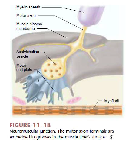
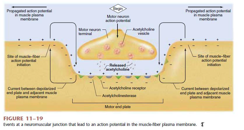
2.4.2.1 Neuromuscular Junction
The Sliding Filament Theory: The contraction of a muscle cell occurs as the thin filaments slide past the thick filaments.
-
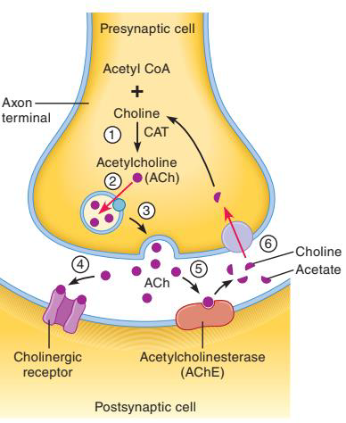
Ach is synthesized in the cytosol of the axon terminals of neurons. Ach 在神经元轴突末端的胞质溶胶中合成。

-
Ach is transported into and stored in synaptic vesicles. Ach 被运输到突触小泡中并储存在其中。
-
An action potential triggers its release by exocytosis into synapse cleft. 动作电位触发其通过胞吐作⽤释放到突触间隙。
-
Ach can bind to receptors on the postsynaptic cell. Ach可以与突触后细胞上的受体结合。
-
Ach can be degraded into choline and acetate by an enzyme called acetylcholinesterase (AChE). 乙酰胆碱可以被一种叫做乙酰胆碱酯酶的酶降解为胆碱和醋酸盐。
-
Choline is taken back into the presynaptic cell and can be used to synthesize more acetylcholine. The acetate diffuses away from the synapse and enters the bloodstream. 胆碱被带回突触前细胞,可⽤于合成更多乙酰胆碱。醋酸盐从突触扩散并进⼊⾎液。
2.4.2.2 Excitation–Contraction Coupling 兴奋‑收缩耦合
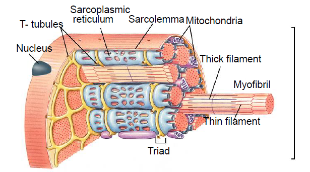
At the end of sarcoplasmic reticulum there are two enlarged regions, known as lateral sacs that are connected to each other by a series of smaller tubular elements. The lateral sacs store the calcium that is released following membrane excitation. 肌浆⽹末端有两个扩大的区域,称为侧囊,它们通过一系列较小的管状元件相互连接。侧囊储存膜兴奋后释放的钙。
A separate tubular structure, the transverse tubule (T tubule), passing between adjacent lateral sacs and eventually joining the plasma membrane The lumen of the T tubule is continuous with the extracellular fluid surrounding the muscle fibre. The membrane of the T tubule is able to propagate action potentials. 横小管(T 小管)是一种独⽴的管状结构,它穿过相邻的侧囊,最终与质膜相连。T 小管的管腔与肌纤维周围的细胞外液相连。T 小管的膜能够传播动作电位。
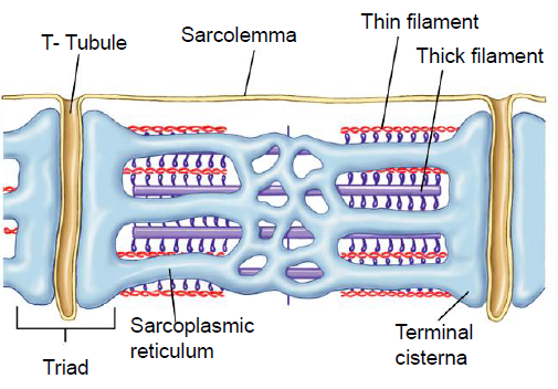
- Once initiated in the plasma membrane,an action potential is rapidly conducted over the surface of the fibre and into its interior by way of the T tubules. 一旦在质膜上启动,动作电位就会迅速传导通过 T 小管从纤维表面进⼊其内部。
- The action potential in a T tubule adjacent to the lateral sacs activates voltage-gated proteins in the T-tubule membrane that are physically or chemically linked to calcium- release channels in the membrane of the lateral sacs. 毗邻侧囊的 T 小管中的动作电位激活 T 小管膜中的电压⻔控蛋白,这些蛋白在物理或化学上与侧囊膜中的钙释放通道相连。
- Depolarization of the T tubule by an action potential thus leads to the opening of the calcium channels in the lateral sacs, allowing calcium to diffuse from the calcium-rich lumen of the lateral sacs into the cytosol. The rise in cytosolic calcium concentration is normally enough to turn on all the cross bridges in the fibre. 动作电位使 T 小管去极化,从而导致侧囊中的钙通道打开,使钙从侧囊富含钙的腔内扩散到细胞质中。细胞质钙浓度的升⾼通常足以打开纤维中的所有横桥。
- A contraction continues until calcium is removed from troponin,and this is achieved by lowering the calcium concentration in the cytosol back to its pre-release level. 收缩持续直至钙从肌钙蛋白中去除,这是通过将细胞溶胶中的钙浓度降低回其释放前的水平来实现的。
- The cytosolic calcium concentration remains elevated,and the contraction continues for some time after a single action potential. 细胞质钙浓度保持升⾼,并且单个动作电位后收缩会持续一段时间。
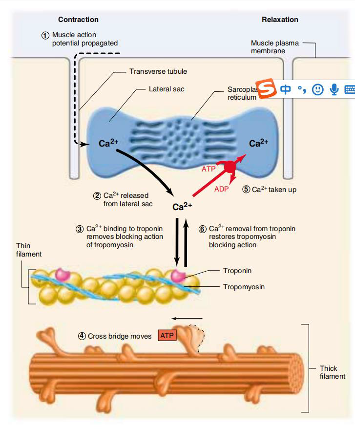
What cytoplasmic Ca^2+^ did? 细胞质 Ca^2+^ 起了什么作⽤?
- An action potential brings about the release of Ca^2+^ from the terminal cisternae of the sarcoplasmic reticulum. 动作电位导致 Ca^2+^ 从肌浆⽹末端池中释放。
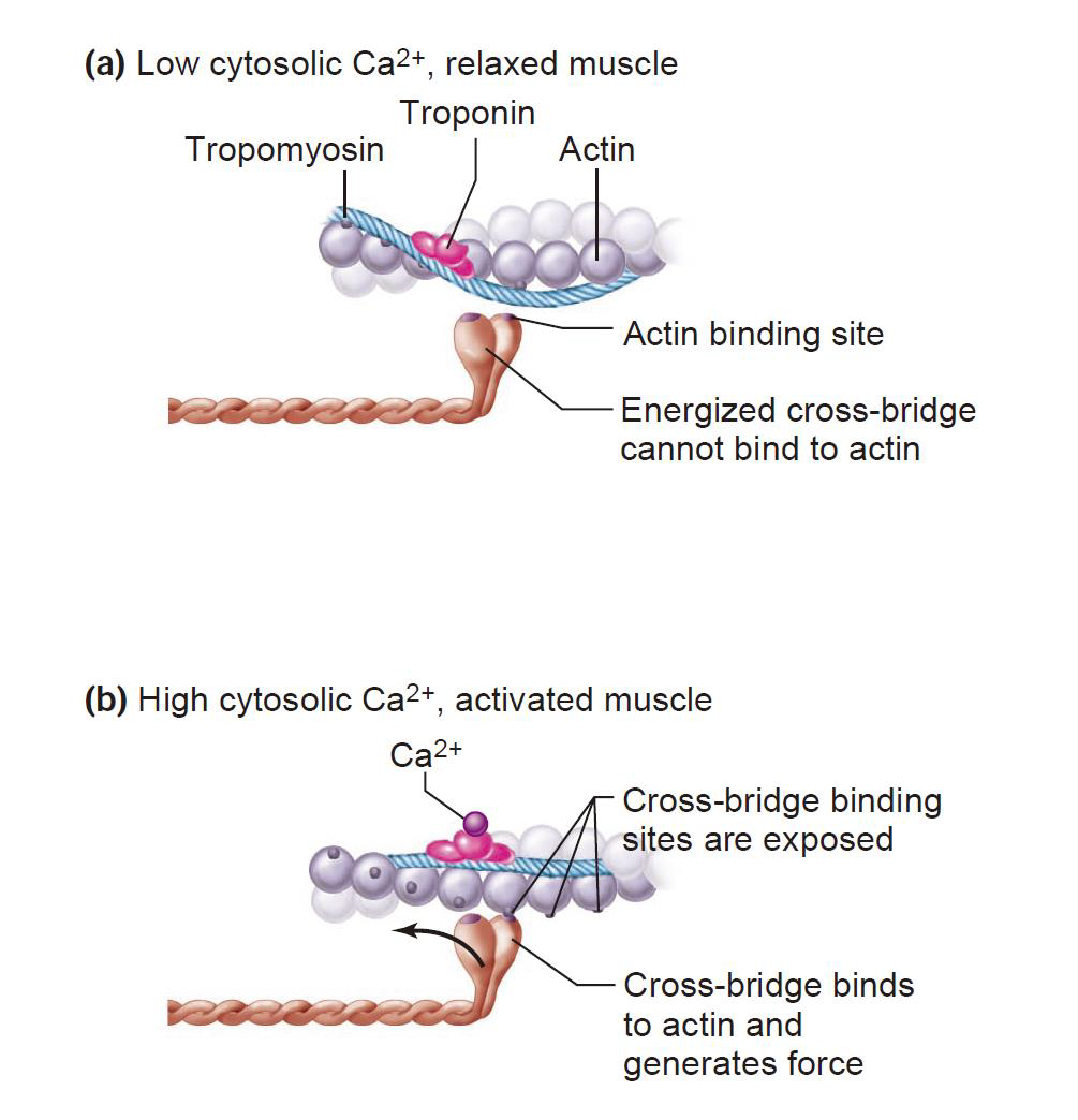
- Ca^2+^ flood into the cytosol and bind to the troponin, causing a change in the conformation of the troponin-tropomyosin complex. Ca^2+^涌⼊细胞质并与肌钙蛋白结合,导致肌钙蛋白‑原肌球蛋白复合物构象发生改变。
- This conformational change exposes the binding sites on actin. 这种构象变化暴露了肌动蛋白上的结合位点。
2.4.2.3 Molecular Mechanisms of Skeletal Muscle Contraction: Sliding-Filament Mechanism
In skeletal muscle myosin molecules are bundled together to form thick filaments. 在⻣骼肌中,肌球蛋白分⼦捆绑在一起形成粗丝。
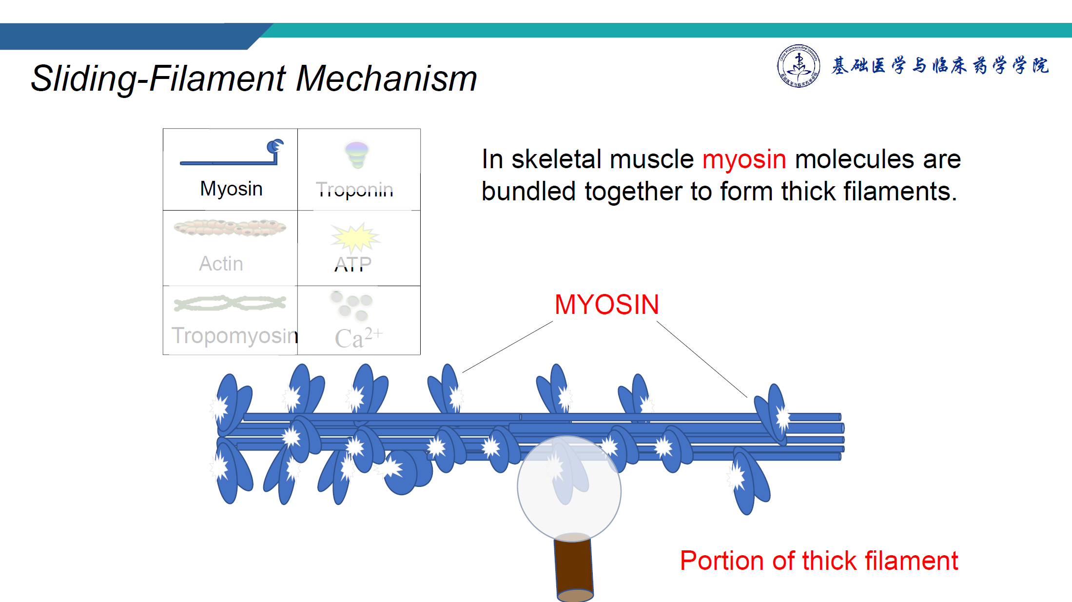
Myosin 肌球蛋白
- The shape of an individual myosin molecule is similar to a golf club with two heads. 单个肌球蛋白分⼦的形状类似于具有两个头的⾼尔夫球杆。
- The heads (cross bridge) have the ability to move back and forth. 头部(横桥)具有前后移动的能⼒。
- The flexing movement of the head provides the 'power for muscle contraction. 头部的屈曲运动为肌⾁收缩提供了动⼒。
- The hinge portion of the tail allows for movement so that the cross bridge can bind to actin (thin filament). 尾部的铰链部分允许运动,以便横桥可以与肌动蛋白(细丝)结合。
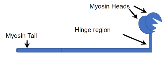
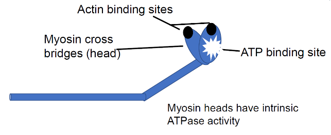
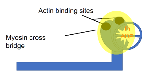
The cross bridge has two important binding sites 肌动蛋白结合位点 ATP结合位点. One site specifically binds ATP. Note the position of the cross bridge. This is called the low-energy conformation 低能构象. The binding of ATP transfers energy to the myosin cross bridge as ATP is hydrolysed into ADP and Pi. The binding site on the myosin cross bridge has a strong attraction for binding to actin.
Actin 肌动蛋白
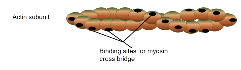
- The thin filament consists of actin subunits arranged into a double helical chain. Each actin subunit has a specific binding site to which myosin cross bridges bind. 细丝由排列成双螺旋链的肌动蛋白亚基组成。每个肌动蛋白亚基都有⼀个特定的结合位点,肌球蛋白交叉桥可以与其结合。
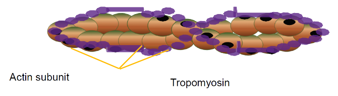
Tropomyosin 原肌球蛋白
The regulatory protein tropomyosin is also part of the thin filament. Tropomyosin entwines around the actin. In the resting muscle,tropomyosin covers the binding sites on the actin subunits and prevents myosin cross bridge binding. 调节蛋白原肌球蛋白也是细丝的⼀部分。原肌球蛋白缠绕在肌动蛋白周围。在静息肌⾁中,原肌球蛋白覆盖肌动蛋白亚基上的结合位点并阻止肌球蛋白横桥结合。
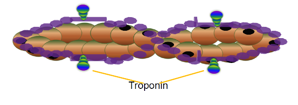
Troponin 肌钙蛋白
To expose the binding sites for binding with myosin, the tropomyosin molecule must be moved aside. This is achieved by a third molecular complex called troponin. Troponin is attached and spaced periodically along the tropomyosin strand.
Calcium ions
Action potentials cause calcium ions to be released from the terminal cisternae, these bind to troponin. A conformational change occurs in the tropomyosin-troponin complex,which moves the tropomyosin strands away from the binding sites.
Cross-bridge cycle 过桥循环
- attachment of the cross-bridge to a thin filament; 将横桥附着在细丝上;
- movement of the cross-bridge, producing tension in the thin filament; 横桥移动,在细丝中产生张⼒;
- detachment of the cross-bridge from the thin filament; 横桥与细丝脱离;
- Energizing the cross-bridge so it can again attach to a thin filament and repeat the cycle. 给横桥通电,使其能够再次附着在细丝上并重复该循环。
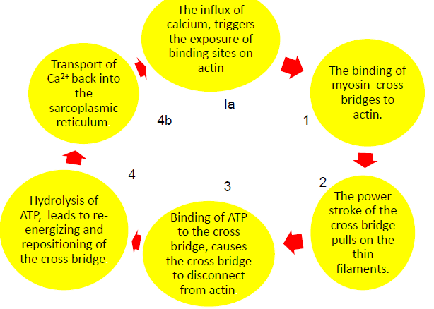
- Binding of Myosin to Actin: When a binding site on actin is exposed, an energised cross bridge can bind to it. The binding of myosin to actin brings about a conformational change in the cross bridge, resulting in the release of inorganic phosphate (Pi). 肌球蛋白与肌动蛋白的结合:当肌动蛋白上的结合位点暴露时,一个带电的横桥可以与其结合。肌球蛋白与肌动蛋白的结合会导致横桥的构象变化,从而释放无机磷酸盐 (Pi)。
- The Power Stroke of the Cross Bridge: The myosin head pivots,pulling the thin filament inward toward the centre of the sarcomere: the power stroke. This conformational change releases ADP from its binding site. Chemical energy of ATP has been transformed into the mechanical energy of movement. 横桥的动力冲程:肌球蛋白头部旋转,将细丝向内拉向肌节中心:动力冲程。这种构象变化会将 ADP 从其结合位点释放出来。ATP 的化学能已转化为运动的机械能。
- Disconnecting the Cross Bridge from Actin: In order to disconnect the cross bridge from actin, and relax the muscle an ATP molecule must bind to its site on the myosin cross bridge. 断开横桥与肌动蛋白的连接:为了断开横桥与肌动蛋白的连接,并放松肌肉,ATP 分子必须结合到肌球蛋白横桥上的位点。
- Re-energising and positioning of the cross bridge: The release of the myosin cross bridge from actin triggers the hydrolysis of the ATP molecule into ADP and Pi. Energy is transferred from ATP to the myosin cross bridge, which returns to its high-energy conformation. 重新给横桥充电并定位:肌球蛋白横桥从肌动蛋白上释放会触发 ATP 分子水解为 ADP 和 Pi。能量从 ATP 转移到肌球蛋白横桥,肌球蛋白横桥恢复到高能构象。
- Myosin Binding Sites on Actin are Hidden: Calcium is actively transported from the cytosol into the sarcoplasmic reticulum by ion pumps. As the calcium is removed,the troponin-tropomyosin complex again covers the binding sited on actin. 肌动蛋白上的肌球蛋白结合位点被隐藏:钙通过离子泵从胞质溶胶主动运输到肌浆网。随着钙被去除,肌钙蛋白-原肌球蛋白复合物再次覆盖肌动蛋白上的结合位点。
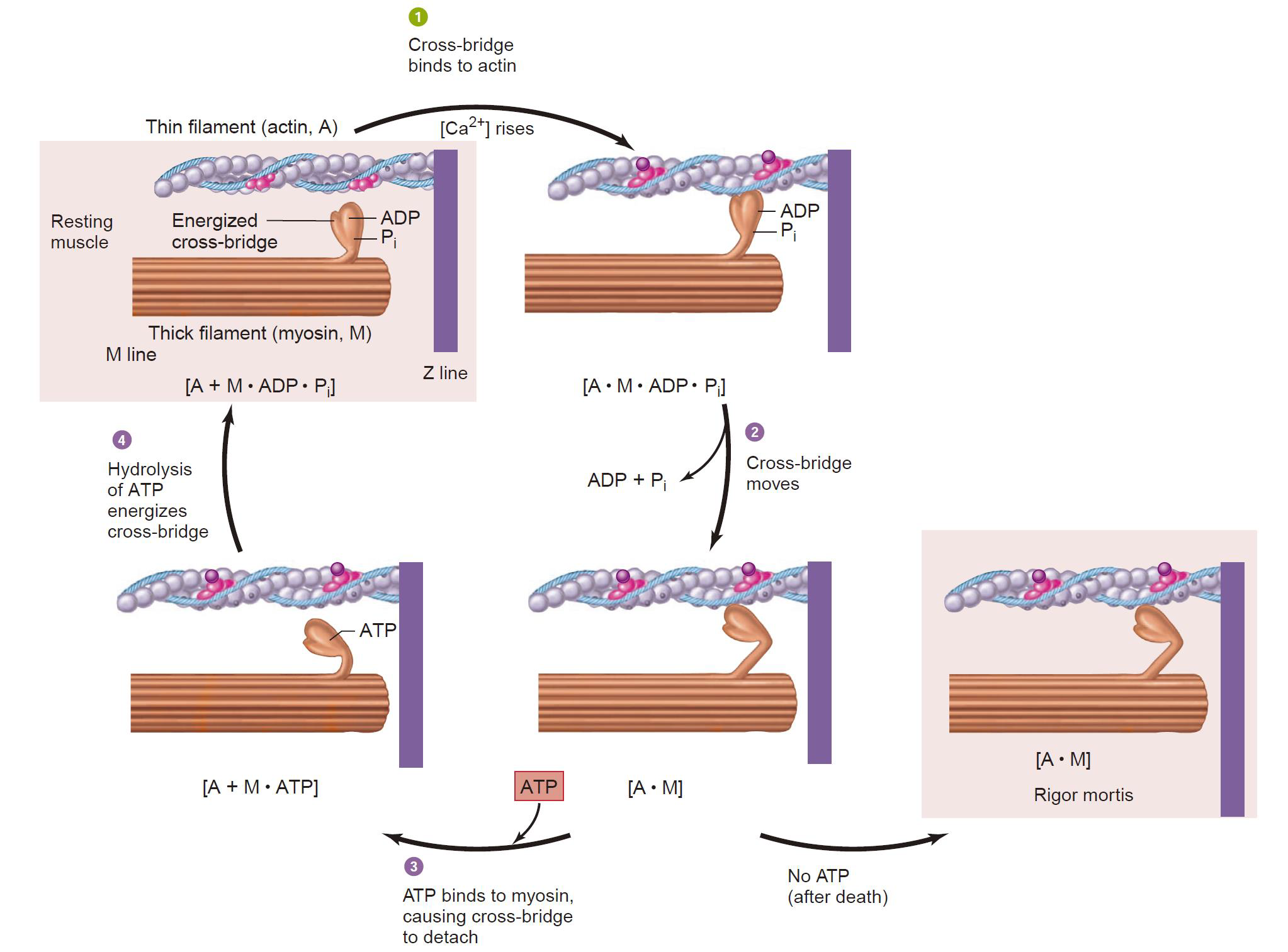
- Multiple Cross Bridge Cycles: Note that during a contraction,all cross bridges are neither bound nor disconnected at the same time 请注意,在收缩期间,所有横桥既不会同时结合也不会同时断开
- Multiple Myofilaments: Several myosin and actin filaments are interacting to demonstrate the sliding filament theory of muscle contraction. Notice that although the sarcomere shortens, the length of each myofilament does not change. However,the width of the H zone changes. 多条肌丝:多条肌球蛋白和肌动蛋白丝相互作用,证实了肌肉收缩的滑动丝理论。请注意,虽然肌节缩短,但每条肌丝的长度没有变化。然而,H 区的宽度发生了变化。

2.4.3 Mechanics of Single-Fiber Contraction
- The force exerted on an object by a contracting muscle is known as muscle tension and the force exerted on the muscle by an object (usually its weight) is the load. 收缩的肌⾁对物体施加的⼒称为肌⾁张⼒,物体(通常是其重量)对肌⾁施加的⼒称为负荷。
- When a muscle develops tension but does not shorten (or lengthen), the contraction is said to be isometric (constant length). 当肌⾁产生张⼒但不会缩短(或拉长)时,这种收缩被称为等长收缩(长度恒定)。
- A contraction in which the muscle shortens or lengthens while the load on the muscle remains constant, is said to be isotonic (constant tension). 当肌⾁负荷保持不变时,肌⾁缩短或伸长收缩,这种收缩被称为等张收缩(恒定张⼒)。
- Muscle tension and load are opposing forces. Whether or not force generation leads to fibre shortening depends on the relative magnitudes of the tension and the load. 肌⾁张⼒与负荷是相反的⼒量,⼒量的产生是否导致纤维缩短,取决于张⼒与负荷的相对大小。
2.4.3.1 Isometric and isotonic contractions
Isometric = same measurement i.e. constant length.
For example, when you exert increasing force say to try to pick up an immovable object. The muscle does not shorten but tension is produced. 例如,当你施加越来越大的⼒量,比如试图拿起一个不可移动的物体时,肌⾁不会缩短,但会产生张⼒。
Isotonic = same tone i.e. same tension.
The muscle shortens but the load against which it contracts is constant, e.g. when you pick up a weight. 肌⾁会缩短,但其收缩所承受的负荷是恒定的,例如当你举起重物时。
- During an isotonic contraction, the cross bridges bound to actin move to their angled positions, causing shortening of the sarcomeres. In contrast, during an isometric contraction, the bound cross bridges are unable to move the thin filaments because of the load on the muscle fibre, but they do exert a force on the thin filaments-isometric tension. 在等张收缩期间,与肌动蛋白结合的横桥会移动到其成⻆度的位置,从而导致肌节缩短。相反,在等长收缩期间,由于肌纤维上的负荷,结合的横桥⽆法移动细丝,但它们会对细丝施加⼒ ‑ 等长张⼒。
- During a lengthening contraction, the cross bridges are pulled backward toward the Z lines by the load while still bound to actin and exerting force. Thus, the chemical changes in the contractile proteins during each type of contraction are the same. 在伸长收缩过程中,横桥在负荷作⽤下向后拉向 Z 线,同时仍与肌动蛋白结合并施加⼒。因此,每种收缩过程中收缩蛋白的化学变
化都是相同的。 - The end result is determined by the magnitude of the load on the muscle.
2.4.3.2 Twitch Contractions
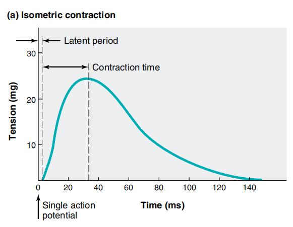
- The mechanical response of a single muscle fibre to a single action potential is known as a twitch. 单个肌⾁纤维对单个动作电位的机械反应称为抽搐。
- Following the action potential, there is an interval of a few milliseconds, known as the latent period. 动作电位之后,有一个几毫秒的间隔,称为潜伏期。
- The time interval from the beginning of tension development at the end of the latent period to the peak tension is the contraction time. 从潜伏期末期开始出现张⼒到张⼒达到峰值的时间间隔为收缩时间。
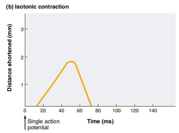
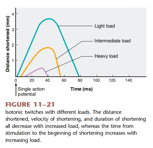
The characteristics of an isotonic twitch depend upon the magnitude of the load being lifted: at heavier loads, the latent period is longer, and the velocity of shortening (distance shortened per unit of time), the duration of the twitch, and the distance shortened are all slower or shorter. 等张抽搐的特征取决于所举起的负荷的大小:负荷越大,潜伏期越长,而缩短速度(单位时间内缩短的距离)、抽搐持续时间和缩短的距离都越慢或越短。
The heavier the load, the longer it takes for the tension to increase to the value of the load, when shortening will begin. If the load on a fibre is increased, eventually a load is reached that the muscle is unable to lift, the velocity and distance of shortening will be zero, and the contraction will become completely isometric. 负荷越重,张⼒增加到负荷值所需的时间就越长,此时肌⾁就会开始缩短。如果纤维上的负荷增加,最终达到肌⾁⽆法举起的负荷,缩短的速度和距离将为零,收缩将完全等长。
2.4.3.3 Frequency-Tension Relation
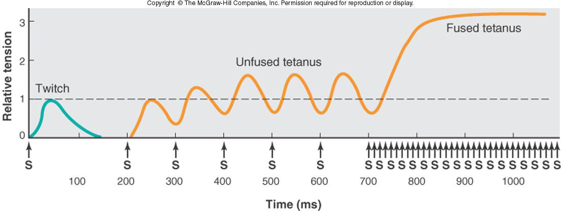
- The increase in muscle tension from successive action potentials occurring during the phase of mechanical activity is known as summation.
- A maintained contraction in response to repetitive stimulation is known as a tetanus (tetanic contraction).
- At low stimulation frequencies, the tension may oscillate as the muscle fibre partially relaxes between stimuli, producing an unfused tetanus (incomplete tetanus). A fused tetanus (complete), with no oscillations, is produced at higher stimulation frequencies.
- Note: depolarisation is conducted along the T- tubules to the SR membrane, within a few milliseconds every sarcomere in a cell contracts simultaneously. 注意:去极化沿着 T 小管传导至 SR 膜,在几毫秒内,细胞中的每个肌节同时收缩。
- Action potentials cannot summate or fuse together because they are all-or-nothing as in nerve.
- However, the contractile responses can summate to form unfused (at around 10 Hz) and fused (at around 100 Hz) tetani.
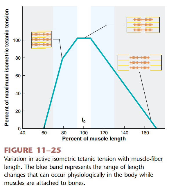
2.4.3.4 Length-Tension Relation
- One can stretch a muscle fibre to various lengths and measure the magnitude of the active tension generated in response to stimulation at each length.
- The amount of active tension developed by a muscle fibre during contraction, and thus its strength, can be altered by changing the length of the fibre before contraction.
- The length at which the fibre develops the greatest isometric active tension is termed the optimal length. 纤维产生最大等长主动张⼒的长度称为最佳长度
2.4.3.5 Whole-Muscle Contraction
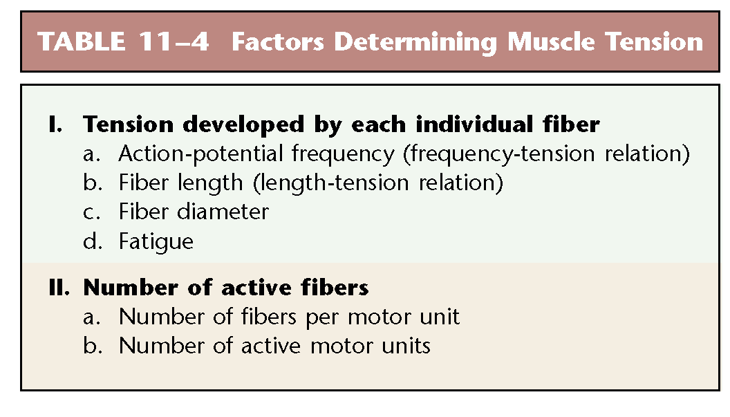
- The total tension a muscle can develop depends upon two factors:
(1) the amount of tension developed by fibre, and (2) the number of fibres contracting at any time. 肌⾁所能产生的总张⼒取决于两个因素:(1)纤维产生的张⼒的大小,(2)随时收缩的纤维数量。 - The number of fibres contracting at any time depends on:
(1) the number of fibres in each motor unit (motor unit size), and (2) the number of active motor units. 任何时候收缩的纤维数量取决于:(1)每个运动单位中的纤维数量(运动单位大小),以及(2)活跃运动单位的数量。 - The process of increasing the number of motor units that are active in a muscle at any given time is called recruitment. It is achieved by increasing the excitatory synaptic input to the motor neurons. The greater the number of active motor neurons, the more motor units recruited, and the greater the muscle tension. 增加肌⾁中某一时刻活跃的运动单位数量的过程称为募集。这是通过增加运动神经元的兴奋性突触输⼊来实现的。活跃的运动神经元数量越多,募集的运动单位就越多,肌⾁张⼒就越大。
Chapter 3 Blood Physiology
3.1 Plasma
General Functions of Blood
- Transportation
• O~2~ & CO~2~
• nutrients and hormones
• metabolic wastes - Regulation
• extracellular fluid pH
• body temperature - Protection
• clotting mechanism 凝⾎机制
• immune defence 免疫防御
Components of Blood
Hematocrit
- 55% plasma
- 45% cells
- 99% RBCs
- < 1% WBCs and platelets
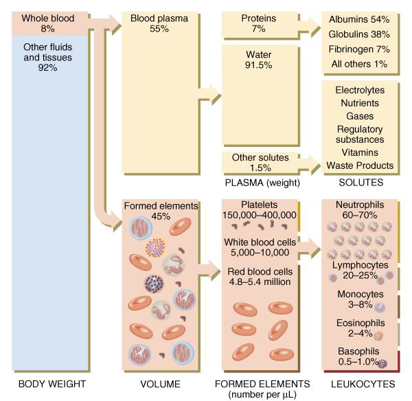
- Percentage of blood occupied by cells
- female normal range: 38 - 46% (average of 42%)
- male normal range: 40 - 54% (average of 46%): testosterone
- Anemia
- not enough RBCs or not enough hemoglobin
- Polycythemia
- too many RBCs (over 65%)
- dehydration, tissue hypoxia, blood doping in athletes
Physical Characteristics of Blood
- Thicker (more viscous) than water and flows more slowly than water
- Temperature of 100.4 degrees F
- pH 7.4 (7.35-7.45)
- 8 % of total body weight
- Blood volume
- 5 to 6 liters in average male
- 4 to 5 liters in average female
- hormonal negative feedback systems maintain constant blood volume and osmotic pressure
Components of Plasma
- Water (>90%) and organic and inorganic substances
- Plasma proteins (65~85g/L)
• Created in liver and confined to bloodstream- albumins: 40~48 g/L: maintain blood osmotic pressure
- globulins: 15~30 g/L
• α1-, α2-, β-, γ- globulins
• Antibodies bind to antigens - fibrinogen: 2~4 g/L: For clotting
- Nutrients
- Metabolic wastes
- Hormones
- Mineral electrolytes
3.2 The Blood Cells
Hematopoiesis: process of blood cells formation is haematopoiesis or hemopoiesis
- Most blood cells types need to be continually replaced: die within hours, days or weeks
- In the embryo
• occurs in yolk sac, liver, spleen, thymus, lymph nodes & red bone marrow 卵黄囊、肝脏、脾脏、胸腺、淋巴结和红骨髓中 - In adult
• occurs only in red marrow of flat bones like sternum, ribs, skull & pelvis and ends of long bones 仅出现在胸骨、肋骨、头骨和骨盆等扁平骨的红骨髓以及⻓骨的末端
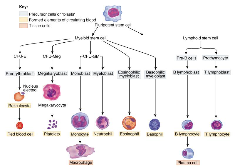
General Function of Blood Cells
- The blood cells are the erythrocytes and the leukocytes, and the cells fragments are the platelets (thrombocytes).
- More than 99% of blood cells are erythrocytes, which transport gas.
- The leukocytes protect against infection and cancer, and the platelets function in blood clotting.
3.2.1 Physiology of Erythrocyte
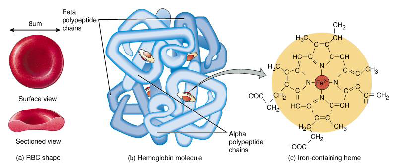
- biconcave discs: 7 ~ 8 μm,
- lack a nucleus and can’t devide
- high Hb concentrations
- lifespan: 80-120 days
3.2.1.1 Physiological characteristic 生理特征
- Permeability of membrane 膜通透性
- Free to gas, like O~2~ , CO~2~ , NO (Isosmotic ≠ Isotonic)
- Deformability 可变形性
- Behaving as elastic bodies, erythrocytes respond to applied pressure (ie, decrease in vessel diameter) by extensive changes in their shape followed by a reversal when the deforming force is removed. 细胞是⼀种弹性体,当受到压力(即⾎管直径减⼩)时,其形状会发生⼤幅度变化,而当变形力消失后,形状又会逆转
- Suspension stability 悬挂稳定性
- greater surface area/volume
- negative charge of RBC membrane 红细胞膜带负电荷
3.2.1.2 Erythrocytes Osmotic Fragility
Osmotic fragility of RBCs is defined as the ease with which the cells burst in hypotonic solutions and is expressed in terms of the concentration of the saline solution in which the cells are haemolyzed. 红细胞的渗透脆性定义为细胞在低渗溶液中破裂的难易程度,以细胞溶⾎的盐溶液浓度来表⽰
Hereditary spherocytosis and thalassemia cause red blood cells to be more fragile than normal, might leading to haemolytic anaemia (anemia AmE).
3.2.1.3 Erythrocyte Sedimentation Rate (ESR) 红细胞沉降率
ESR is a blood test that can show inflammatory activity in the body.
3.2.1.4 Erythrocyte formation 红细胞形成
- The production of erythrocytes : red bone marrow 红骨髓
- Key factor for production: iron and vitamins like folic acid and B~12~
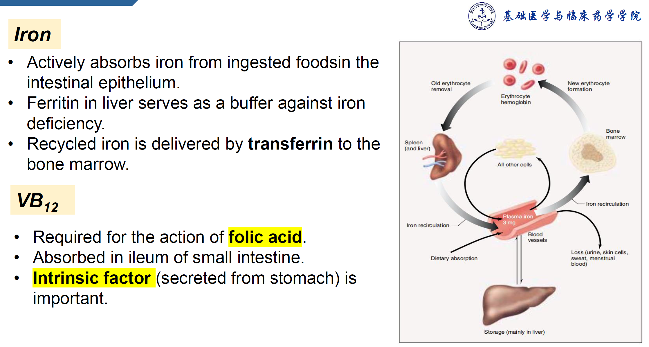
3.2.1.5 Recycling of haemoglobin ⾎红蛋白的回收利⽤
- In liver or spleen 肝脏或脾脏
- globin portion broken down into amino acids & recycled 珠蛋白部分分解成氨基酸并回收
- heme (haem BrE) portion split into iron ($Fe^{3+}$) and biliverdin (green pigment) ⾎红素部分分解为铁($Fe^{3+}$)和胆绿素(绿色色素)
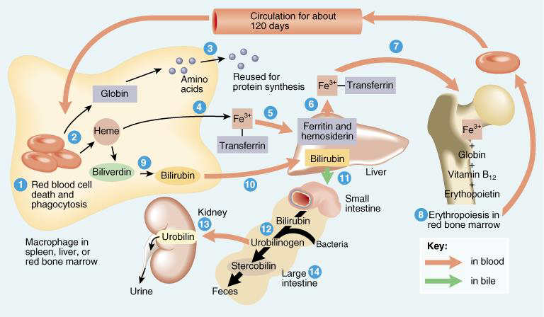
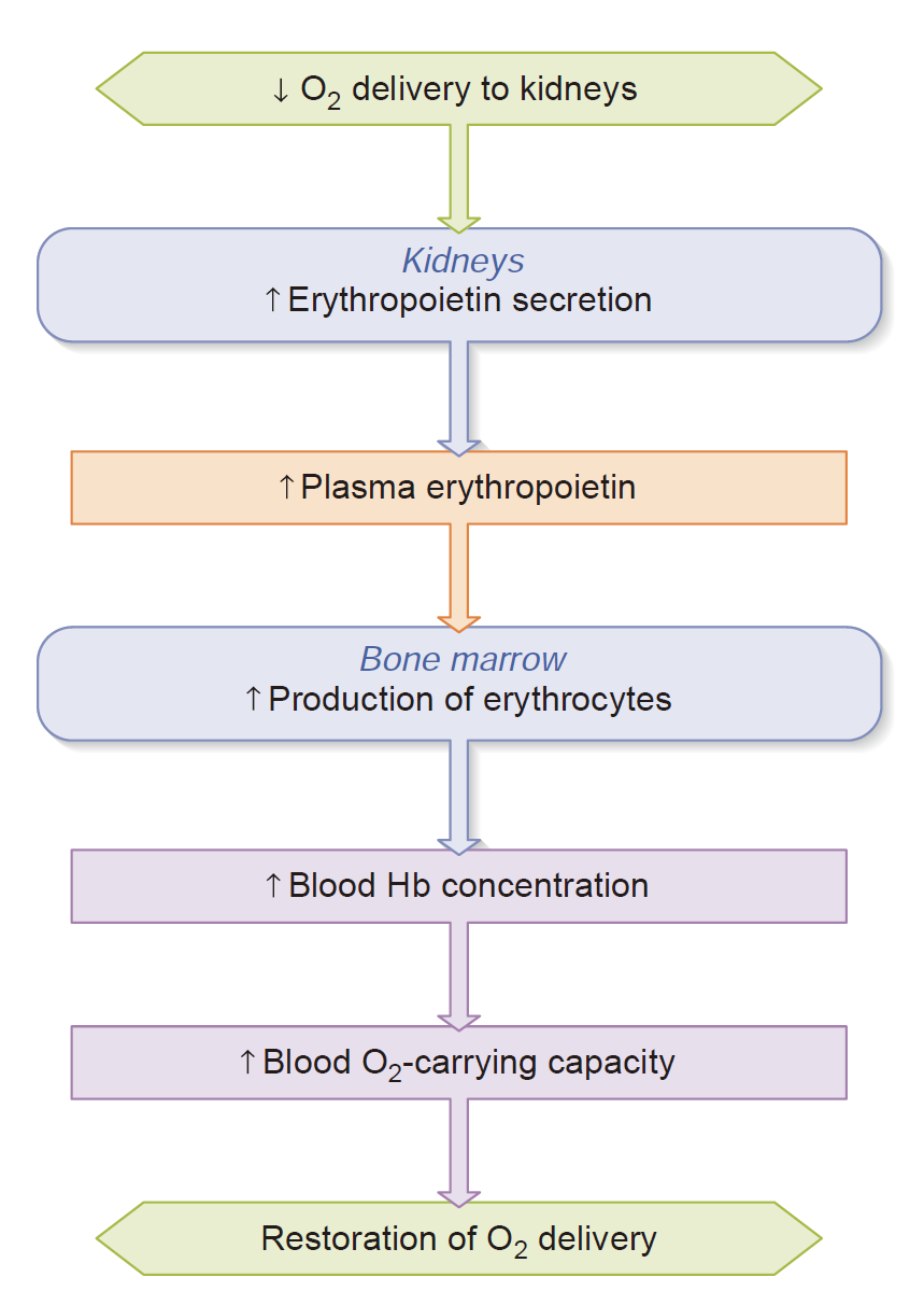
3.2.1.6 Regulation of Erythrocyte Production (erythropoiesis) 红细胞生成的调节(红细胞生成)
- Erythropoietin (EPO) produced by Kidneys (connective-tissue cells in the kidneys, the liver also
secretes this hormone, but to a much lesser extent). 促红细胞生成素 (EPO) 由肾脏产生(肾脏、肝脏中的结缔组织细胞也会分泌这种激素,但程度要⼩得多) - EPO acts on the bone marrow to stimulate the proliferation of erythrocyte progenitor cells and their differentiation into mature erythrocytes. EPO作⽤于骨髓,刺激红细胞祖细胞增殖并分化为成熟红细胞
- Increased EPO responds to tissue hypoxia. EPO 增加可应对组织缺氧
- Testosterone also stimulates the release of erythropoietin. 睾酮还能刺激促红细胞生成素的释放
3.2.1.7 Anemia 贫⾎
Decrease in the absolute quantity of hemoglobin.
- Dietary deficiencies of iron (iron-deficiency anemia), vitamin B12, or folic acid
- Bone marrow failure due to toxic drugs or cancer
- Blood loss from the body (hemorrhage)
- Inadequate secretion of erythropoietin in kidney disease
- Excessive destruction of erythrocytes(for example,sickle-cell disease)
3.2.2 Physiology of Leukocytes
- All WBCs (leukocytes) have a nucleus and no haemoglobin 所有白细胞 (WBC) 都有细胞核,没有⾎红蛋白
- Granular or agranular classification based on presence of cytoplasmic granules made visible by staining 根据染色后可⻅的细胞质颗粒,进行颗粒状或⽆颗粒状分类
- granulocytes are neutrophils, eosinophils or basophils 粒细胞是中性粒细胞、嗜酸性粒细胞或嗜碱性粒细胞
- agranulocytes are monocytes or lymphocytes ⽆粒细胞是单核细胞或淋巴细胞
3.2.2.1 Neutrophil
- Fastest response of all WBC to bacteria 所有白细胞对细菌反应最快的
- Direct actions against bacteria
- release lysozymes which destroy / digest bacteria 释放溶菌酶来破坏/消化细菌
- release defensin proteins that act like antibiotics & poke holes in bacterial cell walls destroying them 释放出像抗生素⼀样的防御素蛋白,在细菌细胞壁上戳洞,摧毁它们
- release strong oxidants (bleach-like, strong chemicals) that destroy bacteria 释放强氧化剂(类似漂白剂的强力化学物质),杀死细菌
3.2.2.2 Monocyte
- Take longer to get to site of infection, but arrive in larger numbers 到达感染部位需要更⻓时间,但到达的人数更多
- Become wandering macrophages, once they leave the capillaries ⼀旦离开⽑细⾎管,就变成游走的巨噬细胞
- Destroy microbes and clean up dead tissue following an infection
3.2.2.3 Lymphocyte
- B cells
• destroy bacteria and their toxins
• turn into plasma cells that produces antibodies - T cells
• attack viruses, fungi, transplanted organs, cancer cells & some bacteria - Natural killer cells
• attack many different microbes & some tumor cells
• destroy foreign invaders by direct attack
3.2.3 Physiology of Platelets (Thrombocytes)
Counts and Morphology
- Disc-shaped, 2 - 4 micron cell fragment
- with no nucleus
- Normal platelet count: 150,000-400,000/drop of blood
Life span
- Short life span (5 to 9 days in bloodstream)
• formed in bone marrow
• few days in circulating blood
• aged ones removed by fixed macrophages in liver and spleen
Function
- Play important roles in hemostasis
- Direct adhesion
- Platelet-derived growth factor (PDGF)
• cause proliferation of vascular endothelial cells, smooth muscle & fibroblasts
to repair damaged vessels
3.3 Hemostasis: The Prevention of Blood Loss
Hemostasis
- Stoppage of bleeding in a quick & localized fashion when blood vessels are damaged
- Prevents hemorrhage (loss of a large amount of blood)
- Methods utilized
• vascular spasm ⾎管收缩
• platelet plug formation ⾎⼩板栓塞形成
• blood clotting (coagulation = formation of fibrin threads) ⾎液凝固(凝固 = 纤维蛋白丝形成)
3.3.1 Vascular Spasm
- Damage to blood vessel produces stimulates pain receptors ⾎管损伤会刺激疼痛感受器
- Reflex contraction of smooth muscle of small blood vessels ⼩⾎管平滑肌反射性收缩
- Can reduce blood loss for several hours until other mechanisms can take over
- Only for small blood vessel or arteriole 仅适⽤于⼩⾎管或⼩动脉
3.3.2 Platelet Plug Formation
- Adhesion to collagen: vWF
- Release: a variety of chemical agents via secretory vesicles 通过分泌囊泡释放各种化学物质
• Alpha-granules: clotting factors, PDGF
• Dense granules: ADP, ATP, Ca^2+^, serotonin, fibrin-stabilizing factor, & enzymes that produce thromboxane A2. ADP、ATP、Ca^2+^、⾎清素、纤维蛋白稳定因子和产生⾎栓素 A2 的酶 - Aggregation: new platelets adhered to the old ones due to the platelet release reaction
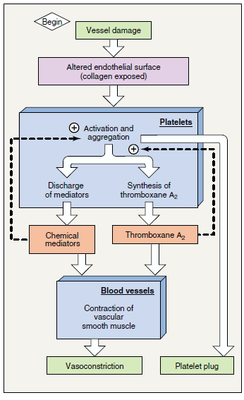
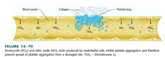
- Why is the platelet plug limited to damage endothelium?
- Inhibitory factors for platelet aggregation are released in intact endothelium. 完整的内皮会释放⾎⼩板聚集的抑制因子
3.3.3 Blood Coagulation: Clot Formation
- Blood drawn from the body thickens into a gel 从⾝体抽出的⾎液变稠变成凝胶
- gel separates into liquid (serum) and a clot of insoluble fibres (fibrin) in which the cells are trapped 凝胶分离成液体(⾎清)和⼀团不溶性纤维(纤维蛋白),细胞被困在其中
- If clotting occurs in an unbroken vessel is called a thrombosis 如果未破裂的⾎管中发生凝⾎,则称为⾎栓形成
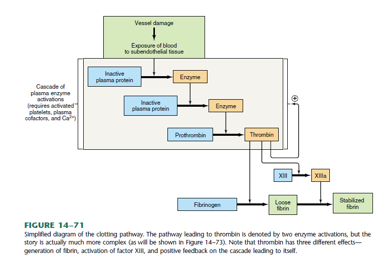
- Substances required for clotting are Ca^2+^, enzymes synthesized by liver cells and substances released by platelets or damaged tissues
- Clotting is a cascade of reactions in which each clotting factor activates the next in a fixed sequence resulting in the formation of fibrin threads 凝⾎是⼀系列反应,其中每个凝⾎因子按固定顺序激活下⼀个凝⾎因子,导致纤维蛋白丝的形成
- prothrombinase & Ca^2+^ convert prothrombin into thrombin 凝⾎酶原酶和Ca^2+^将凝⾎酶原转化为凝⾎酶
- thrombin converts fibrinogen into fibrin threads 凝⾎酶将纤维蛋白原转化为纤维蛋白丝
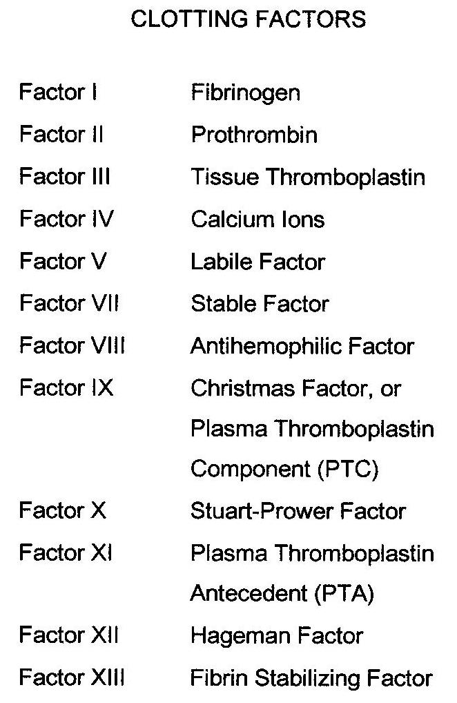
3.3.3.1 Clotting factors
- There are many clotting factors in blood.
- The clotting factors work one after the other. At the end of the chain, bleeding stops.
- If one is missing or does not work, clots will not form properly and bleeding will continue. 如果缺少⼀个或者不起作⽤,⾎凝块就⽆法正常形成,出⾎就会持续
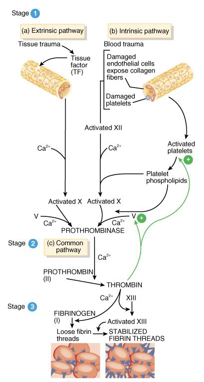
3.3.3.2 Stage of blood coagulation
- Prothrombinase is formed by either the intrinsic or extrinsic pathway 凝⾎酶原酶由内在或外在途径形成
- Prothrombin is converted to thrombin 凝⾎酶原转化为凝⾎酶
- Final common pathway produces fibrin threads 最后的共同通路产生纤维蛋白丝
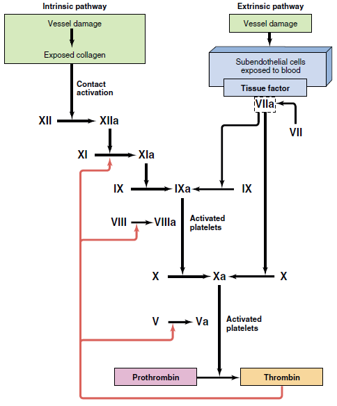
Extrinsic Pathway 外在途径
- Damaged tissues leak tissue factor (thromboplastin) into bloodstream 受损组织泄漏组织因子(凝⾎活酶)进入⾎液
- Prothrombinase forms in seconds 凝⾎酶原酶在数秒内形成
- In the presence of Ca^2+^, clotting factor X combines with V to form prothrombinase ,凝⾎因子 X 与 V 结合形成凝⾎酶原酶
Intrinsic Pathway 内在途径
- Activation occurs
- endothelium is damaged & platelets come in contact with collagen of blood vessel wall 内皮受损,⾎⼩板与⾎管壁胶原蛋白接触
- platelets damaged & release phospholipids ⾎⼩板受损并释放磷脂
- Requires several minutes for reaction to occur 反应需要⼏分钟才能发生
- Substances involved: Ca^2+^ and clotting factors XII, X and V
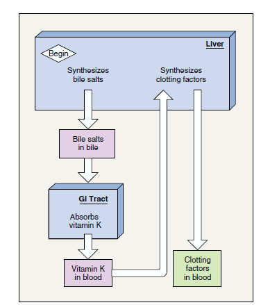
3.3.3.3 Role of Vitamin K in Clotting
- Normal clotting requires adequate vitamin K 正常凝⾎需要⾜够的维生素 K
- fat soluble vitamin absorbed if lipids are present 脂溶性维生素在脂质存在的情况下被吸收
- absorption slowed if bile release is insufficient 如果胆汁释放不⾜,吸收就会减慢
- Required for synthesis of 4 clotting factors by hepatocytes 肝细胞合成 4 种凝⾎因子所需
- factors II (prothrombin), VII, IX and X
- Produced by bacteria in large intestine 由⼤肠中的细菌产生
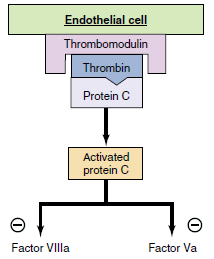
3.3.4 Anticlotting system
- tissue factor pathway inhibitor (TFPI) 组织因子途径抑制剂 (TFPI) 抑制 X~a~ 因子
• Inactivates factor X~a~ - Thrombomodulin receptor ⾎栓调节蛋白受体 抑制 V~a~ 因子和 XIII~a~ 因子
• Inactivates factor V~a~ and XIII~a~ - antithrombin III 抗凝⾎酶III 抑制凝⾎酶和其他因子
• Inactivates thrombin and other factors
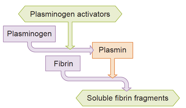
3.3.4.1 Fibrinolytic system 纤溶系统
The fibrinolytic (or thrombolytic) system is the principal effector of clot removal. 纤维蛋白溶解(或⾎栓溶解)系统是清除⾎凝块的主要效应器
- Fibrinolytic system dissolves small, inappropriate clots & clots at a site of a completed repair 纤维蛋白溶解系统溶解⼩的、不适当的⾎凝块和已完成修复部位的⾎凝块
- fibrinolysis is dissolution of a clot 纤维蛋白溶解是⾎凝块的溶解
- Inactive plasminogen is incorporated into the clot ⽆活性的纤溶酶原被掺入⾎凝块中
- activation occurs because of factor XII and thrombin 由于因子 XII 和凝⾎酶而发生激活
- plasminogen becomes plasmin (fibrinolysin) which digests fibrin threads 纤溶酶原变成纤溶酶(纤维蛋白溶酶),可消化纤维蛋白丝
3.3.4.2 Anticlotting Drugs
- Anticoagulants suppress or prevent blood clotting 抗凝剂抑制或预防⾎液凝固
- heparin: administered during hemodialysis and surgery 肝素:⾎液透析和手术期间使⽤
- warfarin (Coumadin): antagonist to vitamin K so blocks synthesis of clotting factors, slower than heparin 华法林(Coumadin):维生素 K 拮抗剂,可阻止凝⾎因子的合成,速度比肝素慢
- stored blood in blood banks treated with citrate phosphate dextrose (CPD) that removes Ca^2+^ ⾎库中储存的⾎液经过柠檬酸磷酸葡萄糖 (CPD) 处理
- Thrombolytic agents are injected to dissolve clots 注射溶栓剂来溶解⾎栓
- directly or indirectly activate plasminogen 直接或间接激活纤溶酶原
- streptokinase or tissue plasminogen activator (t-PA) 链激酶或组织纤溶酶原激活剂 (t‑PA)
3.4 Blood Groups and Blood Transfusion
3.4.1 ABO Blood Groups
- Based on 2 glycolipid iso-antigens called A and B found on the surface of
RBCs
• display only antigen A – blood type A
• display only antigen B – blood type B
• display both antigens A & B – blood type AB
• display neither antigen – blood type O - Plasma contains isoantibodies or agglutinins to the A or B antigens not found in your blood
• anti-A antibody reacts with antigen A
• anti-B antibody reacts with antigen B
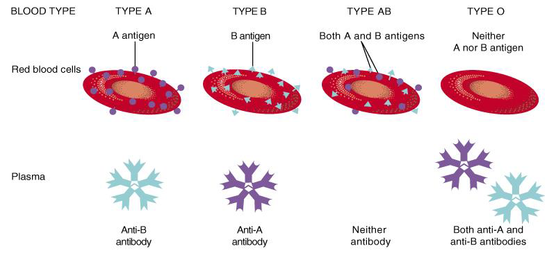
RBC surfaces are marked by genetically determined glycoproteins & glycolipids
- agglutinogens or iso-antigens
- distinguishes at least 24 different blood groups
• ABO, Rh, Lewis, Kell, Kidd and Duffy systems
3.4.2 Typing and Cross-Matching Blood
- Mixing of incompatible blood causes agglutination (visible clumping)
• formation of antigen-antibody complex that sticks cells together
• not the same as blood clotting - Typing involves testing blood with known antisera that contain antibodies A, B or Rh+
- Cross-matching is to test by mixing donor cells with recipient’s serum
- Screening is to test recipient’s serum against known RBC’s having known antigens
Transfusion and Transfusion Reactions
-
Transfer of whole blood, cells or plasma into the bloodstream of recipient
- used to treat anaemia or severe blood loss
-
Incompatible blood transfusions
- antigen-antibody complexes form between plasma antibodies & “foreign proteins” on donated RBC’s (agglutination)
- donated RBCs become leaky (complement proteins) & burst
- loose haemoglobin causes kidney damage
-
Problems caused by incompatibility between donor’s cells and recipient’s plasma
-
Donor plasma is too diluted to cause problems
Chapter 5 Respiratory Physiology
5.1 Pulmonary Ventilation and Lung Mechanics
5.1.1 Organization of the Respiratory System
5.1.1.1 The Respiratory Tract 呼吸道
- Consists of a conducting portion
• From nasal cavity to terminal bronchioles - Consists of a respiratory portion
• The respiratory bronchioles and alveoli
5.1.1.2 Conducting zone 传导区
- The conducting zone extends from the top of the trachea to the beginning of the respiratory bronchioles; it contains no alveoli and there is no gas exchange with the blood. 传导区从⽓管顶部延伸至呼吸性细⽀⽓管的起始处;它不包含肺泡,并且不与⾎液进行⽓体交换
- Trachea, Bronchi, larger bronchioles-allows air flow
- Have cartilage rings - robust will not collapse 有软骨环‑坚固不会塌陷
- Cells: Ciliated epithelium; Goblet cells; Smooth muscle cells 细胞:纤⽑上皮;杯状细胞;平滑肌细胞
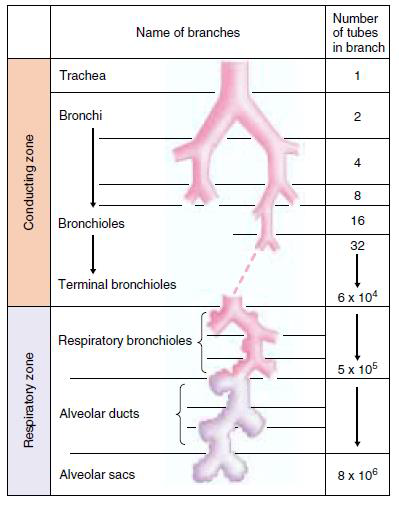

5.1.1.3 Respiratory zone 呼吸区
- The respiratory zone, which extends from the respiratory bronchioles down, contains alveoli and is the region where gases exchange with the blood. 呼吸区从呼吸性细⽀⽓管向下延伸,包含肺泡,是⽓体与⾎液交换的区域
- Cells: Type I, Type II
- For gases to exchange efficiently:
• Alveoli walls must be very thin (<1 μm)
• Surface area must be very great (about 35 times the surface area of the body) - Most of the air-facing surfaces of the wall are lined by a continuous layer, one cell thick, of flat epithelial cells termed type I alveolar cells. ⼤部分⾯向空⽓的壁表⾯都覆盖有⼀层连续的扁平上皮细胞,厚度只有⼀个细胞,称为 I 型肺泡细胞
- Interspersed between these cells are thicker specialized cells termed type II alveolar cells that produce a detergent-like substance, surfactant. 这些细胞之间散布着更厚的特殊细胞,称为 II 型肺泡细胞,产生⼀种类似洗涤剂的物质:表⾯活性剂。
- The respiratory membrane is composed of 6 layers:
- a layer of fluid containing surfactant that lines the alveolus and reduces the surface tension; ⼀层含有表⾯活性剂的液体,排列在肺泡上,减少表⾯紧张;
- the alveolar epithelium; 肺泡上皮;
- an epithelial basement membrane; 上皮基底膜;
- a thin interstitial space between the alveolar epithelium and the capillary membrane; 肺泡上皮和⽑细⾎管膜之间的薄间质空间;
- a capillary basement membrane; ⽑细⾎管基底膜;
- the capillary endothelial membrane. ⽑细⾎管内皮膜
5.1.1.4 Alveoli 肺泡
- Are air-filled pockets within the lungs
- Where all gas exchange takes place
- Inspiration (inhalation) is the movement of air from the external environment through the airways into the alveoli during breathing.
- Expiration (exhalation) is movement in the opposite direction. An inspiration and expiration constitute a respiratory cycle.

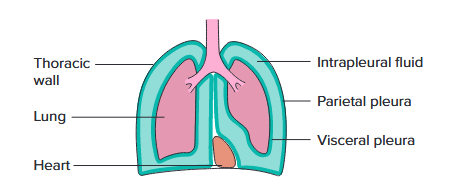
5.1.1.5 Relation of the Lungs to the Thoracic (Chest) Wall
The Pleura 胸膜
- Consists of two layers:
• Parietal pleura
• Visceral pleura - Pleural fluid
• Lubricates space between two layers
• the total volume is only a few milliliters
5.1.2 Ventilation and Lung mechanics
5.1.2.1 Ventilation
- External respiration - the exchange of gases between the body and the environment. ⾝体与环境之间的⽓体交换
- Gas transport - O~2~ is transported from lungs to the tissues and CO~2~ is transported in the opposite direction by blood circulation. O~2~ 从肺部运输到组织,CO~2~ 通过⾎液循环以相反的方向运输。
- Gas diffusion – gas is transported over short distances of a few micrometers—e.g., through cell membranes and other physiological barriers. ⽓体在⼏微米的短距离内运输;例如,通过细胞膜和其他生理屏障
- Internal respiration - the exchange of gas occurs between the blood and cells of tissues and O~2~ utilization in the cells. ⾎液和组织细胞之间发生⽓体交换,细胞中利⽤ O~2~
- Ventilation - the exchange of gas between the atmosphere and alveoli. ⼤⽓与肺泡之间的⽓体交换
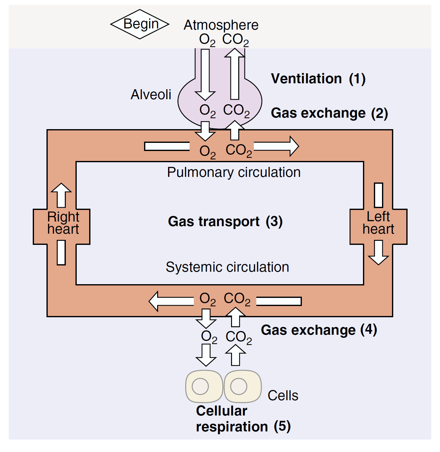
Pulmonary Ventilation
• Is the physical movement of air in and out of respiratory tract 是空⽓进出呼吸道的物理运动
• Provides alveolar ventilation
$$
𝐹 = (𝑃_{𝑎𝑙𝑣} − 𝑃_{𝑎𝑡𝑚})/𝑅
$$
$F$: air flow
$P_{alv}$: alveolar pressure
$P_{atm}$: atmospheric pressure
$R$: resistance
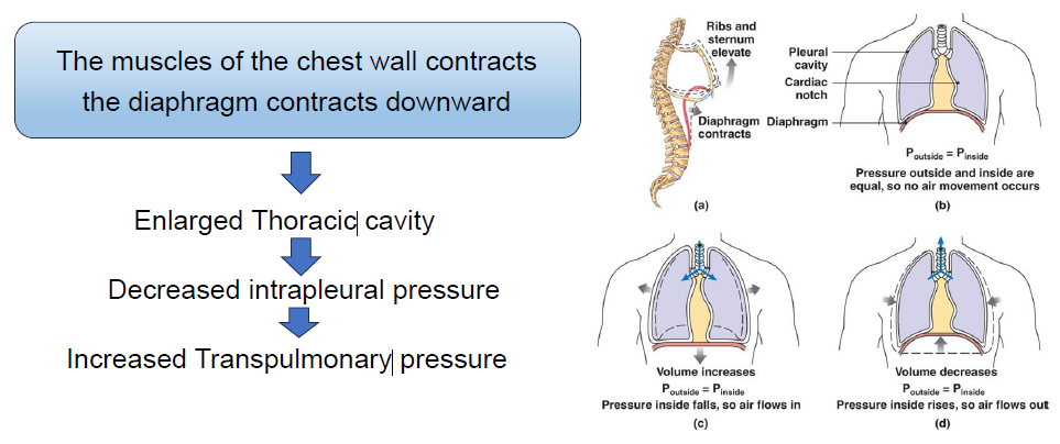
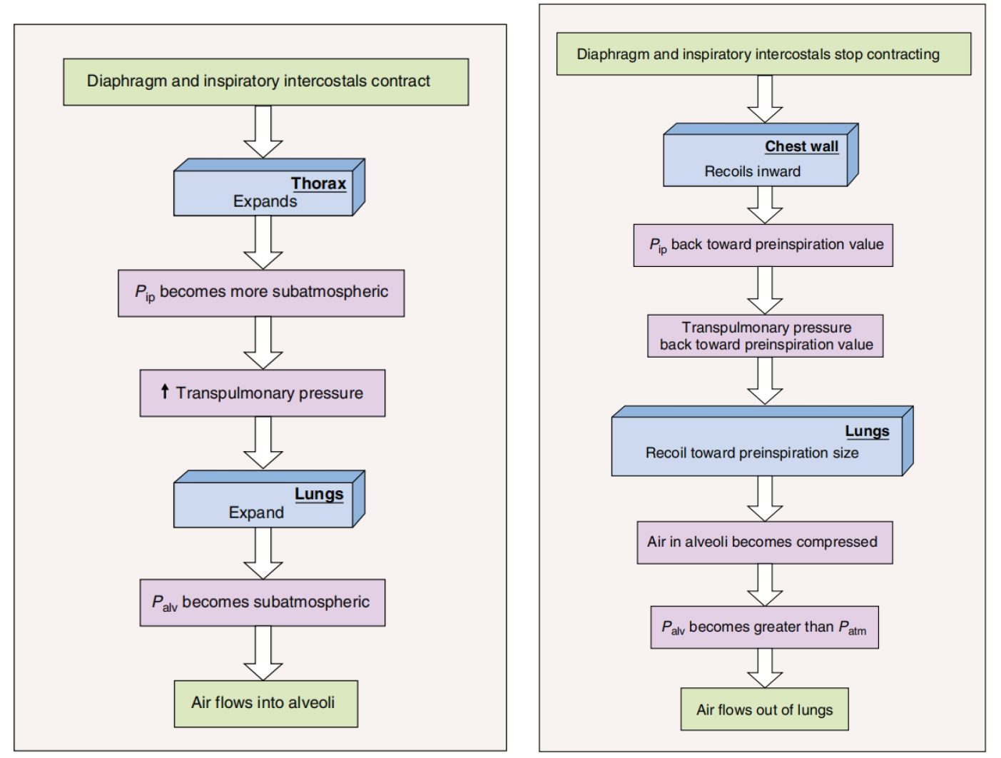
- During ventilation, air moves into and out of the lungs because the alveolar pressure is alternately made less than and greater than atmospheric pressure.
- The lungs are passive elastic structures—like balloons—and their volume, therefore, depends upon:
- the difference in pressure—termed the transpulmonary pressure—between the inside and the outside of the lungs; 肺内和肺外的压力差,称为跨肺压以及肺部外部;
- how stretchable the lungs are. 肺部的伸展性如何
$$
\text{Transpulmonary Pressure}=P_{alv}-P_{ip}
$$
Transpulmonary pressure
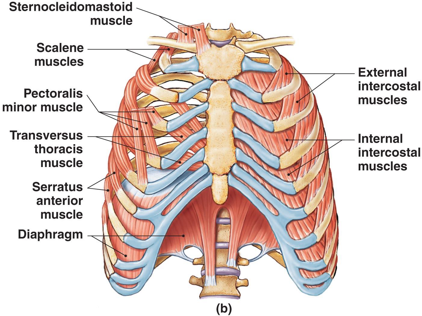
5.1.2.2 The Respiratory Muscles
- The diaphragm 横膈膜
- External intercostal muscles of the ribs 肋骨外肋间肌
- Accessory respiratory muscles:
• activated when respiration increases significantly 辅助呼吸肌:当呼吸显著增加时激活
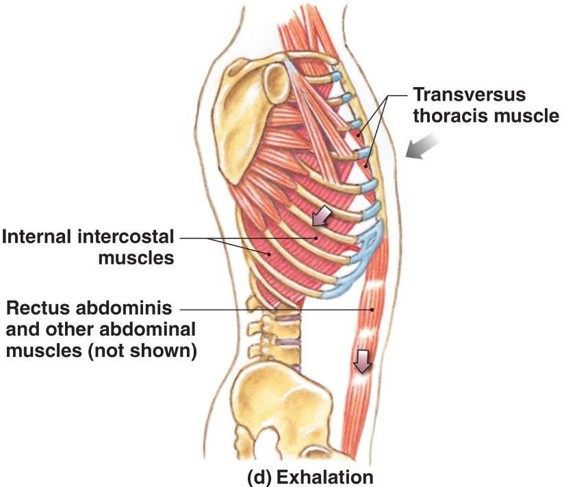
Muscles of Active Exhalation 主动呼⽓的肌⾁
- Internal intercostal and transversus thoracis muscles
• Depress the ribs 压低肋骨 - Abdominal muscles
• Compress the abdomen 挤压腹部
• Force diaphragm upward 迫使横膈膜向上
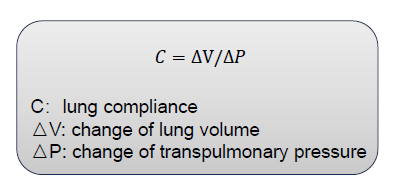
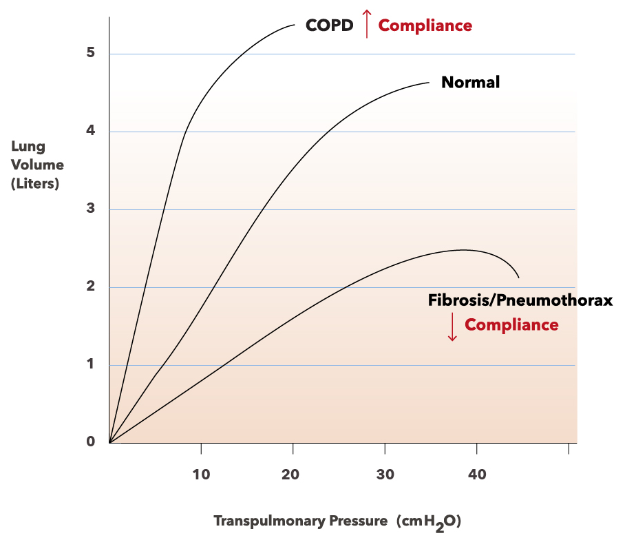
Lung Compliance 肺顺应性
- An indicator of expandability
- Low compliance requires greater force
- High compliance requires less force
Factors That Affect Compliance
- Connective tissue structure of the lungs 肺的结缔组织结构
- Level of surfactant production 表⾯活性剂生产水平
- Mobility of the thoracic cage 胸廓的活动性
5.1.2.3 Inspiratory and Expiratory
Inhalation: Always active
Exhalation: Active or passive
How does airway resistance change? ⽓道阻力如何变化?
- Airway resistance to airflow is normally very small ⽓道对⽓流的阻力通常很⼩
- Physical factor: transpulmonary pressure and lateral traction 物理因素:跨肺压和侧向牵引
- Neuroendocrine and paracrine factors: epinephrine, leukotrienes 神经内分泌和旁分泌因子:肾上腺素、白三烯
5.1.3 Lung Volumes and Capacities
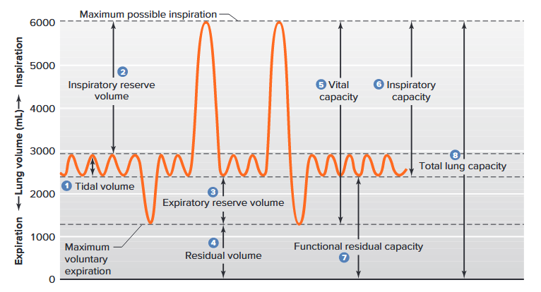
- Tidal volume (TV): the volume of air entering the lungs during a single inspiration. The tidal volume during normal quiet breathing is termed the resting tidal volume and is approximately 500ml. 单次吸⽓时进入肺部的空⽓量。正常安静呼吸时的潮⽓量称为静息潮⽓
- Inspiratory reserve volume (IRV): the maximal amount of air that can be increased above TV during deepest inspiration (3000ml). 最深吸⽓时可增加至 TV 以上的最⼤空⽓量
- Expiratory reserve volume (ERV): maximal extra volume of air that can be expired by forceful expiration after the end of a normal tidal expiration (900-1200ml) 正常潮⽓量结束后⽤力呼⽓所能呼出的最⼤额外空⽓量
- Residual volume (RV): after a maximal active expiration, approximately 1200ml of air still remains in the lungs. 最⼤主动呼⽓后,肺内仍残留约1200ml空⽓
- Vital capacity (VC): the maximal volume of air that a person can expire after a maximal inspiration. ⼀个人最⼤程度吸⽓后能够呼出的最⼤空⽓量
- Inspiratory capacity (IC): is equal to the tidal volume plus the inspiratory reserve volume. 等于潮⽓量加上补吸⽓量
- Functional residual capacity (FRC): After expiration of a resting tidal volume, the lungs still contain a very large volume of air. 静息潮⽓量呼出后,肺部仍然含有⼤量空⽓
- Total lung capacity (TLC): Maximum amount of air that can be inhaled after a normal tidal expiration. 正常潮⽓呼⽓后可吸入的最⼤空⽓量
- Forced expiratory volume in 1 sec (FEV~1~): In 1s, the person takes a maximal inspiration
and then exhales maximally as fast as possible. 在1秒内,人以最⼤程度吸⽓,然后尽可能快地最⼤程度呼⽓
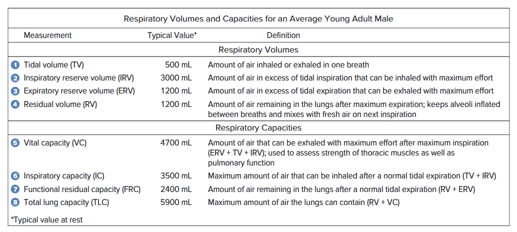
5.1.4 Dead Space and Alveolar Ventilation
5.1.4.1 Minute Ventilation
The total ventilation per minute—the minute ventilation ($\dot{V}_E$)—is equal to the tidal volume multiplied by the respiratory rate as shown in equation 13–6. (The dot above the letter $V$ indicates per minute.) 每分钟总通⽓量(分钟通⽓量)等于潮⽓量乘以呼吸频率
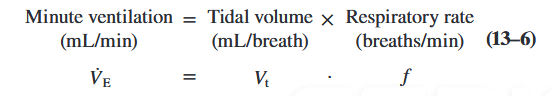
5.1.4.2 Dead Space
Dead space is the volume of inspired air that does not take part in gas exchange. There are two reasons why this occurs. 死腔是指吸入的空⽓中未参与⽓体交换的部分
- The first is due to the anatomy of the airways themselves. Anatomic dead space: Part of atmospheric air entering the respiratory system during each inspiration never reaches the alveoli but is merely moved in and out of the airways. 第⼀是⽓道本⾝的解剖结构。解剖死腔:每次吸⽓时进入呼吸系统的部分⼤⽓空⽓从未到达肺泡,而只是进出⽓道。
- The second component of dead space occurs because some fresh inspired air is not used for gas exchange with the blood even though it reaches the alveoli. This is because some alveoli may, for various reasons, have little or no blood supply. This volume of air is known as alveolar dead space. It is quite small in healthy persons but may be very large with lung disease. As we shall see, local mechanisms that match air and blood flows minimize the alveolar dead space. 死腔的第二个组成部分是因为一些新鲜吸入的空气即使到达肺泡也没有用于与血液进行气体交换。这是因为一些肺泡可能由于各种原因很少或没有血液供应。该体积的空气称为肺泡死腔。它在健康人中相当小,但在患有肺部疾病时可能非常大。正如我们将看到的,匹配空气和血液流动的局部机制最大限度地减少了肺泡死腔。
The sum of the anatomical and alveolar dead spaces is known as the physiological dead space. 解剖学死腔和肺泡死腔的总和称为生理死腔。
5.1.4.3 Alveolar Ventilation
The total volume of fresh air entering the alveoli per minute is called the alveolar ventilation ($\dot{V}_A$). 每分钟进入肺泡的新鲜空⽓总量称为肺泡通⽓量。
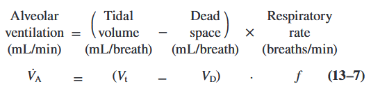
5.2 Exchange of Gases in Alveoli and Tissues
5.2.1 Foundational Principles of Gas Exchange: Diffusion of Gases
5.2.1.1 Partial Pressure of Gases
- A mixture of gases exert a pressure
- An individual gas exerts a partial pressure in the mixture
- Each gas contributes to the total pressure (Dalton’s law)
5.2.1.2 Diffusion of Gases in Liquids
Henry’s Law
- When gas under pressure comes in contact with liquid
• Gas dissolves in liquid until equilibrium is reached - At a given temperature
• Amount of a gas in solution is proportional to partial pressure of that gas
Gas Content
- The actual amount of a gas in solution (at given partial pressure and temperature) depends on the solubility of that gas in that particular liquid
Solubility in Body Fluids
- CO~2~ is very soluble
- O~2~ is less soluble
- N2 has very low solubility
5.2.2 Exchange Between Alveoli and Blood
Efficiency of Gas Exchange Due to:
- Substantial differences in partial pressure across the respiratory membrane 呼吸膜两侧分压存在显著差异
- Distances involved in gas exchange are short ⽓体交换距离较短
- O~2~ and CO~2~ are lipid soluble O~2~ 和 CO~2~ 可溶于脂溶性
- Total surface area is large 总表⾯积⼤
- Blood flow and airflow are coordinated ⾎流和⽓流协调
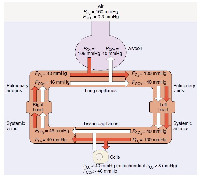
Ventilation versus perfusion 通⽓与灌注
- Matching the blood flow to the parts of the lung where the oxygen is 将⾎流与肺部氧⽓充⾜的部位相匹配
- Ventilation / perfusion ratio 通⽓/灌注比
- Not equal across the lung (because of where the heart is). Can change due to metabolism 肺部不同(因为⼼脏的位置不同),可能因新陈代谢而改变
- Can change with disease-asthma, pulmonary hypertension 可能因哮喘、肺动脉⾼压等疾病而改变
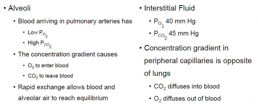
5.3 Transport of Oxygen and Carbon Dioxide in the Blood
5.3.1 Transport of Oxygen in the Blood
The oxygen is present in two forms:
- dissolved in the plasma and erythrocyte cytosol 溶解于⾎浆和红细胞胞质中
- reversibly combined with haemoglobin molecules in the erythrocytes 与红细胞中的⾎红蛋白分子可逆地结合
As predicted by Henry’s law, the amount of oxygen dissolved in blood is directly proportional to the $P_{O_2}$ of the blood. 根据亨利定律预测,血液中溶解的氧气量与血液中的 $P_{O_2}$ 成正比。
5.3.1.1 Hemoglobin
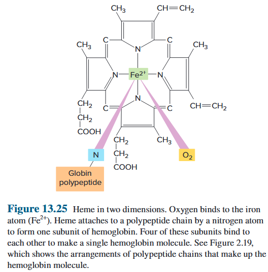
Each hemoglobin molecule is a protein made up of four subunits bound together. Each subunit consists of a molecular group known as heme and a polypeptide attached to the heme. The four polypeptides of a hemoglobin molecule are collectively called globin. Each of the four heme groups in a hemoglobin molecule (Figure 13.25) contains one atom of iron ($Fe^{2+}$), to which molecular oxygen binds. Because each iron atom shown in Figure 13.25 can bind one molecule of oxygen, a single hemoglobin molecule can bind four oxygen molecules (see Figure 2.19 for the quaternary structure of hemoglobin). 每个血红蛋白分子都是由四个结合在一起的亚基组成的蛋白质。每个亚基由称为血红素的分子组和附着在血红素上的多肽组成。血红蛋白分子的四种多肽统称为珠蛋白。血红蛋白分子中的四个血红素基团(图 13.25)中的每一个都含有一个铁原子 ($Fe^{2+}$),分子氧与其结合。因为图13.25所示的每个铁原子可以结合一个氧分子,所以单个血红蛋白分子可以结合四个氧分子(血红蛋白的四级结构见图2.19)。
deoxyhemoglobin - oxyhemoglobin

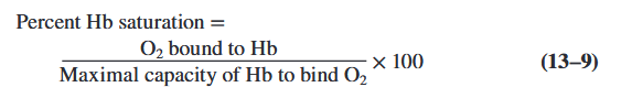
5.3.1.2 Hemoglobin Saturation ⾎红蛋白饱和度
The percentage of haem units in a haemoglobin molecule
5.3.1.2.1 Effect of partial pressure of O~2~

Oxygen–Hemoglobin Saturation Curve
- Is a curve rather than a straight line
• Because Hb changes shape each time a molecule of O~2~ is bound - Each O~2~ bound makes next O~2~ binding easier
- Allows Hb to bind O~2~ when O~2~ levels are low
Oxygen Reserves
- O~2~ diffuses
• From peripheral capillaries (high $P_{O_2}$)
• Into interstitial fluid (low $P_{O_2}$) - Amount of O~2~ released depends on interstitial $P_{O_2}$
- Up to 3/4 may be reserved by RBCs 最多 3/4 可以由 RBC 保留
5.3.1.2.2 Effect of Carbon Monoxide
- CO from burning fuels
• Binds strongly to hemoglobin
• Takes the place of O~2~
• Can result in carbon monoxide poisoning
5.3.1.2.3 Effect of pH and temperature (normal pH 7.4, 37℃)
- When pH drops or temperature rises
• More oxygen is released 当 pH 值下降或温度升⾼时 释放更多氧气 - When pH rises or temperature drops
• Less oxygen is released 当 pH 值上升或温度下降时 释放的氧气减少
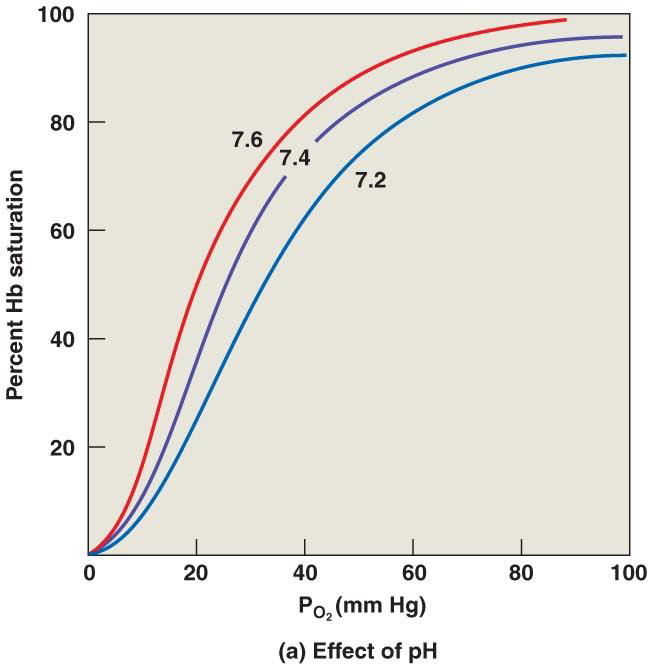
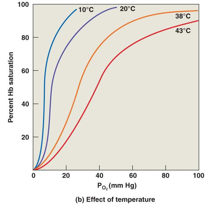
5.3.1.2.4 The Bohr Effect
- Is the effect of pH on hemoglobin-saturation curve
- Caused by CO~2~
• CO~2~ diffuses into RBC
• An enzyme, called carbonic anhydrase, catalyzes reaction with H~2~O ⼀种称为碳酸酐酶的酶催化与 H~2~O 的反应
• Produces carbonic acid (H~2~CO~3~) - Carbonic acid (H~2~CO~3~)
• Dissociates into hydrogen ion (H^+^) and bicarbonate ion (HCO~3~^-^) - Hydrogen ions diffuse out of RBC, lowering pH 氢离子从红细胞中扩散出来,降低 pH 值
5.3.1.2.5 Effect of 2,3-bisphosphoglycerate (DPG) 2,3‑双磷酸⽢油酸
- RBCs generate ATP by glycolysis 红细胞通过糖酵解产生 ATP
• Forming lactic acid and DPG 形成乳酸和DPG - DPG directly affects O~2~ binding and release
• More DPG, more oxygen released DPG 越多,释放的氧气越多
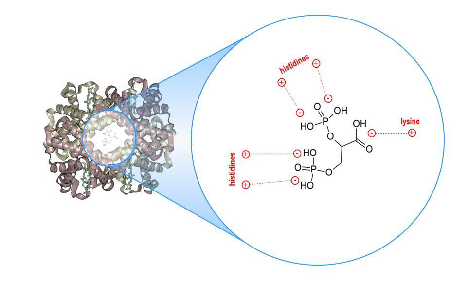
5.3.2 Transport of Carbon Dioxide in Blood
- Physically dissolved in the blood (10%) 物理溶解
- Bound to haemoglobin (25-30%) 与⾎红蛋白结合
- bicarbonate (60-65%) 碳酸氢盐
Bicarbonate Ions
- Move into plasma by an exchange mechanism (the chloride shift) that takes in Cl^-^ ions without using ATP 过交换机制(氯离子转移)进⼊⾎浆,⽆需使⽤ ATP 即可吸收 Cl^‑^ 离子
Hydrogen Ions
- Deoxyhemoglobin has a much greater affinity for H^+^ 脱氧⾎红蛋白对 H^+^ 的亲和⼒更强
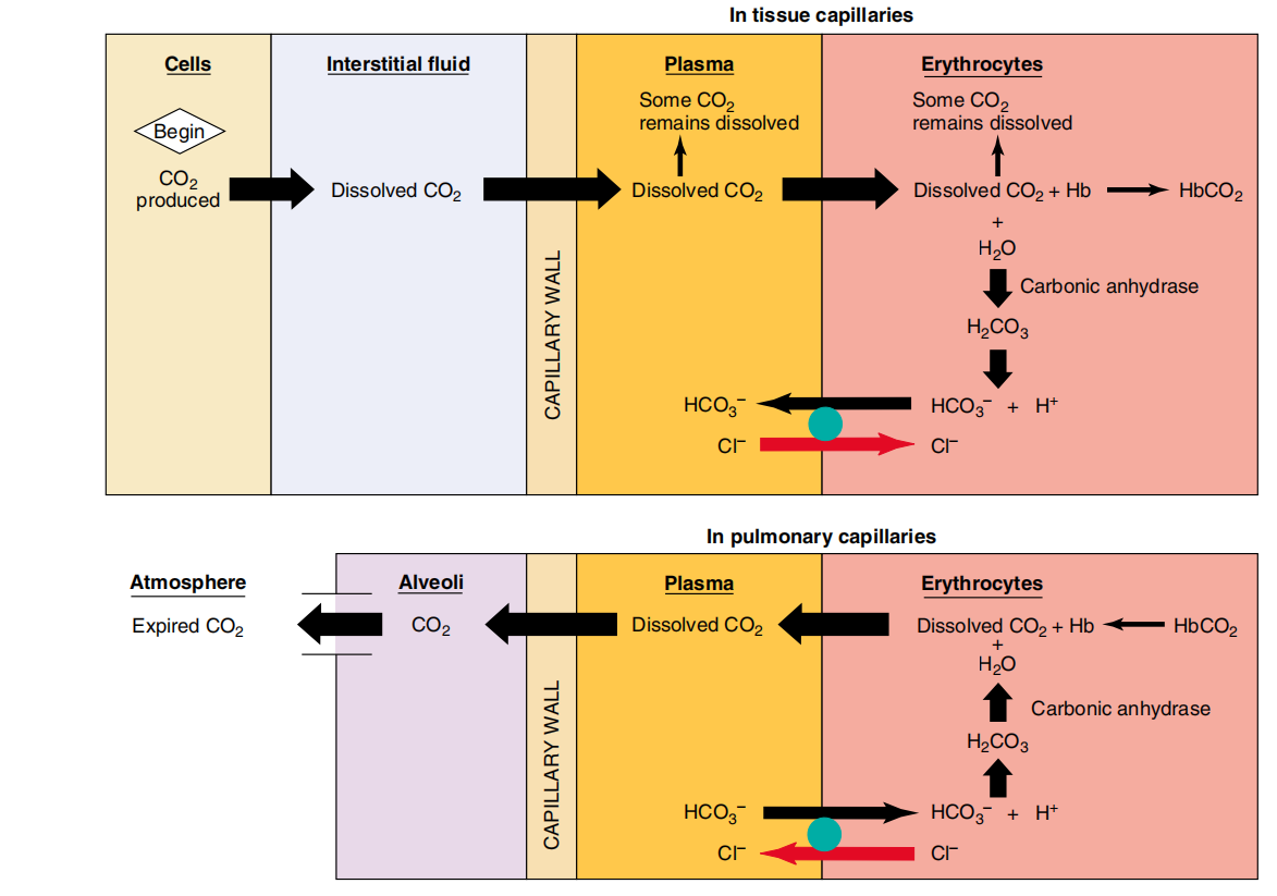
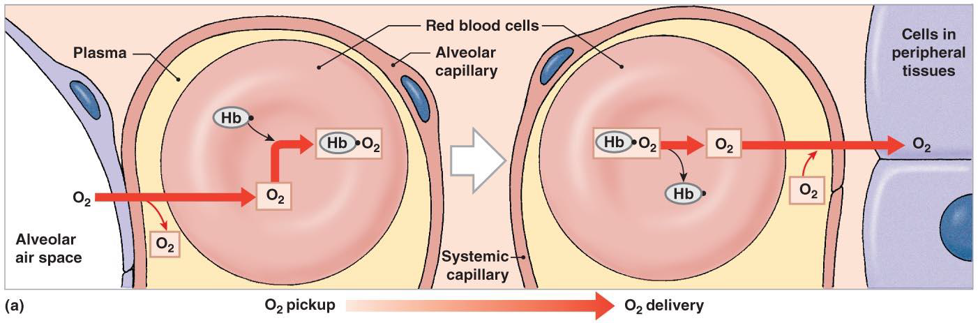
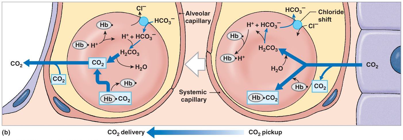
5.4 Control of Respiration
5.4.1 Neural Generation of Rhythmic Breathing
The respiratory centers: Three pairs of nuclei in the reticular formation of medulla oblongata and pons 呼吸中枢:延髓和脑桥⽹状结构中的三对核
脊髓:联系高位呼吸中枢和呼吸肌的中继站
Respiratory rhythmicity centers of the medulla oblongata: set the pace of respiration 延髓的呼吸节律中⼼:设定呼吸的节奏
- Dorsal respiratory group (DRG): The neurons of the dorsal respiratory group (DRG) primarily fire during inspiration and have input to the spinal motor neurons that activate respiratory muscles involved in inspiration—the diaphragm and inspiratory intercostal muscles. The primary inspiratory muscle at rest is the diaphragm, which is innervated by the phrenic nerves. 背侧呼吸群 (DRG) 的神经元主要放电吸气过程中,并输入脊髓运动神经元,激活参与吸气的呼吸肌——膈肌和吸气肋间肌。静止时的主要吸气肌是膈肌,由膈神经支配。
- Inspiratory center
- Functions in quiet and forced breathing
- Ventral respiratory group (VRG): The ventral respiratory group (VRG) is the other main complex of neurons in the medullary respiratory centre. The respiratory rhythm generator is located in the pre-Bötzinger complex of neurons in the upper part of the VRG. 腹侧呼吸群(VRG)是延髓呼吸中枢的另一个主要神经元复合体。呼吸节律发生器位于 VRG 上部神经元的前 Bötzinger 复合体中。
- Inspiratory and expiratory center
- Functions only in forced breathing
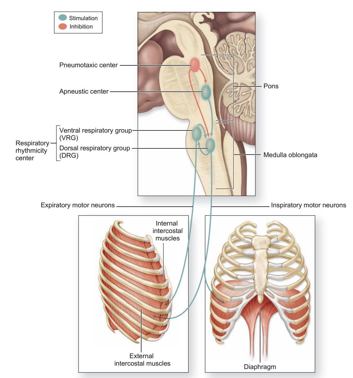
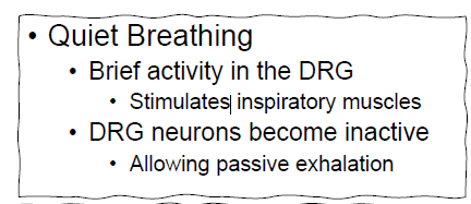
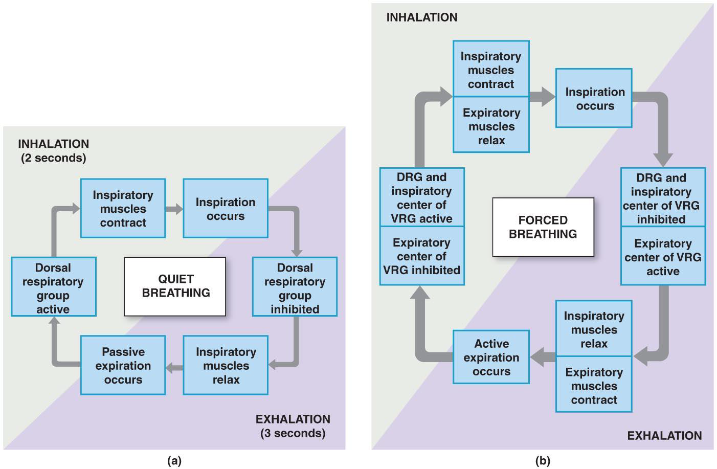
During increases in breathing, the inspiratory and expiratory motor neurons and muscles are not activated at the same time but, rather, alternate in function. 在呼吸增加的过程中,吸气和呼气运动神经元和肌⾁不会同时激活,而是交替发挥作⽤
- The Apneustic and Pneumotaxic Centers of the Pons 脑桥的长吸中枢和呼吸调整中枢
• Paired nuclei that adjust output of respiratory rhythmicity centers 调节呼吸调整中枢输出的成对核
• Regulating respiratory rate and depth of respiration 调节呼吸频率和呼吸深度 - Apneustic Centre 长吸中枢
• Provides continuous stimulation to its DRG center 使吸气延长 - Pneumotaxic Centers 呼吸调整中枢
• Inhibit the apneustic centers 抑制长吸中枢
• Promote passive or active exhalation 促进被动或主动呼气
The Hering-Breuer Reflexes
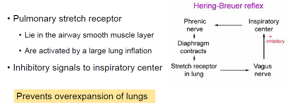
5.4.2 Chemoreceptor Reflex of Breathing
5.4.2.1 Chemoreceptors
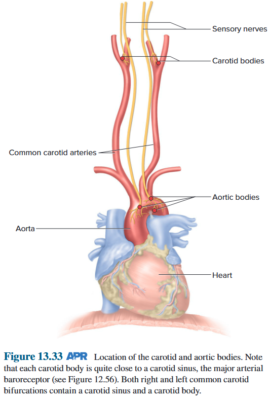
Peripheral chemoreceptor 外周化学感受器
- Carotid body 颈动脉体
- The glossopharyngeal nerve (Cranial nerve IX)
- Stimulated by changes in blood pH or $P_{O_2}$
- Aortic body 主动脉体
- The vagus nerve (Cranial nerve X)
- Stimulated by changes in blood pH or $P_{O_2}$
The peripheral chemoreceptors, located high in the neck at the bifurcation of the common carotid arteries and in the thorax on the arch of the aorta (Figure 13.33), are called the carotid bodies and aortic bodies, respectively. In both locations, they are quite close to, but distinct from, the arterial baroreceptors and are in intimate contact with the arterial blood. The carotid bodies, in particular, are strategically located to monitor oxygen supply to the brain. 外周化学感受器位于颈部高位颈总动脉分叉处和胸腔主动脉弓处(图 13.33),分别称为颈动脉体和主动脉体。在这两个位置,它们都非常接近但又不同于动脉压力感受器,并且与动脉血液密切接触。特别是颈动脉体的位置非常重要,可以监测大脑的氧气供应。
Central chemoreceptor 中枢化学感受器
- Respiratory centre of medulla oblongata 延髓呼吸中枢
- ventrolateral surface
- monitor cerebrospinal fluid 监测脑脊液
- respond to pH and $P_{CO_2}$ change
The central chemoreceptors are located in the medulla and, like the peripheral chemoreceptors, provide excitatory synaptic input to the medullary inspiratory neurons. They are stimulated by an increase in the H^+^ concentration of the brain’s extracellular fluid. As we will see later, such changes result mainly from changes in blood $P_{CO_2}$. 中枢化学感受器位于髓质中,与外周化学感受器一样,为髓质吸气神经元提供兴奋性突触输入。它们受到大脑细胞外液 H^+^ 浓度增加的刺激。正如我们稍后将看到的,这种变化主要是由血液 $P_{CO_2}$ 的变化引起的。
5.4.2.2 Control of ventilation by $P_{O_2}$
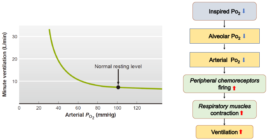
5.4.2.3 Control of ventilation by $P_{CO_2}$
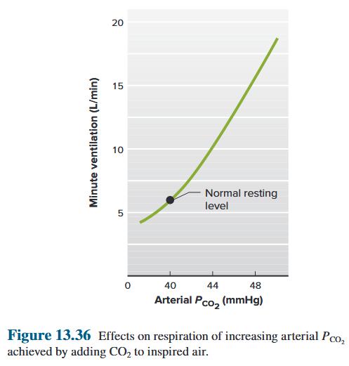
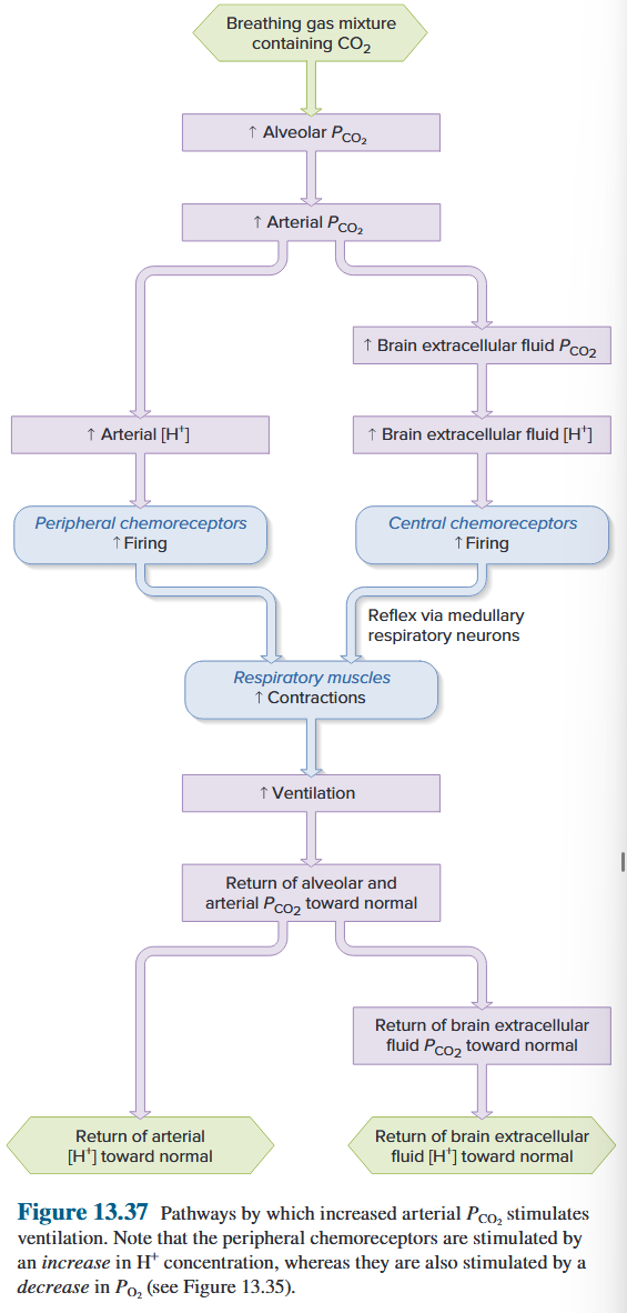
5.4.2.4 Control Ventilation by $H^+$
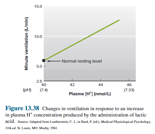
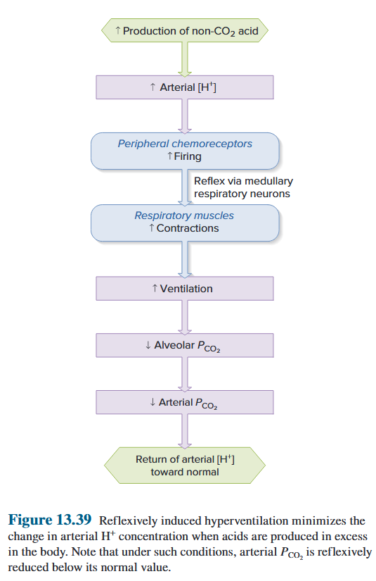
Chapter 4 Cardiovascular Physiology
The Cardiovascular system (CVS) serves to provide rapid transport of nutrients to the tissues in the body and allow rapid removal of waste products.
Main functions of CVS
- Rapid transport of nutrients (oxygen, amino acids, glucose, fatty acids, water, etc.) 快速运输营养物质(氧气、氨基酸、葡萄糖、脂肪酸、⽔等)
- Removal of waste products of metabolism (carbon dioxide, urea, creatinine, etc.) 清除代谢废物(二氧化碳、尿素、肌酐等)
- Hormonal control, by transporting hormones to their target organs and by secreting its own hormones (e.g. atrial natriuretic peptide) 激素控制,通过将激素输送到⽬标器官并分泌其⾃身激素(例如⼼房利钠肽)
- Temperature regulation, by controlling heat distribution between the body core and the skin (e.g. arteriovenous shunt). 温度调节,通过控制身体核⼼和皮肤
- Host defense, transporting immune cells, antigen and other mediators (e.g. antibody). 宿主防御,运输免疫细胞、抗原和其他介质(例如抗体)
Circulatory system
- The Heart (a biological pump)
- Blood vessels or vascular system (interconnected tubes)
- Blood (a fluid connective tissue)
Circulatory system consists of the pulmonary circulation and the systemic circulation. As depicted by the color change from blue to red, blood becomes fully oxygenated (red) as it flows through the lungs and then loses some oxygen (red to blue) as it flows through the other organs and tissues.
The systemic circulation: Left ventricle → Aorta → Systemic arteries → Capillaries → Systemic veins → Right atrium
The pulmonary circulation: Right ventricle → Pulmonary arteries → Lungs → Pulmonary capillaries → Pulmonary veins → Left atrium
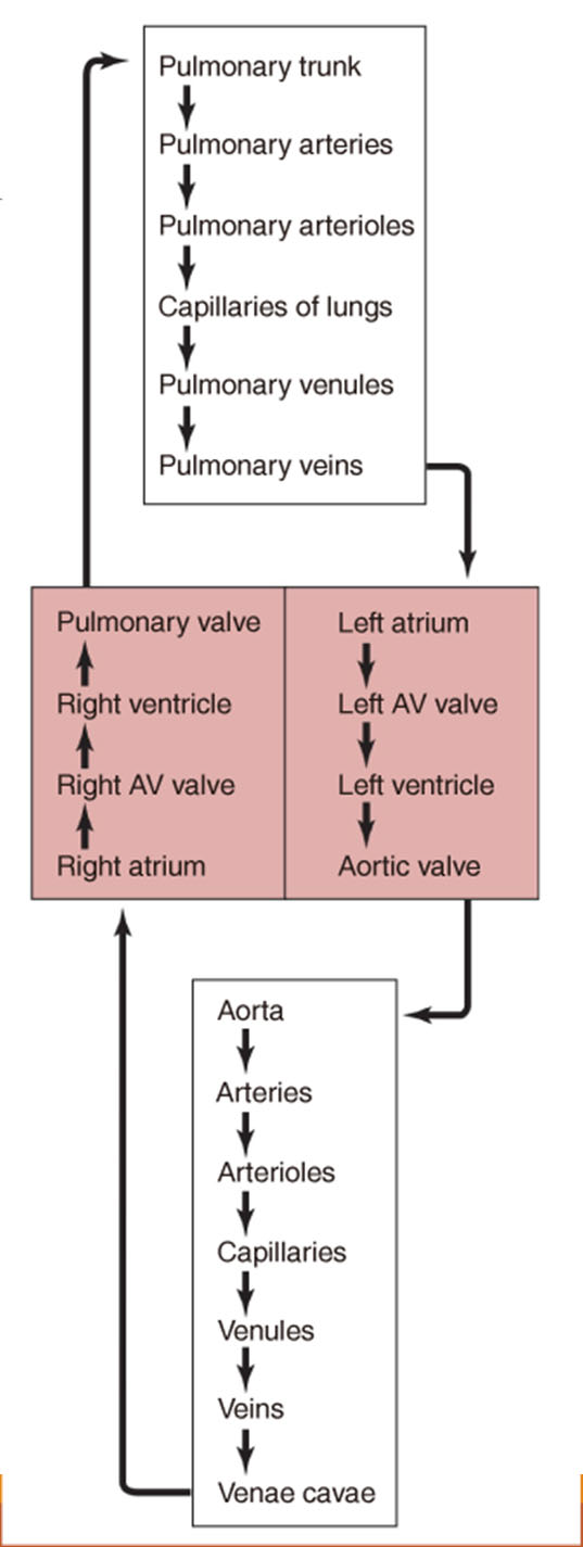
4.1 The heart
4.1.1 Anatomy [Review]
The heart is a muscular organ enclosed in a protective fibrous sac, the pericardium, and located in the left of chest. A fibrous layer is also closely affixed to the heart and is called the epicardium (心外膜). ⼼脏是⼀个肌⾁器官,位于胸腔左侧,被⼀个保护性的纤维囊(⼼包)包裹着。还有⼀层纤维层紧密贴合在⼼脏上,称为⼼外膜。
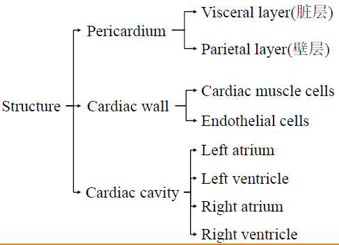
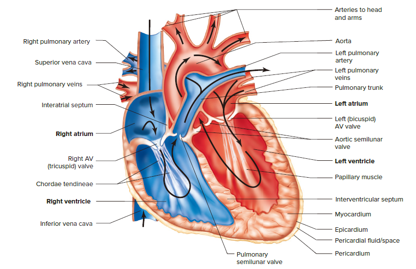
Pericardium
- Visceral layer (脏层)
- Parietal layer (壁层)
Cardiac wall
The wall of the heart, the myocardium, is composed primarily of cardiac muscle cells. The inner surface of the cardiac chambers, as well as the inner wall of all blood vessels, is lined by a thin layer of cells known as endothelial cells, or endothelium. ⼼脏壁,即⼼肌,主要由⼼肌细胞组成。⼼腔内表⾯以及所有⾎管内壁均覆盖⼀层薄薄的细胞,称为内皮细胞或内皮
Cardiac cavity
- Left atrium
- Left ventricle
- Right atrium
- Right ventricle
- Atrial septum: separate two atriums
- Interventricular septum: separate two ventricles
Valves
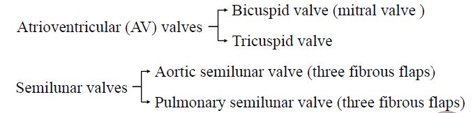
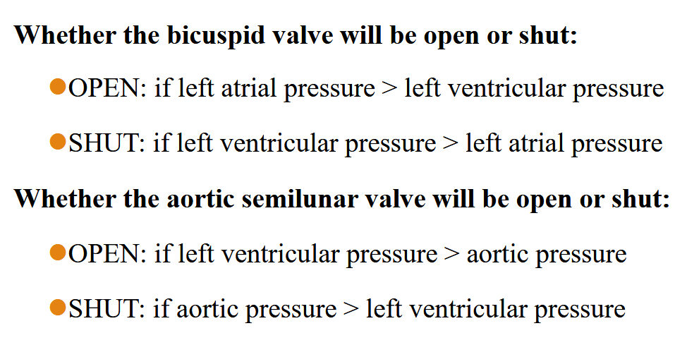
Function of valves:
- Prevent backward flow of blood 防⽌⾎液回流
- Open and close in response to pressure gradient across the valve 根据阀⻔上的压⼒梯度打开和关闭
Innervation 支配
Sympathetic nerve → Norepinephrine → β-adrenergic receptor → Entire heart 交感神经 → 去甲肾上腺素 → β‑肾上腺素受体 → 整个⼼脏
Vagus nerve → Acetylcholine → M-muscarinic receptor → Atria 迷⾛神经 → ⼄酰胆碱 → M‑毒蕈碱受体 → ⼼房
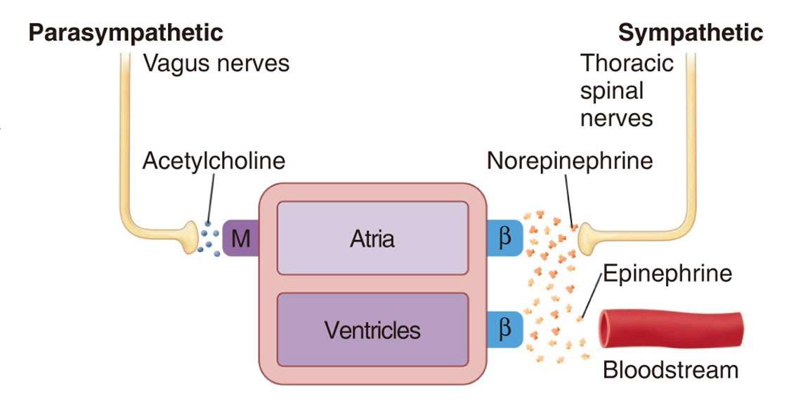
Blood Supply
Heart receives their blood supply via arteries that branch from the aorta. The arteries supplying the myocardium are the coronary arteries, and the blood flowing through them is the coronary blood flow. The coronary arteries exit from behind the aortic valve cusps in the very first part of the aorta and lead to a branching network of small arteries, arterioles, capillaries, venules, and veins similar to those in other organs. Most of the cardiac veins drain into a single large vein, the coronary sinus, which empties into the right atrium. ⼼脏通过从主动脉分支出来的动脉获得⾎液供应。供应⼼肌的动脉是冠状动脉,流经冠状动脉的⾎液是冠状动脉⾎流。冠状动脉从主动脉最开始部分的主动脉瓣瓣尖后⾯出来,通向与其他器官类似的⼩动脉、⼩动脉、毛细⾎管、⼩静脉和静脉的分支⽹络。大多数⼼脏静脉流⼊⼀条大静脉,即冠状窦,再流⼊右⼼房。
4.1.2 Mechanical Events of the Cardiac Cycle
心脏的一次收缩和舒张构成的一个机械活动周期,称为心动周期(cardiac cycle)。在一个心动周期中,心房和心室的机械活动都可分为收缩期(systole)和舒张期(diastole)。由于心室在心脏泵血活动中起主要作用,故心动周期通常是指心室的活动周期。Consists of systole (contraction) and then diastole (relaxation) of both atria followed by systole and then diastole of both ventricles.
A typical heart rate is 72 beats/min, and each cardiac cycle lasts approximately 0.8s.
ventricular contraction: 0.3s
ventricular relaxation: 0.5s
atrial contraction: 0.1s
atrial relaxation: 0.7s
Atrial contraction occurs at the end of diastole, after most of the ventricular filling has taken place. The ventricle receives blood throughout most of diastole, not just when the atrium contracts. ⼼房收缩发生在舒张末期,即⼼室充盈大部分时间之后。⼼室在大部分舒张期都会接受⾎液,而不仅仅是在⼼房收缩时
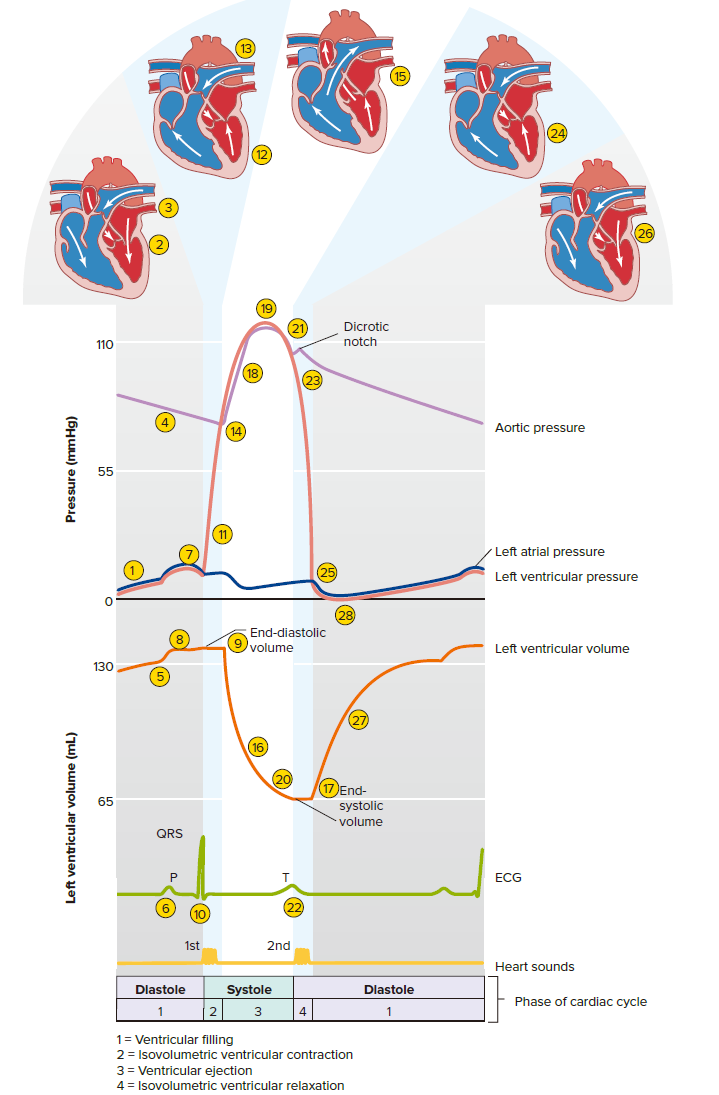
4.1.2.1 Mid-Diastole to Late Diastole
对心室活动周期而言,心房收缩期(period of atrial systole)实际上是前一周期的舒张末期。心房收缩前,心脏处于全心舒张期,此时处于半月瓣关闭、房室瓣开启状态,血液从静脉经心房流入心室,使心室不断充盈。在全心舒张期内,回流入心室的血液量约占心室总充盈量的75%。全心舒张期之后是心房收缩期,历时0.1秒,心房壁较薄、收缩力不强,由心房收缩推动进入心室的血液通常只占心室总充盈量的25%左右。心房收缩时,心房内压和心室内压都轻度升高,但由于大静脉的心房入口处环形肌也收缩,再加上血液向前的惯性,所以虽然大静脉和心房交接处没有瓣膜,心房内的血液很少会反流入大静脉。
- The left atrium and ventricle are both relaxed, but atrial pressure is slightly higher than ventricular pressure because the atrium is filled with blood that is entering from the veins. 左⼼房和左⼼室均放松,但⼼房压⼒略⾼于⼼室压⼒,因为⼼房充满了从静脉进⼊的⾎液。
- The AV valve is held open by this pressure difference, and blood entering the atrium from the pulmonary veins continues on into the ventricle. AV 瓣膜在压⼒差的作⽤下保持打开,⾎液从肺静脉继续进⼊⼼室。
- The aortic valve is closed because the aortic pressure is higher than the ventricular pressure. 由于主动脉压⼒⾼于⼼室压⼒,主动脉瓣关闭。
- The aortic pressure is slowly decreasing because blood is moving out of the arteries and through the vascular system. 主动脉压⼒缓慢下降,因为⾎液正在流出动脉。
- In contrast, ventricular pressure is increasing slightly because blood is entering the relaxed ventricle from the atrium. 相反,⼼室压⼒略有增加,因为⾎液进⼊放松的⼼室与⼼房。
- Near the end of diastole, the SA node discharges and the atria depolarize, as signified by the P wave of the ECG. 舒张末期,窦房结放电,⼼房去极化,表现为⼼电图的 P 波。
- Atrium contraction of the causes atrial pressure increasing. ⼼房收缩引起⼼房压⼒升⾼。
- The elevated atrial pressure forces a small additional volume of blood into the ventricle. ⼼房压⼒升⾼迫使少量额外的⾎液进⼊⼼室。
- The end of ventricular diastole, so the amount of blood in the ventricle at this time is called the end-diastolic volume (EDV). ⼼室舒张末期,此时⼼室内的⾎量称为舒张末期容积(EDV)。
4.1.2.2 Systole 心缩期
- isovolumic contraction period 等容收缩期
- ejection period 射血期
- period of rapid ejection 快速射血期 70% 0.1s
- period of reduced ejection 减慢射血期 30% 0.15s
-
From the AV node, the wave of depolarization passes into and throughout the ventricular tissue—as signified by the QRS complex of the ECG—and this triggers ventricular
contraction. 从房室结开始,去极化波进⼊并穿过⼼室组织(如⼼电图的 QRS 复合波所⽰),从而引发⼼室收缩。 -
As the ventricle contracts, ventricular pressure increases rapidly; almost immediately, this pressure exceeds the atrial pressure. 当⼼室收缩时,⼼室压⼒迅速增加;几乎立即,这压⼒超过⼼房压⼒。
-
This change in pressure gradient forces the AV valve to close, thus preventing the backflow of blood into the atrium. 压⼒梯度的变化迫使 AV 瓣膜关闭,从而防⽌回流⾎液进⼊⼼房。
-
Because the aortic pressure still exceeds the ventricular pressure at this time, the aortic valve remains closed and the ventricle cannot empty despite its contraction. For a brief time, then, all valves are closed during this phase of isovolumetric ventricular contraction. 由于此时主动脉压⼒仍超过⼼室压⼒,主动脉瓣保持关闭状态,⼼室尽管收缩,但⽆法排空。因此,在等容⼼室收缩的这⼀阶段,所有瓣膜都会短暂关闭。
-
This brief phase ends when the rapidly increasing ventricular pressure exceeds aortic pressure. 当快速增加的⼼室压⼒超过主动脉压⼒。
-
The pressure gradient now forces the aortic valve to open, and ventricular ejection begins. 现在压⼒梯度迫使主动脉瓣打开,⼼室射⾎开始。
-
The ventricular volume curve shows that ejection is rapid at first and then slows down. ⼼室容积曲线表现为射⾎起初较快,随后减慢。
-
The amount of blood remaining in the ventricle after ejection is called the end-systolic volume (ESV). 射⾎后⼼室内剩余的⾎液量称为收缩末期⾎流量体积。
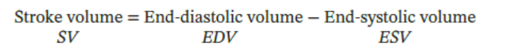
-
As blood flows into the aorta, the aortic pressure increases along with the ventricular pressure. 当⾎液流⼊主动脉时,主动脉压⼒随着⼼室压⼒的增加而增加。
-
Peak ventricular and aortic pressures are reached before the end of ventricular ejection. 在⼼室射⾎结束之前,⼼室和主动脉压⼒达到峰值。
-
This force reduction is evidenced by the reduced rate of blood ejection during the last part of systole. 这种⼒量的减少可以通过收缩期最后阶段⾎液喷射率的降低来证明。
-
The volume and pressure in the aorta decrease as the rate of blood ejection from the ventricles becomes slower than the rate at which blood drains out of the arteries into the tissues. 由于心室射血速度低于动脉血液流入组织的速度,主动脉的容积和压力会下降。
4.1.2.3 Early Diastole
- T wave of the ECG corresponds to ventricular repolarization. 心电图的 T 波对应于心室复极化。
- As the ventricles relax, the ventricular pressure decreases below aortic pressure. The change in the pressure gradient forces the aortic valve to close. 随着心室舒张,心室压力降至主动脉压力以下。压力梯度的变化迫使主动脉瓣关闭。
- The AV valve also remains closed because the ventricular pressure is still higher than atrial pressure. For a brief time, then, all valves are again closed during this phase of isovolumetric
ventricular relaxation. AV 瓣膜也保持关闭状态,因为心室压力仍然高于心房压力。然后,在等容心室舒张的这一阶段,所有瓣膜都会短暂地再次关闭。 - This phase ends as the rapidly decreasing ventricular pressure decreases below atrial pressure. 当快速下降的心室压力降至心房压力以下时,这一阶段结束。
- This change in pressure gradient results in the opening of the AV valve. 这种压力梯度的变化导致 AV 瓣膜打开。
- Venous blood that had accumulated in the atrium since the AV valve closed flows rapidly into the ventricles. 自 AV 瓣膜关闭以来,在心房中积聚的静脉血迅速流入心室。
- The rate of blood flow is enhanced during this initial filling phase by a rapid decrease in ventricular pressure. 在这个初始充盈阶段,心室压力的快速下降提高了血流速度。
-
isovolumic relaxation period 等容舒张期
射血后,心室开始舒张,室内压下降,主动脉内的血液向心室方向反流,推动半月瓣使之关闭;但此时室内压仍高于房内压,故房室瓣仍处于关闭状态,心室又暂时成为一个封闭的腔。从半月瓣关闭至房室瓣开启前的这一段时间内,心室舒张而心室的容积并不改变。
-
period of ventricular filling 心室充盈期
随着心室肌的舒张,室内压进一步下降,当室内压下降到低于房内压时,心房内的血液冲开房室瓣进入心室。
- period of rapid filling 快速充盈期 2/3 0.11s
- period of reduced filling 减慢充盈期 0.22s
| AV valves | Semilunar valves | |
|---|---|---|
| Isovolumetric ventricular contraction 等容⼼室收缩 | closed | closed |
| Ventricular ejection ⼼室射⾎ | closed | open |
| Isovolumetric ventricular relaxation 等容⼼室舒张 | closed | closed |
| Ventricular filling ⼼室充盈 | open | closed |
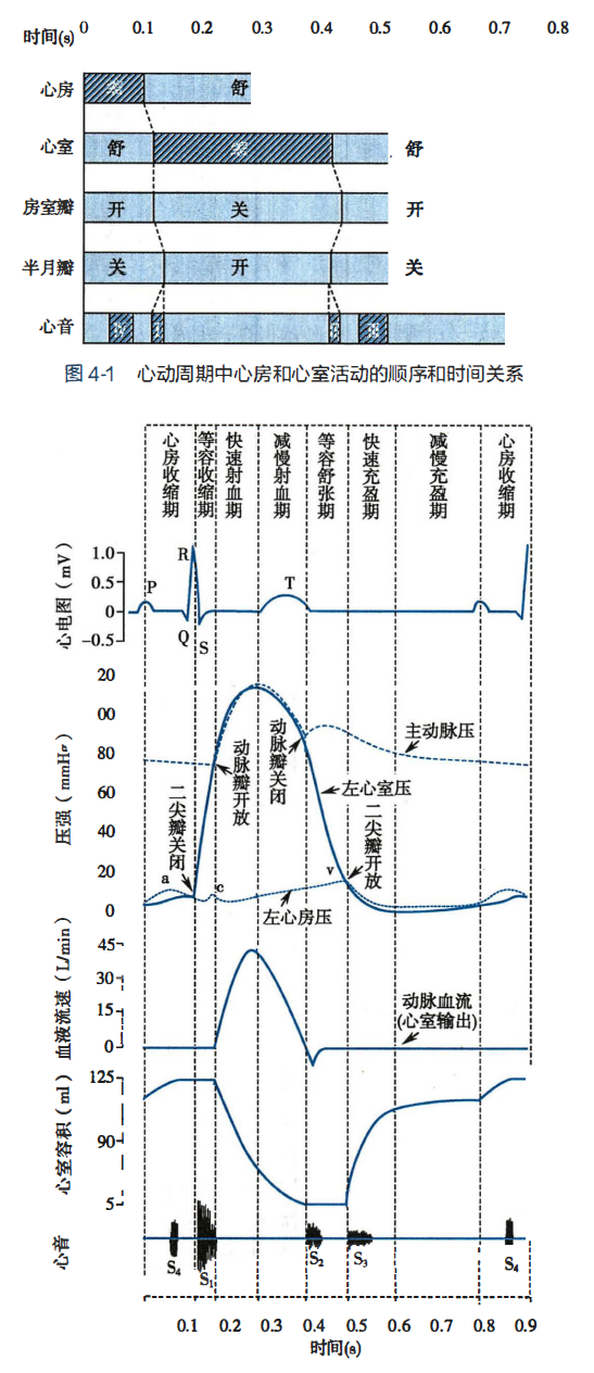
4.1.3 The Cardiac Output
Cardiac output (CO): 一侧心室一次心脏搏动所射出的血液量,称为每搏输出量(stroke volume),简称搏出量。一侧心室每分钟射出的血液量,称为心输出量(cardiac output),也称每分输出量或心排出量。The volume of blood each ventricle pumps as a function of time, usually expressed in liters per minute.
正常成年人在安静状态下,左心室舒张期末容积(end-diastolic volume,EDV)约125ml,收缩期末容积(end-systolic volume,ESV)约55ml,两者差值即为搏出量,约70ml(60~80ml)。左、右两侧心室的心输出量基本相等。心输出量等于心率与搏出量的乘积。心输出量与机体的新陈代谢水平相适应,可因性别、年龄及其他生理情况的不同而不同。如果心率为75次/分,搏出量为70ml,则心输出量约为5L/min。一般健康成年男性在安静状态下的心输出量为4.5~6.0L/min。
The cardiac output can be calculated by multiplying the heart rate (HR)—the number of beats per minute—and the stroke volume (SV)—the blood volume ejected by each ventricle with each beat:
$$
CO = HR \times SV
$$
Ejection fraction (EF): 搏出量占心室舒张期末容积的百分比,称为射血分数(ejection fraction)。One way to quantify contractility is through the ejection fraction (EF), defined as the ratio of stroke volume (SV) to enddiastolic volume (EDV):
$$
EF = SV / EDV
$$
一侧心室每分钟射出的血液量,称为心输出量(cardiac output),也称每分输出量或心排出量。左、右两侧心室的心输出量基本相等。心输出量等于心率与搏出量的乘积。
心输出量与机体的新陈代谢水平相适应,可因性别、年龄及其他生理情况的不同而不同。如果心率为75次/分,搏出量为70ml,则心输出量约为5L/min。一般健康成年男性在安静状态下的心输出量为4.5~6.0L/min。
The average total blood volume is about 5.5 L, nearly all the blood is pumped around the circuit once each minute. During periods of exercise in well-trained athletes, the cardiac output may reach 35 L/min; the entire blood volume is pumped around the circuit almost seven times per minute! Even untrained individuals can reach cardiac outputs of 20-25 L/min during exercise. 平均总血量约为5.5升,几乎所有血液每分钟被泵入回路一次。训练有素的运动员运动时,心输出量可达35L/min;整个血液量几乎每分钟在回路中泵送七次!即使未经训练的人在运动时也能达到 20-25 L/min 的心输出量。
4.1.3.1 Control of heart rate
The slope of phase 4 depolarization in SA nodal cells determines heart rate. SA node is normally under the constant influence of nerves and hormones. SA 节点细胞第 4 阶段去极化的斜率决定了心率。SA 节点通常受到神经和激素的持续影响。
Sympathetic neurous: increase
parasympathetic neurous: decrease
In the resting state, more parasympathetic activity to the heart than sympathetic, so the normal resting heart rate of about 70 beats/min is well below the inherent rate of 100 beats/min. 在静息状态下,副交感神经对心脏的活动比交感神经多,因此正常的静息心率约为 70 次/分钟,远低于固有的 100 次/分钟。
Sympathetic stimulation increases the slope of the pacemaker potential by increasing Na^+^ entering the cell. This causes the SA node cells to reach threshold more rapidly and the heart rate to increase. Increasing parasympathetic input has the opposite effect-the slope of the pacemaker potential decreases due to a reduction in the inward current. Threshold is therefore reached more slowly, and heart rate decreases. Parasympathetic stimulation also hyperpolarizes the plasma membranes of SA node cells by increasing their permeability to K^+^. The pacemaker potential thus starts from a more negative value and has a reduced slope.
交感神经刺激通过增加进入细胞的 Na^+^ 来增加起搏器电位的斜率。这导致 SA 节点细胞更快地达到阈值并且心率增加。增加副交感神经输入会产生相反的效果——起搏器电位的斜率由于内向电流的减少而降低。因此,达到阈值的速度会更慢,心率也会降低。副交感神经刺激还通过增加 SA 节点细胞对 K^+^ 的通透性,使 SA 节点细胞的质膜超极化。因此,起搏器电位从更负的值开始,并且斜率减小。

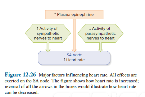
Epinephrin 肾上腺素 (Adrenal medulla 肾上腺髓质)
Norepinephrine 去甲肾上腺素
Acetylcholine 甲状腺激素
4.1.3.2 Control of Stroke Volume
The ventricles do not completely empty themselves during contraction. Therefore, a more forceful contraction can produce an increase in stroke volume by causing greater emptying. Dominant factors influencing stroke volume: 收缩期间心室不会完全排空。因此,更有力的收缩可以通过引起更大的排空来增加每搏输出量。影响每搏输出量的主要因素:
- end-diastolic volume (preload) 舒张期末容积(前负荷)
- sympathetic regulation 交感神经调节
- afterload: how hard the heart must work to eject blood 后负荷
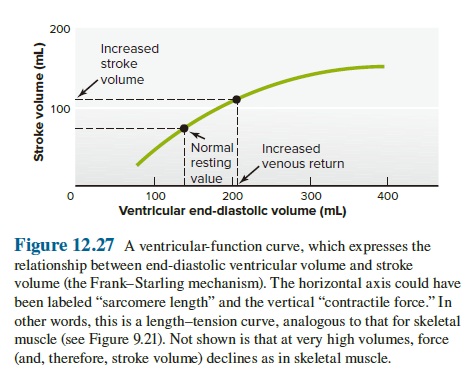
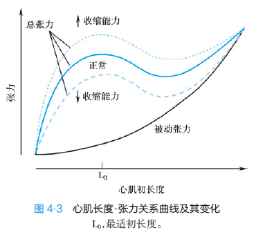
-
changes in the end-diastolic volume (the volume of blood in the ventricles just before contraction, sometimes referred to as the preload)
- the ventricle contracts more forcefully during systole when it has been filled to a greater degree during diastole. When all other factors being equal, the stroke volume increases as the end-diastolic volume increases. 当心室在舒张期被更大程度地充盈时,心室在收缩期收缩得更剧烈。当所有其他因素相同时,每搏输出量随着舒张末期容量的增加而增加。
前负荷可使骨骼肌在收缩前处于一定的初长度。对中空、近似球形的心脏来说,心室肌的初长度取决于心室舒张期末的血液充盈量,换言之,心室舒张期末容积相当于心室的前负荷。异长自身调节的生理学意义:异长自身调节的主要生理学意义是对搏出量的微小变化进行精细的调节,使心室射血量与静脉回心血量之间保持平衡,从而使心室舒张期末容积和压力保持在正常范围内。
-
changes in the magnitude of sympathetic nervous system input to the ventricles
- the increased sympathetic nerve stimulation or epinephrine will enhance contraction and stroke volume. 增加交感神经刺激或肾上腺素会增强收缩和每搏输出量
- 心肌不受副交感神经支配。
Area Affected Sympathetic Nerves (Norepinephrine on β-Adrenergic Receptors) Parasympathetic Nerves (ACh on Muscarinic Receptors) SA node heart rate ↑ heart rate ↓ AV node conduction rate ↑ conduction rate ↓ Atrial muscle contractility ↑ contractility ↓ Ventricular muscle contractility ↑ —— -
changes in afterload (i.e., the arterial pressures against which the ventricles pump)
- An increased arterial pressure tends to reduce stroke volume. The arterial pressure constitutes a “load” that contracting ventricular muscle must work against when it is ejecting blood. The greater the load, the less contracting muscle fibers can shorten at a given contractility. In the normal heart, several inherent adjustments minimize the overall influence of arterial pressure on stroke volume. However, in the sections on high blood pressure and heart failure, we will see that alterations in vascular resistance and long-term elevations of arterial pressure can weaken the heart and thereby influence stroke volume. 动脉压增加往往会减少每搏输出量。动脉压构成一种“负荷”,收缩的心室肌在喷射血液时必须对抗该“负荷”。负荷越大,在给定的收缩力下,收缩的肌纤维可以缩短得越少。在正常心脏中,一些固有的调整可以最大限度地减少动脉压对每搏输出量的总体影响。然而,在高血压和心力衰竭部分,我们会看到血管阻力的改变和动脉压的长期升高会削弱心脏,从而影响每搏量。
心室收缩时,必须克服大动脉血压,才能将血液射入动脉内。因此,大动脉血压是心室收缩时所遇到的后负荷。在心肌初长度、收缩能力和心率都不变的情况下,如果大动脉血压增高,等容收缩期室内压的峰值将增高,结果使等容收缩期延长而射血期缩短,射血期心室肌缩短的程度和速度都减小,射血速度减慢,搏出量减少;反之,大动脉血压降低,则有利于心室射血。
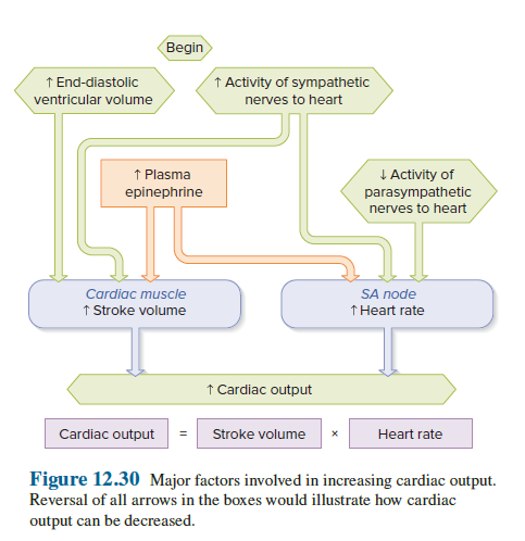
4.1.2 Electrophysiology of Cardiac Muscle
根据组织学和电生理学特点,可将心肌细胞分成工作细胞(working cell)和自律细胞(autorhythmic cell),前者包括心房肌和心室肌,它们有稳定的静息电位,主要执行收缩功能。后者主要包括窦房结细胞和浦肯野细胞,它们组成心内特殊传导系统,大多没有稳定的静息电位,并可自动产生节律性兴奋。
根据心肌细胞动作电位去极化的快慢及其产生机制,又可将心肌细胞分成快反应细胞(fast response cell)和慢反应细胞(slow response cell)。快反应细胞包括心房、心室肌和浦肯野细胞,其动作电位的特点是去极化速度和幅度大,兴奋传导速度快,复极过程缓慢并且可分成几个时相,因而动作电位时程很长。慢反应细胞包括窦房结和房室结细胞,其动作电位特点是去极化速度和幅度小,兴奋传导速度慢,复极过程缓慢而没有明确的时相区分。快反应细胞和慢反应细胞在某些实验条件或病理情况下,可发生转变。
Fast response non-autorhythmic cell: Ventricular and atrial myocardium
Fast response autorhythmic cell: Purkinje cells
Slow response non-autorhythmic cell: Atrioventricular node
Slow response autorhythmic cell: Sinoatrial node
4.1.2.1 Myocardial Cell Action Potentials (Fast response non-autorhythmic cell)
Phase 0: Na^+^ enters
Phase 1: K^+^ exits
Phase 2: K^+^ exits || Ca^2+^ enters
Phase 3: K^+^ exits
Phase 4: Na^+^-K^+^ ATPase
The depolarizing phase of the AP is due mainly to the opening of voltage-gated Na+ channels. Sodium ion entry depolarizes the cell and sustains the opening of more Na+ channels in positive feedback fashion. AP 的去极化阶段主要是由于电压门控 Na+ 通道的打开。钠离子的进入使细胞去极化,并以正反馈方式维持更多 Na+ 通道的开放。
4.1.2.2 Nodal Cell Action Potentials (Slow response autorhythmic cell)
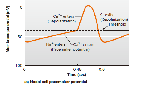
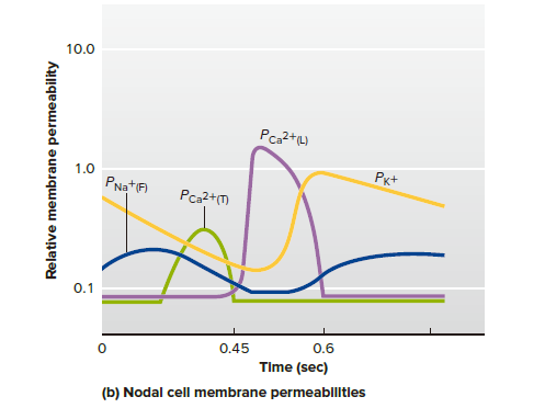
Phase 0: Ca^2+^ enters (mainly)
Phase 1: -
Phase 2: -
Phase 3: K^+^ exits
Phase 4: Na^+^ enters, Ca^2+^ enters, less K^+^ exits over time
The SA node cell does not have a steady resting potential but, instead, undergoes a slow depolarization. This gradual depolarization is known as a pacemaker potential; it brings the membrane potential to threshold, at which point an AP occurs. SA 节点细胞不具有稳定的静息电位,而是经历缓慢的去极化。这种逐渐去极化被称为起搏电位;它将膜电位带到阈值,此时发生 AP。
The depolarizing phase is caused not by Na^+^ but rather by Ca^2+^ influx that is slowly than voltage-gated Na+ channels, and AP propagate is more slowly along nodal-cell membranes than in other cardiac cells. This explains the slow transmission of cardiac excitation through the AV node. 去极化阶段不是由 Na^+^ 引起的,而是由 Ca^2+^ 流入引起的,Ca^2+^ 流入比电压门控 Na^+^ 通道慢,并且 AP 沿着结细胞膜传播比在其他心肌细胞中更慢。这解释了心脏兴奋通过房室结传输缓慢的原因。
This gradual depolarization is known as a pacemaker potential; it brings the membrane potential to threshold, at which point an AP occurs.
4.1.3 Myocardial Characteristics
- Excitability 兴奋性
- Autorhythmicity 自律性
- Conductivity 传导性
- Contractility 收缩性
Non-autorhythmic cell: 1.3.4.
Autorhythmic cell: 1.2.3.
4.1.3.1 Excitability
心肌细胞每产生一次兴奋,其膜电位将发生一系列规律性变化,兴奋性也因之而产生相应的周期性变化。这种周期性变化,使心肌细胞在不同时期内对重复刺激表现出不同的反应特性,从而对心肌兴奋的产生和传导,甚至对收缩反应产生重要影响。
Excitability: The ability to produce electrical signals that can transmit information between different regions of the membrane. 产生可在膜的不同区域之间传递信息的电信号的能力。
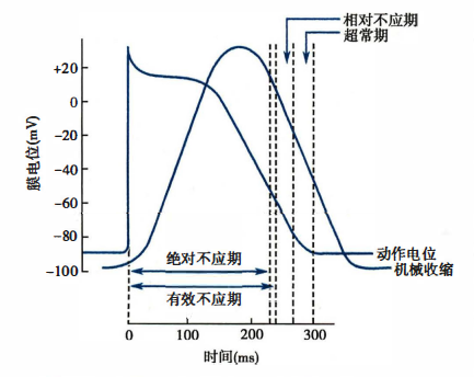
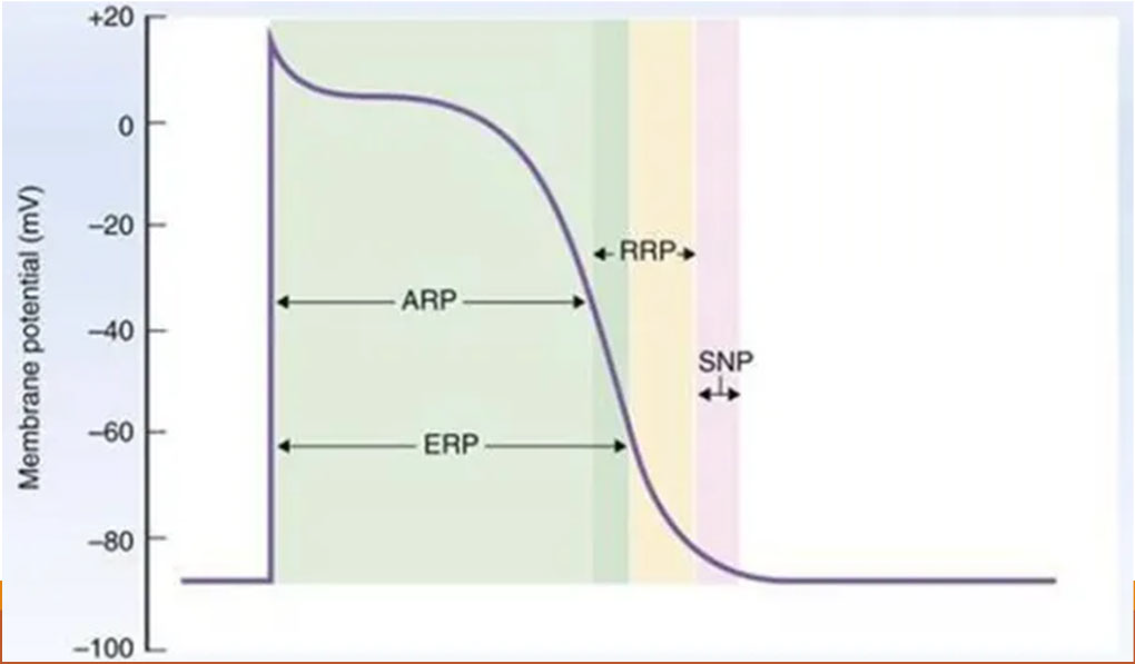
Effective refractory period (ERP) : Absolute refractory period (ARP) + Local response period
Absolute refractory period (ARP)
- 从0期去极化开始到复极化3期膜电位达-55mV这一段时间内,无论给予多强的刺激,都不会引起心肌细胞产生去极化反应,此段时期称为绝对不应期(absolute refractory period, ARP) 。从复极至-55mV继续复极至-60mV的这段时期内,若给予阈上刺激虽可引起局部反应,但仍不会产生新的动作电位,这一时期称为局部反应期(local response period) 。上述两段时期合称为有效不应期(effective refractory period, ERP) 。此期内心肌细胞兴奋性的暂时缺失或极度下降是由于钠通道完全失活或尚未恢复到可以被激活的备用状态的缘故。但兴奋性的下降是可逆的。心肌的ERP特别长,是兴奋性变化的重要特点。
Relative refactory period (RRP)
- 从膜电位复极化-60mV至-80mV这段时间内,若给予阈上刺激,可使心肌细胞产生动作电位,此期称为相对不应期(relative refractory period , RRP) 。此期已有相当数量的钠通道复活到备用状态,但在阈刺激下激活的钠通道数量仍不足以产生使膜去极化达阈电位的内向电流,故需加强刺激强度方能引起一次新的兴奋。
Supranormal period (SNP)
- 心肌细胞继续复极,膜电位由-80mV 恢复到-90mV 这一段时期,其膜电位值虽低于静息电位,但钠通道已基本恢复到可被激活的备用状态,且膜电位水平与阈电位接近,故一个低于阈值的刺激即可引起一次新的动作电位,此即超常期(supranormal period, SNP) 。
Cardiac muscle is incapable of undergoing summation of contractions like that occurring in skeletal muscle, and this is a very good thing. If a prolonged, tetanic contraction were to occur in the heart, it would cease to function as a pump because the ventricles can adequately fill with blood only while they are relaxed. The inability of the heart to generate tetanic contractions is the result of the long absolute refractory period of cardiac muscle, defined as the period during and following an action potential when an excitable membrane cannot be re-excited. As in the case of neurons and skeletal muscle fibers, the main mechanism is the inactivation of Na^+^ channels. 该绝对不应期定义为动作电位期间和之后可兴奋膜无法重新兴奋的时期。与神经元和骨骼肌纤维的情况一样,主要机制是 Na^+^ 通道失活。
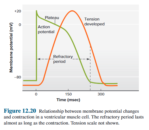
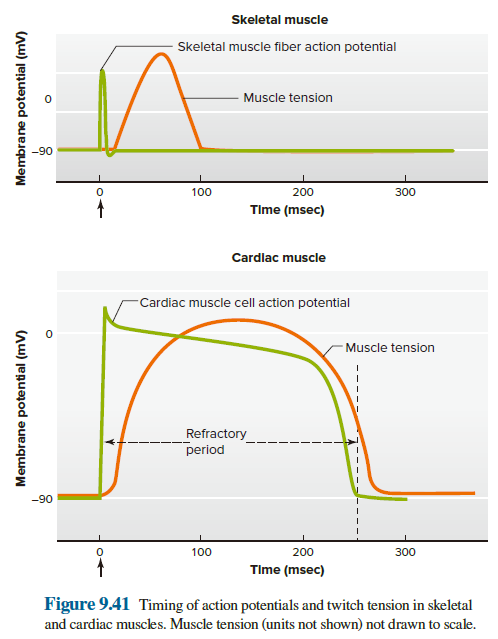
The absolute refractory period of skeletal muscle is much shorter (2 to 4 msec) than the duration of contraction (20 to 100 msec), so a second action potential can be elicited while the contraction resulting from the first action potential is still under way (see Figure 9.10). In contrast, because of the prolonged, depolarized plateau in the cardiac muscle action potential, the absolute refractory period of cardiac muscle lasts almost as long as the contraction (approximately 250 msec), and the muscle cannot be re-excited multiple times during an ongoing contraction. 骨骼肌的绝对不应期(2至4毫秒)比收缩持续时间(20至100毫秒)短得多,因此当第一个动作电位引起的收缩仍在进行时,可以引发第二个动作电位。相反,由于心肌动作电位的去极化平台时间较长,心肌的绝对不应期几乎与收缩一样长(约250毫秒),并且在持续的收缩过程中,不能多次重新兴奋。
在正常情况下,当窦房结产生的每一次兴奋传到心房肌和心室肌时,心房肌和心室肌前一次兴奋的不应期均已结束,因此能不断产生新的兴奋,于是,整个心脏就能按照窦房结的节律进行活动。如果在心室肌的有效不应期后,下一次窦房结兴奋到达前,心室受到一次外来刺激,则可提前产生一次兴奋和收缩,分别称为期前兴奋(premature excitation) 和期前收缩(premature systole) 。(形成二联律、三联律) 期前兴奋也有其自身的有效不应期,当紧接在期前兴奋后的一次窦房结兴奋传到心室时,如果正好落在期前兴奋的有效不应期内,则此次正常下传的窦房结兴奋将不能引起心室的兴奋和收缩,即形成一次兴奋和收缩的“ 脱失"'须待再下一次窦房结的兴奋传来时才能引起兴奋和收缩。这样,在一次期前收缩之后往往会出现一段较长的心室舒张期,称为代偿间歇(compensatory pause),然后再恢复窦性节律。但窦性心率较慢,下一次窦房结的兴奋也可在期前兴奋的有效不应期结束后才传到心室,在这种情况下,代偿性间歇将不会出现。
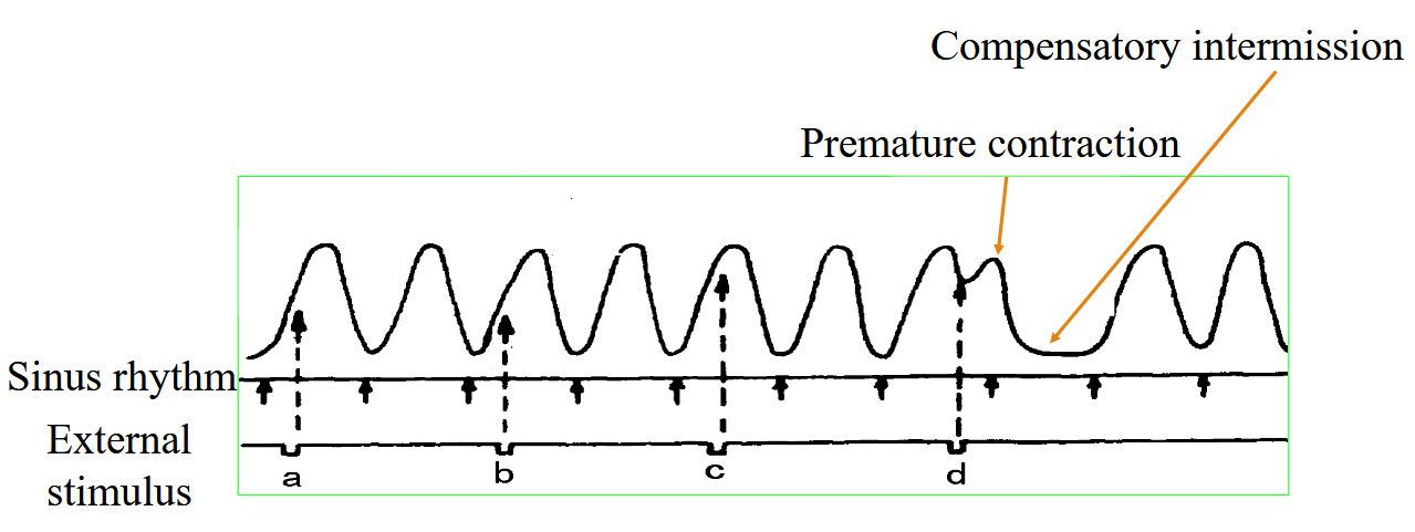
4.1.3.2 Autorhythmicity
Autorhythmicity: The capacity for spontaneous, rhythmic self-excitation. ⾃发、有节奏的⾃我兴奋的能⼒。
Sinoatrial node (90-100 beats/min) > Atrioventricular node (40-60 beats/min) > Purkinje fibres (20-40 beats/min)
Pacemaker: Sinoatrial node
Ectopic pacemakers: Atrioventricular node, Purkinje fibres
The pacemaker potential provides the SA node with automaticity, the capacity for spontaneous, rhythmic self-excitation. The slope of the pacemaker potential—that is, how quickly the membrane potential changes per unit time—determines how quickly threshold is reached and the next action potential is elicited. The inherent rate of the SA node—the rate exhibited in the absence of any neural or hormonal input to the node—is approximately 100 depolarizations per minute. Because other cells of the conducting system have slower inherent pacemaker rates, they normally are driven to threshold by action potentials from the SA node and do not manifest their own rhythm.
However, they can do so under certain circumstances and Purkinje network, no longer driven by the SA node, begin to initiate excitation at their own inherent rate and become the pacemaker for the ventricles. Their rate is quite slow, generally 25 to 40 beats/min. Therefore, when the AV node is disrupted, the ventricles contract completely out of synchrony with the atria, which continue at the higher rate of the SA node. Under such conditions, the atria are less effective because they are often contracting when the AV valves are closed. (房室传导阻滞)
起搏器电位为窦房结提供了自律性,即自发、节律性自激的能力。起搏器电位的斜率(即每单位时间膜电位变化的速度)决定了达到阈值和引发下一个动作电位的速度。 SA 节点的固有速率约为每分钟 100 次去极化。 由于传导系统的其他细胞具有较慢的固有起搏器速率,因此它们通常被来自 SA 节点的动作电位驱动到阈值,并且不会表现出它们的心率。自己的节奏。然而,他们可以在某些情况下这样做,并且浦肯野网络不再由 SA 节点驱动,开始以其自身固有的速率启动兴奋,并成为心室的起搏器。它们的速度相当慢,一般为25至40次/分钟。因此,当房室结受到干扰时,心室收缩与心房完全不同步,而心房结以较高的速率继续收缩。在这种情况下,心房的效率较低,因为当房室瓣膜关闭时它们经常收缩。
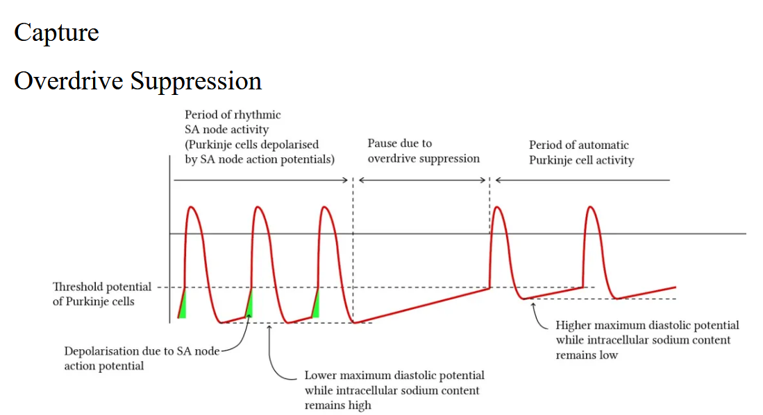
4.1.3.3 Conductivity
Conductivity: The action potential initiated in the sinoatrial node spreads throughout the myocardium, passing from cell to cell by way of gap junctions :在⼼房结发起的动作电位传遍整个⼼肌,通过间隙连接从⼀个细胞传递到另⼀个细胞
Purkinje fibres (4m/s) → Bundle of His (2m/s) → Ventricular myocardium (1m/s) → Atrial myocardium (0.4m/s) → Atrioventricular node (0.02m/s)
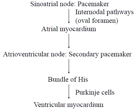
Atrioventricular node:
- Conducts action potential to ventricles
- Delays action potential to allow atria to complete contraction before ventricles contract 延迟动作电位,使⼼房在⼼室收缩之前完成收缩
Purkinje fibres:
- Large-diameter conduction cells
- Rapidly distribute the impulse throughout much of the ventricles
- Contact with ventricular myocardial cells, and spread the action potential. 与⼼室⼼肌细胞接触,并传播动作电位
4.1.3.4 Contractility
Contractility: Triggered by depolarization of the plasma membrane. 由质膜去极化引发
The heart is a dual pump in that the left and right sides of the heart pump blood separately-but simultaneously-into the systemic and pulmonary vessels. Efficient pumping of blood requires that the atria contract first, followed almost immediately by the ventricles (Synchronous contraction). The initial excitation of one cardiac cell therefore eventually results in the excitation of all cardiac cells (intercalated disc). 有效泵送血液需要心房首先收缩,然后心室几乎立即收缩。与骨骼肌和许多平滑肌一样,心肌的收缩是由质膜的去极化触发的。间隙连接将心肌细胞互连并允许动作电位从一个细胞传播到另一个细胞。一个心肌细胞的初始兴奋最终会导致所有心肌细胞的兴奋。
4.1.4 Electrocardiogram (ECG)
The electrocardiogram (ECG, also abbreviated EKG—the k is from the German elektrokardiogramm) is a tool for evaluating the electrical events within the heart. When action potentials occur simultaneously in many individual (contractile) myocardial cells, currents are conducted through the body fluids around the heart and can be detected by recording electrodes at the surface of the skin. 心电图(ECG,也缩写为 EKG,k 源自德语 elektrokardiogramm)是评估心脏内电活动的工具。当动作电位在许多单独的(收缩性)心肌细胞中同时出现时,电流通过心脏周围的体液传导,并且可以通过皮肤表面的记录电极来检测。
ECG is not a direct record of the changes in membrane potential across individual cardiac muscle cells. Instead, it measures the currents generated in the extracellular fluid by the changes occurring simultaneously in many cardiac cells. ECG is a powerful tool for diagnosing certain types of heart disease. 心电图并不是单个心肌细胞膜电位变化的直接记录。相反,它通过许多心脏细胞同时发生的变化来测量细胞外液中产生的电流。心电图是诊断某些类型心脏病的有力工具。
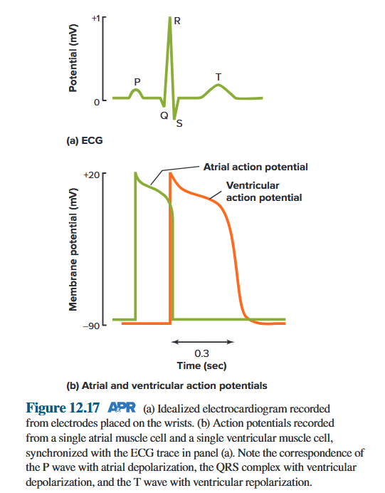
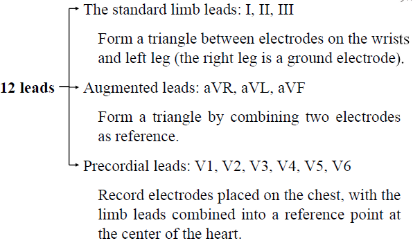
ECG
- Waves
- P wave: atrial depolarization 心房去极化
- QRS wave: ventricular depolarization 心室去极化
- T wave: ventricular repolarization 心室复极化
- Intervals
- P-R interval - PR间期代表由窦房结产生的兴奋经由心房、房室交界和房室束到达心室并引起心室肌开始兴奋所需要的时间,故也称为房室传导时间。当发生房室传导阻滞时,PR间期延长。临床上将房室传导功能分为正常、一度阻滞(PR间期延长,无心室漏搏)、二度阻滞(PR可以正常或延长,有心室漏搏)和三度阻滞(心房和心室搏动互不相关,各按自己频率搏动,PP间期<RR间期,P波与QRS波群无关系,PR间期不固定)
- R-T interval
- Q-T interval - QT 间期是指从 QRS 波起点到 T 波终点的时程,代表心室开始去极化到完全复极化所经历的时间。QT 间期的长短与心率成反变关系,心率愈快, QT 间期愈短。QT 间期延长易引起早后去极,并可能诱发严重的室性心律失常——尖端扭转型室性心动过速。
- P-P interval (R-R interval)
- Segment
- P-R segment - PR段是指从P波终点到QRS波起点之间的时段,心电图中所描记到的PR段通常出现在基线水平上。PR段反映兴奋通过心房后在向心室传导过程中的电位变化,由于兴奋在通过房室交界区时的传导非常慢,形成的综合电位很小,故在P波之后曲线便回到基线水平,从而形成PR段。
- S-T segment - ST 段是指从QRS 波群终点到T 波起点之间的线段。由千ST 段代表心室各部分细胞均处千去极化状态(相当于动作电位的平台期),各部分之间的电位差很小。正常时 ST 段应与基线平齐,常描记为一段水平线(等电位线)。心肌缺血或损伤时 ST 段会出现异常压低或抬高。
4.2 The Vascular System
Arteries, Veins and Capillaries
| Functions of Endothelial Cells |
|---|
| Serve as a physical lining in heart and blood vessels to which blood cells do not normally adhere 充当心脏和血管中血细胞通常不粘附的物理衬里 |
| Serve as a permeability barrier for the exchange of nutrients, metabolic end products, and fluid between plasma and interstitial fluid; regulate transport of macromolecules and other substances 充当血浆和间质液之间营养物质、代谢终产物和液体交换的渗透性屏障;调节大分子和其他物质的运输 |
| Secrete paracrine agents that act on adjacent vascular smooth muscle cells, including vasodilators such as prostacyclin and nitric oxide (endothelium-derived relaxing factor [EDRF]), and vasoconstrictors such as endothelin-1 分泌作用于邻近血管平滑肌细胞的旁分泌剂,包括前列环素和一氧化氮(内皮源性舒张因子 [EDRF])等血管舒张剂,以及内皮素-1等血管收缩剂 |
| Mediate angiogenesis (new capillary growth) 介导血管生成(新毛细血管) |
| Have a central function in vascular remodeling by detecting signals and releasing paracrine agents that act on adjacent cells in the blood vessel wall 通过检测信号并释放作用于血管壁邻近细胞的旁分泌剂,在血管重塑中发挥核心功能 |
| Contribute to the formation and maintenance of extracellular matrix 有助于细胞外基质的形成和维持 |
| Produce growth factors in response to damage 产生生长因子以应对损伤 |
| Secrete substances that regulate platelet clumping, clotting, and anticlotting 分泌调节血小板聚集、凝血的物质和抗凝血 |
| Synthesize active hormones from inactive precursors (Chapter 14) 从非活性前体合成活性激素 |
| Extract or degrade hormones and other mediators (Chapters 11, 13) 提取或降解激素和其他介质 |
| Secrete cytokines during immune responses (Chapter 18) 在免疫反应期间分泌细胞因子 |
| Influence vascular smooth muscle proliferation in the disease atherosclerosis (Chapter 12, Section 12.24) 影响动脉粥样硬化疾病中的血管平滑肌增殖 |
4.2.1 Functional Classifications of Blood Vessels
| Morphological | Physiological Categories | Main Physiological Functions |
|---|---|---|
| Large arteries | Windkessel vessels | Buffer the arterial blood pressure and drive the blood flow continuously |
| Middle arteries | Distribution vessels | Transport the blood into the organs and tissues |
| Small arteries, arterioles | Precapillary resistance vessels | Maintain the arterial blood pressure and control the blood flow of the organs and tissues |
| Precapillary sphincters | Precapillary sphincters | Control the opening or closing of the capillaries |
| Capillaries | Exchange vessels | Are the exchange place between the blood and interstitial fluid |
| Venules | Postcapillary resistance vessels | Affect the capillary pressure and the filtration |
| Veins | Capacitance vessels | Contain 60-70% of circulating blood volume |
| Arteriovenous anastomosis | Shunt vessels | Regulate the body temperature |
4.2.2 Arteries
4.2.2.1 Arterial Blood Pressure
Blood pressure (BP): the pressure of circulating blood on the walls of blood vessels. 循环⾎液对⾎管壁的压⼒
Artery blood pressure: the pressure exerted by the blood within the arteries, usually refers to the pressure in the aorta. 动脉内⾎液产生的压⼒,通常指主动脉内的压⼒
Mechanism of BP:
- Sufficiency of blood in closed loop 闭环⾎液充足
- Heart contraction ⼼脏收缩
- Peripheral resistance 外周阻⼒
The pressure inside the artery depends on:
- the volume of blood ⾎容量
- how easily the vessel can stretch ⾎管伸展的难易程度
- Elasticity of aorta and large arteries 主动脉和大动脉的弹性贮器作用
Systolic duration: only about one-third of the stroke volume leaves the arteries. The rest of the stroke volume remains in the arteries, distending them and increasing the arterial pressure. When ventricular contraction ends, the stretched arterial walls recoil passively like a deflating balloon. 收缩期:只有大约三分之一的每搏输出量离开动脉。其余的每搏输出量保留在动脉中,使动脉扩张并增加动脉压。当心室收缩结束时,拉伸的动脉壁像泄气的气球一样被动地回缩。
Diastolic duration: blood continues to be driven into the arterioles, and the arterial volume and pressure slowly decrease. The next ventricular contraction occurs while the artery walls are still stretched by the remaining blood. Therefore, the arterial pressure does not decrease to zero. 舒张期:血液继续被推入小动脉,动脉容量和压力缓慢下降。当动脉壁仍被剩余的血液拉伸时,发生下一次心室收缩。因此,动脉压不会降至零。
A volume of blood equal to only about one-third of the stroke volume leaves the arteries during systole. The rest of the stroke volume remains in the arteries during systole, distending them and increasing the arterial pressure. When ventricular contraction ends, the stretched arterial walls recoil passively like a deflating balloon, and blood continues to be driven into the arterioles during diastole. 在收缩期离开动脉的血液量仅相当于每搏输出量的三分之一。其余的每搏量在收缩期间保留在动脉中,使动脉扩张并增加动脉压。当心室收缩结束时,拉伸的动脉壁像泄气的气球一样被动地回缩,并且血液在舒张期间继续被推入小动脉。
Systolic arterial pressure (SAP): The maximum arterial pressure reached during peak ventricular ejection 心室射血峰值时达到的最大动脉压
Diastolic arterial pressure (DAP): The minimum arterial pressure occurs just before ventricular ejection begins 最小动脉压发生在心室射血开始之前
Pulse pressure (SAP-DAP): The difference between systolic pressure and diastolic pressure (120 − 80 = 40 mmHg in the example 收缩压和舒张压之间的差值(示例中为 120 − 80 = 40 mmHg)
Mean arterial pressure (MAP): The average pressure during the cycle, referred to as the mean arterial pressure (MAP) 周期内的平均压力,称为平均动脉压(MAP) $MAP=DP+\frac{1}{3}(SP-DP)$
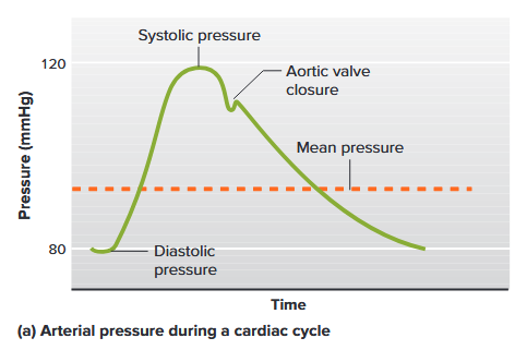
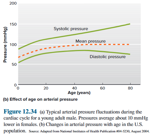
The pressure changes that occur along the rest of the systemic and pulmonary circulations. By the time the blood has completed its journey back to the atrium in each circuit, most of the pressure
originally generated by the ventricular contraction has dissipated. 其余体循环和肺循环中发生的压力变化。当血液在每个回路中完成返回心房的旅程时,最初由心室收缩产生的大部分压力已经消散。
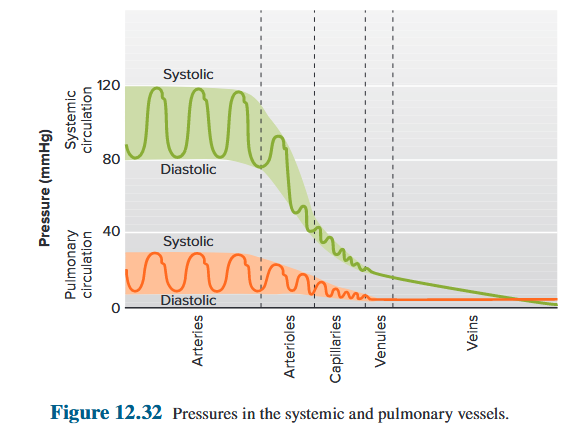
The roles of arterioles 小动脉:
- The arterioles in individual organs are responsible for determining the relative blood flows to those organs at any given mean arterial pressure. 各个器官中的小动脉负责确定在任何给定的平均动脉压下流向这些器官的相对血流量。
- The arterioles, all together, are the major factor in determining mean arterial pressure (MAP) itself. 小动脉是决定平均动脉压本身的主要因素。
4.2.2.2 Factors Affecting Arterial Blood Pressure
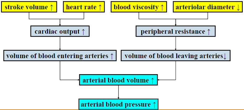
| SAP | DAP | Pulse pressure | BP | |
|---|---|---|---|---|
| Cardiac output ↑ 心输出量 |
↑↑ | ↑ | ↑ | ↑ |
| Heart rate ↑ 心率 |
↑ | ↑↑ | ↓ | ↑ |
| Peripheral resistance ↑ 外周阻⼒ |
↑ | ↑↑ | ↓ | ↑ |
| Elasticity of aorta and large artery ↓ 主动脉及大动脉弹性 |
↑ | ↓ | ↑↑ | ↑ |
| Mean circulation filling pressure ↓ 平均循环充盈压⼒ |
↓↓ | ↓ | ↓↓ | ↓ |
4.2.2.3 Abnormal Blood Pressure
Abnormally low BP (hypotension): leads to shock, anoxia and tissue destruction. 血压异常低(低血压):导致休克、缺氧和组织破坏。
Chronically elevated BP (hypertension): causes damage because important vessels (especially those of the heart, brain, kidneys and retina) are injured. 长期血压升高(高血压):造成损害,因为重要血管(尤其是心脏、大脑、肾脏和视网膜的血管)会受伤。
4.2.3 Veins
Definition: any of the tubular branching vessels that carry blood from the capillaries toward the heart. 任何一种将血液从毛细血管输送到心脏的管状分支血管。
Function:
- low-resistance conduits 低阻力导管
- maintaining peripheral venous pressure and venous return to the heart 维持外周静脉压和静脉回流到心脏
To aid venous return 帮助静脉回流:
- Skeletal muscle pump
- Respiratory pump
- Venoconstriction - contraction of smooth muscle in wall of vein (sympathetic stimulation) 静脉收缩‑静脉壁平滑肌收缩(交感神经刺激)
The walls of veins are thinner and much more compliant than those of arteries. Thus, veins can accommodate large volumes of blood with a relatively small increase in internal pressure. Approximately 60% of the total blood volume is present in the systemic veins, but the venous pressure is only about 10 mmHg on average. (In contrast, the systemic arteries contain less than 15% of the blood, at a pressure of nearly 100 mmHg.) 静脉壁比动脉壁更薄且更顺应。因此,静脉可以容纳大量的血液,而内部压力的增加相对较小。大约60%的总血量存在于全身静脉中,但静脉压平均只有10毫米汞柱左右。 (相比之下,在压力接近 100 mmHg 的情况下,全身动脉的血液含量不足 15%。)
Venous smooth muscle contraction, the skeletal muscle pump, and the respiratory pump all work to facilitate venous return and thereby enhance cardiac output by the same amount. 静脉平滑肌收缩、骨骼肌泵和呼吸泵均起作用以促进静脉回流,从而使心输出量增加相同量。
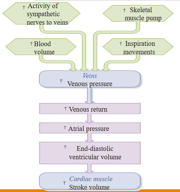
4.2.4 Microcirculation
Distribution: widest
Wall: thinnest
Diameter: smallest
Velocity: slowest
4.2.4.1 Capillaries
Approximately 5% of the total circulating blood is flowing through the capillaries, which performing the ultimate purpose of the entire exchange of nutrients, metabolic end products, and cell secretions. The capillaries permeate every tissue of the body except the cornea. Because most cells are no more than 0.1 mm from a capillary, diffusion distances are very small and exchange is highly efficient. An adult has an estimated 25,000 miles (40,000 km) of capillaries, each individual capillary being only about 1 mm long with an inner diameter of about 8 μm, just wide enough for an erythrocyte.
大约 5% 的循环血液流经毛细血管,其最终目的是实现营养物质、代谢终产物和细胞分泌物的全部交换。毛细血管渗透到除了角膜之外的身体的每个组织。由于大多数细胞距毛细管不超过 0.1 毫米,因此扩散距离非常小,交换效率很高。一个成年人估计有 25,000 英里(40,000 公里)的毛细血管,每个毛细血管长约 1 毫米,内径约 8 微米,宽度刚好足以容纳红细胞。
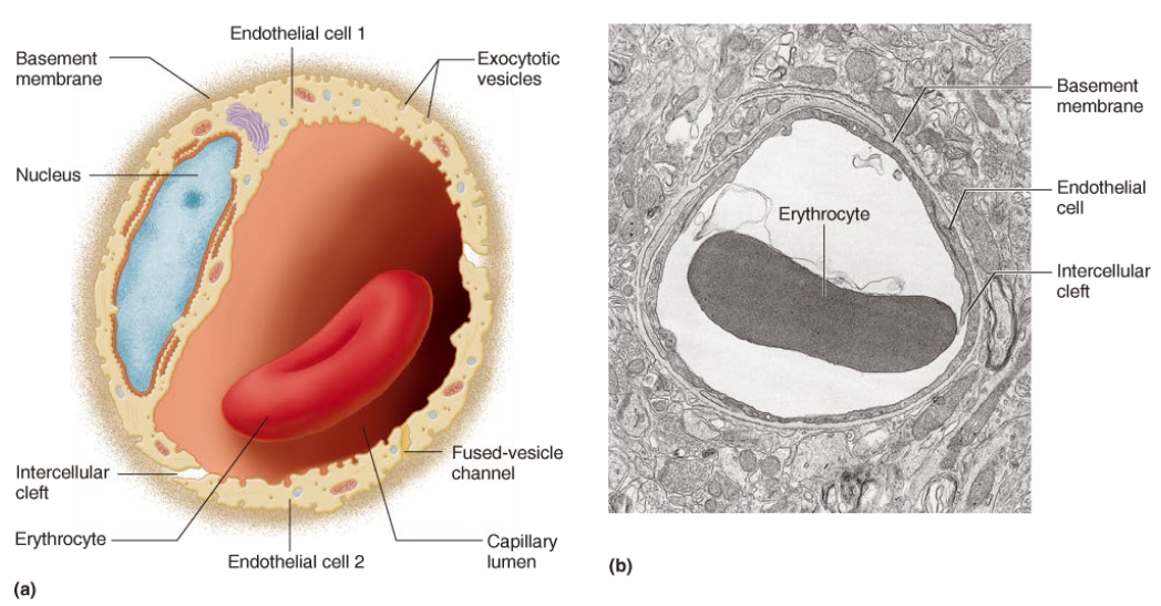
Typical capillary is a thin-walled tube of endothelial cells and one layer thick resting on a basement membrane, without any surrounding smooth muscle or elastic tissue. The endothelial cells generally contain large numbers of endocytotic and exocytotic vesicles, and sometimes these fuse to form continuous fused-vesicle channels across the cell.
典型的毛细血管是由内皮细胞组成的薄壁管,厚度为一层,位于基底膜上,周围无任何平滑肌或弹性组织。内皮细胞通常含有大量内吞和外排囊泡,有时这些囊泡融合形成贯穿细胞的连续融合囊泡通道。
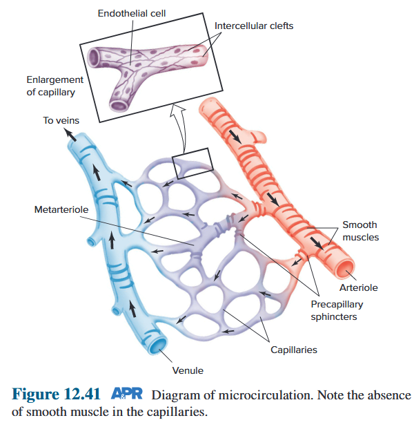
4.2.4.2 Arteriolar and Capillary Network
Microcirculation: the blood circulation between the arteriole and venule.
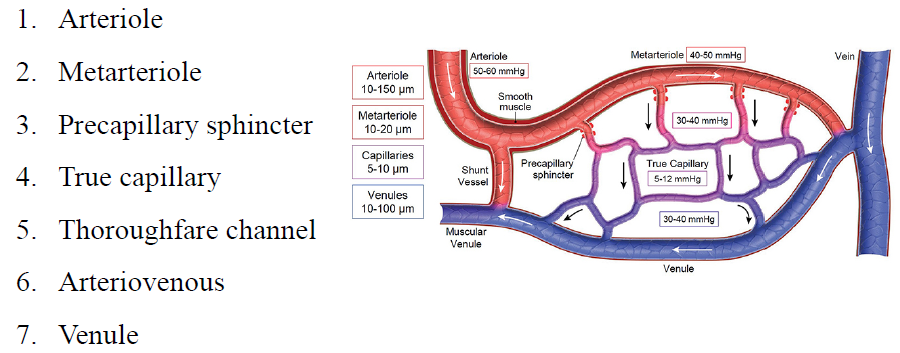
Three pathway of microcirculation
- Circuity pathway: Nutrient pathway 循环通路:营养通路
- Thoroughfare channel: maintain the flow rate of pathway 通道:保持循环血流量
- Arteriovenous shunt: Regulate body temperature 动静脉短路:调节体温
4.2.4.3 Microcirculation Local Controls
Local control of organ blood flow in response to
- increases in metabolic activity 代谢活动增加
- decreases in blood pressure ⾎压下降
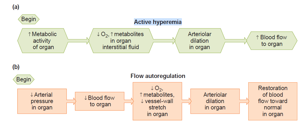
4.2.5 Formation of Interstitial Fluid
The constituents of interstitial fluid are almost the same as plasma except for much lower concentrations of proteins. Most of interstitial fluid is gelatinous, which cannot flow freely. The substrate of gel consists of collagen fibre bundles and proteoglycan filaments. 组织液的成分与⾎浆基本相同,但蛋白质浓度低得多。组织液大部分呈胶状,不能⾃由流动。胶状基质由胶原纤维束和蛋白聚糖组成。
4.2.5.1 Mechanisms of Interstitial Fluid Formation
Favoring fluid movement out of the capillary: 有利于液体流出毛细⾎管
- $P_c$ (capillary blood hydrostatic pressure); 毛细⾎管⾎液静⽔压
- $π_{IF}$ (osmotic force due to interstitial fluid protein concentration). 由于间质液蛋白质浓度而产生的渗透⼒
Favoring fluid movement into the capillary: 有利于液体进⼊毛细⾎管
- $P_{IF}$ (interstitial hydrostatic pressure); 间质静⽔压⼒
- $π_c$ (osmotic force due to plasma protein concentration); 由于⾎浆蛋白浓度引起的渗透⼒
$$
NFP=P_C+\pi_{IF}-P_{IF}-\pi_C
$$
Outward forces are arbitrarily assigned positive values, so a positive net filtration pressure favors filtration, whereas a negative pressure indicates that net absorption of fluid will occur. interstitial fluid hydrostatic pressure (P~IF~) is approximately zero. 向外的力被任意指定为正值,因此正的净过滤压力有利于过滤,而负的压力则表明将发生流体的净吸收。间质液静水压 (P~IF~) 大约为零。
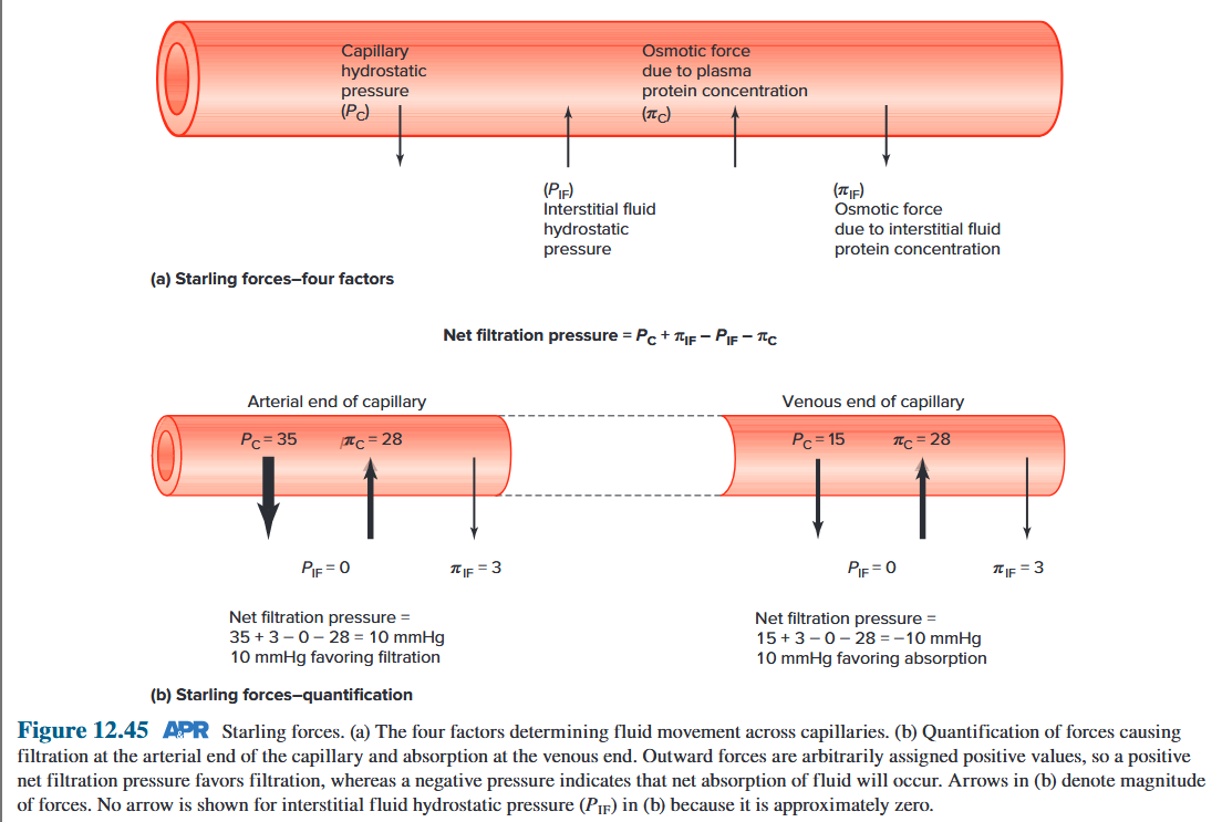
4.2.5.2 Factors Affecting Interstitial Fluid Formation 影响间质液形成的因素
- Capillary blood hydrostatic pressure 毛细血管血液静水压
- Effective colloid osmotic pressure 有效胶体渗透压
- Permeability of capillary wall 毛细血管壁通透性
- Lymphatic drainage 淋巴回流
4.2.6 Lymph Circulation
The lymphatic system is a network of small organs (lymph nodes) and tubes (lymphatic vessels or simply “lymphatics”) through which lymph flows. The lymphatic system is not technically part of the circulatory system, but it is described in this chapter because its vessels provide a route for the movement of interstitial fluid to the circulatory system. 淋巴系统是一个由小器官(淋巴结)和管道(淋巴管或简称“淋巴管”)组成的网络,淋巴液(一种来自间质液的液体)通过该网络流动。从技术上讲,淋巴系统并不属于循环系统的一部分,但其血管为间质液移动到循环系统提供了一条路线。
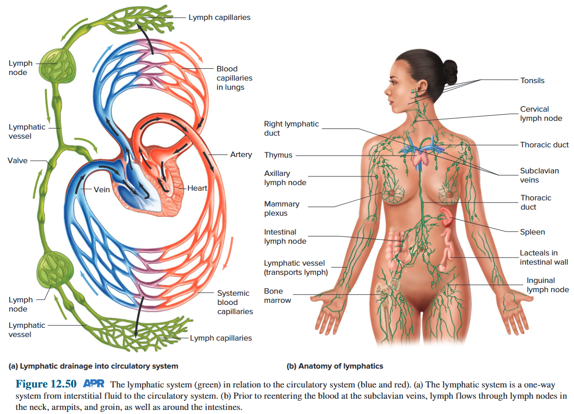
interstitial fluid → lymphatic capillaries → lymph nodes → lymphatic vessels → two large lymphatic ducts → Veins near the junction of the jugular and subclavian veins
间质液 → 毛细淋巴管 → 淋巴结 → 淋巴管 → 两条大淋巴管 → 颈静脉与锁骨下静脉交界处的静脉
Valves at junctions between lymphatic ducts and vascular vessel permit only one-way flow from lymphatic ducts into the veins. Thus, the lymphatic vessels carry interstitial fluid to the cardiovascular system. 淋巴管与⾎管连接处的瓣膜只允许淋巴管单向流⼊静脉。因此,淋巴管将间质液输送到⼼⾎管系统。
Function:
- Drain excess interstitial fluid to vascular system. 将多余的间质液引流至⾎管系统
- Drain fat absorbed from the gastrointestinal tract reaches the blood. 排出从胃肠道吸收的脂肪并进⼊⾎液
- Help cancer cells spread original organ to other parts of the body.
- 回收组织蛋白质
- 清除细菌,参与免疫
4.3 Regulation of Cardiovascular Activity
4.3.1 Neural Regulation
4.3.1.1 homeostatic control
homeostatic control systems (reflex arc):
An internal environmental variable → Receptors → Afferent pathways → An integrating center → Efferent pathways → Effectors
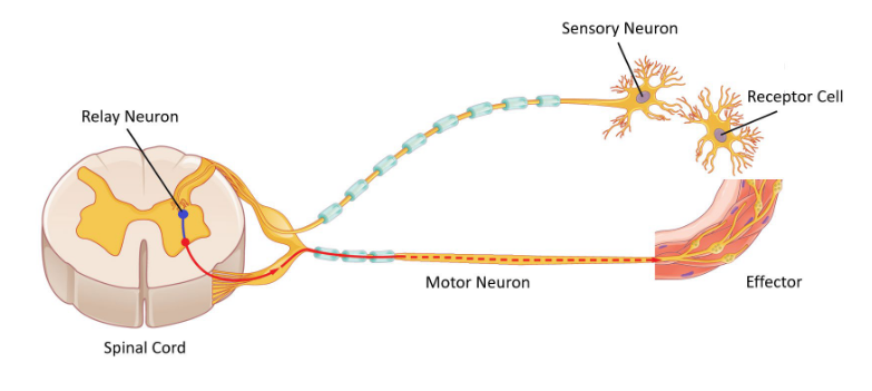
Homeostasis is essential for health and survival. Without a homeostatic control system operating to maintain blood pressure, the tissues of the body would quickly die if pressure decreases significantly. The major cardiovascular variable being regulated is the mean arterial pressure in the systemic circulation. Arterial pressure is the driving force for blood flow through all the organs except the lungs. Maintaining arterial pressure is therefore a prerequisite for ensuring adequate blood flow to these organs. 体内平衡对于健康和生存至关重要。如果没有稳态控制系统来维持血压,如果压力显着下降,身体组织很快就会死亡。 受到调节的主要心血管变量是体循环中的平均动脉压。动脉压是血液流经除肺外所有器官的驱动力。因此,维持动脉压是确保足够的血液流向这些器官的先决条件。
To maintain arterial blood pressure at about 120/80 mmHg in the face of changing demands for blood flow to different organs.
MAP= CO × TPR
MAP: Mean systemic arterial pressure
CO: Cardiac output
TPR: Total peripheral resistance
A pump pushes fluid into a container at the rate of 1 L/min. At steady state, fluid also leaves through the outflow tubes at a total rate of 1 L/min. Therefore, the height of the fluid column (Δ P), which is the driving pressure for outflow, remains stable. 泵以 1 升/分钟的速度将液体推入容器。 在稳定状态下,流体也以 1 L/min 的总速率通过流出管流出。因此,作为流出驱动压力的液柱高度(ΔP)保持稳定。
When the radius of vessel is increasing, its resistance is reducing. The total outflow for the system immediately becomes greater than 1 L/min, and more fluid leaves the reservoir than enters from the pump. 当船舶半径增大时,其阻力减小。系统的总流出量立即变得大于 1 L/min,并且离开储液器的流体多于从泵进入的流体。
The volume and height of the fluid column begin to decrease until a new steady state between inflow and outflow is reached. At any given pump input, a change in total outflow resistance must produce changes in the volume and height (pressure) in the reservoir. 液柱的体积和高度开始减小,直到达到流入和流出之间的新稳定状态。在任何给定的泵输入下,总流出阻力的变化必然会导致储液器中的体积和高度(压力)发生变化。
On the right, outflow tube 1 has been opened, while tubes 2 to 4 have been simultaneously constricted. The increased resistance in tubes 2 to 4 compensates for the decreased resistance in tube 1. Therefore, total resistance remains unchanged, and the reservoir pressure is unchanged. Therefore, it is the total arteriolar resistance that influences systemic arterial blood pressure. 在右侧,流出管 1 已打开,同时管 2 至 4 已收缩。管 2 至 4 中增加的阻力补偿了管 1 中减少的阻力。因此,总阻力保持不变,并且储层压力不变。因此,总动脉阻力影响全身动脉血压。
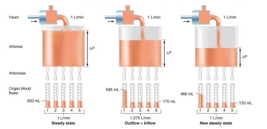
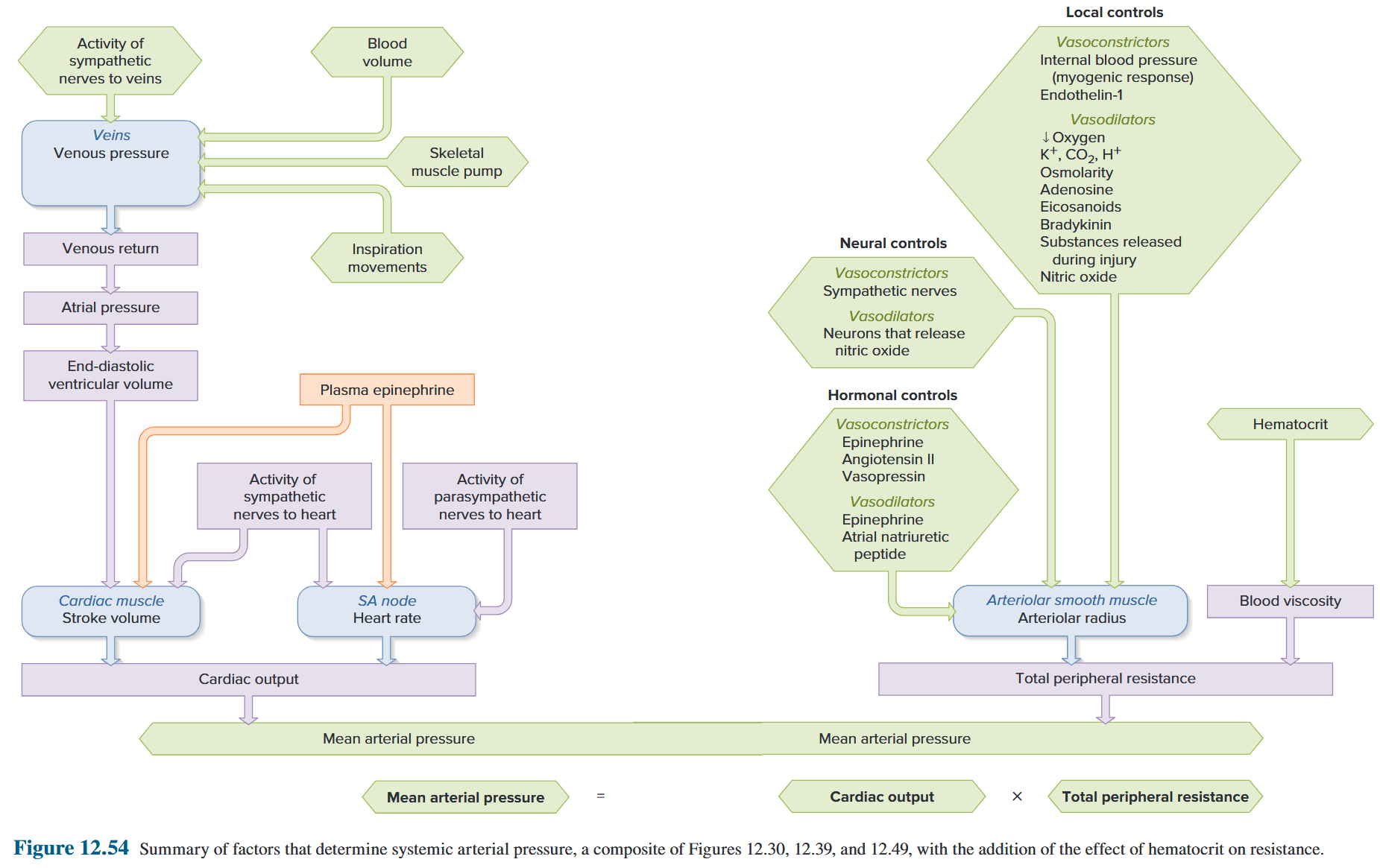
Bleed results in significant blood loss (hemorrhage) leads to a decrease in mean arterial pressure. Conversely, any deviation in mean arterial pressure, such as that occurring during hemorrhage, will elicit homeostatic reflexes so that cardiac output and/or total peripheral resistance will change in the direction required to minimize the initial change in arterial pressure. 出血会导致大量失血(出血),从而导致平均动脉压下降。相反,平均动脉压的任何偏差,例如出血期间发生的偏差,都会引起稳态反射,使得心输出量和/或总外周阻力将沿最小化动脉压初始变化所需的方向变化。
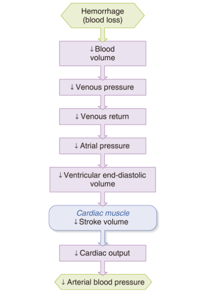
4.3.1.2 Baroreceptor reflexes (depressor reflex, sino-aortic reflex)
The reflexes that homeostatically regulate arterial pressure originate primarily with arterial receptors that respond to changes in pressure. 稳态调节动脉压⼒的反射主要源⾃对压⼒变化作出反应的动脉受体
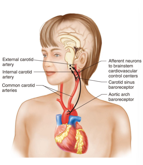
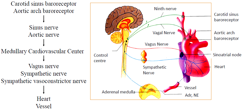
BP ↑ → sinus nerve (+) / aortic nerve (+) → sympathetic centre (-) / parasympathetic centre (+) / sympathetic vasoconstrictor centre (-) → sympathetic nerve (-) / parasympathetic nerve (+) / sympathetic vasoconstrictor nerve (-)
血压↑ → 窦神经 (+) / 主动脉神经 (+) → 交感神经中枢 (-) / 副交感神经中枢 (+) / 交感血管收缩中枢 (-) → 交感神经 (-) / 副交感神经 (+) / 交感血管收缩神经 (-)
heart: heart rate ↓ / (contraction ↓ → strike volume ↓) → cardiac output ↓ → BP ↓
心脏:心率 ↓ / (收缩 ↓ → 搏动量 ↓) → 心输出量 ↓ → 血压 ↓
vessel: arteriolar contraction ↓ → peripheral resistance ↓ → BP ↓
血管:小动脉收缩 ↓ → 外周阻力 ↓ → 血压 ↓
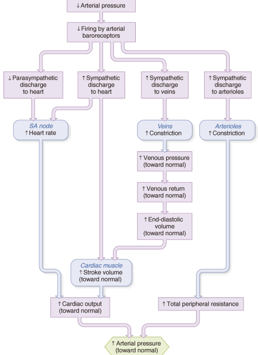
The baroreceptor reflex functions primarily as a short-term regulator of arterial blood pressure. It is activated instantly by any blood pressure change and functions to restore blood pressure rapidly toward normal. However, if arterial pressure remains increased from its normal set point for more than a few days, the arterial baroreceptors adapt to this new pressure and decrease their frequency of action potential firing at any given pressure. 压力感受器反射主要起短期动脉血压调节器的作用。血压一有变化,压力感受器反射就会立即激活,并起到迅速将血压恢复正常的作用。但是,如果动脉压力持续几天高于正常设定点,动脉压力感受器就会适应这种新压力,并降低在任何给定压力下动作电位发射的频率。
4.3.2 Humoral Regulation
4.3.2.1 Renin-Angiotensin-Aldosterone System (RAAS)
肾素-血管紧张素-醛固酮系统
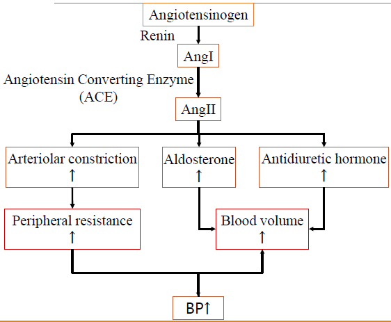
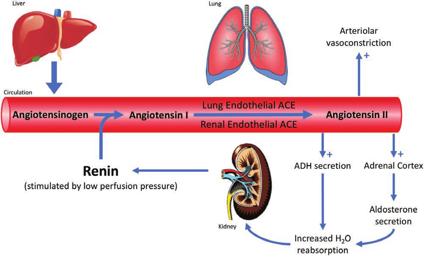
Aldosterone: 保钠保水排钾 尿量减少
血管紧张素转换酶(ACE)抑制剂:普利类
血管紧张素受体(AT~1~)阻断剂:沙坦类
4.3.2.2 Epinephrine (E) and Norepinephrine (NE)
| Epinephrine (E) | Norepinephrine (NE) | |
|---|---|---|
| Resource | Adrenal medulla 肾上腺髓质 |
Adrenal medulla, sympathetic nerve 肾上腺髓质、交感神经 |
| Function | Cardiotonic 强心剂 | Vasopressor 升压药 |
| Heart | β~1~ receptor: heart rate ↑, myocardial contractility ↑, cardiac output ↑, blood pressure ↑ 心率↑、心肌收缩力↑、心输出量↑、血压↑ |
β~1~ receptor: heart rate ↓, myocardial contractility ↑, cardiac output ↑, blood pressure ↑ 心率↓、心肌收缩力↑、心输出量↑、血压↑ |
| Vessel | β~2~ receptor: vasoconstriction on skin and visceral, while vasodilation on skeletal muscle, heart and liver 皮肤和内脏血管收缩,骨骼肌、心脏和肝脏血管舒张 |
α~1~ receptor: vasoconstriction on all organs except for coronary artery, peripheral resistance ↑, DAP ↑ 除冠状动脉外所有器官血管收缩,外周阻力↑,DAP↑ |
| Smooth muscle | Dilation on gastrointestinal tract and bronchus 胃肠道和支气管扩张 |
Less than Epinephrine |
| Metabolism | blood glucose ↑, lipolysis ↑, oxygen consumption, heat production ↑ 血糖↑、脂肪分解↑、耗氧量、产热量↑ |
Less than Epinephrine |
4.3.2.3 Blood Volume and Long-term Regulation of AP
The major mechanism for long-term regulation occurs through the blood volume. Blood volume is a major determinant of arterial pressure because it influences venous pressure, venous return, enddiastolic volume, stroke volume, and cardiac output. Thus, increased blood volume increases
arterial pressure. 长期调节的主要机制是通过血容量实现的。血容量是动脉压的主要决定因素,因为它会影响静脉压、静脉回流、舒张末期容积、每搏输出量和心输出量。因此,血容量增加会导致动脉压升高。
An increase in blood pressure for any reason causes a decrease in blood volume, which tends to bring the blood pressure back down. An increase in the blood volume for any reason increases the blood pressure, which tends to bring the blood volume back down. 任何原因引起的血压升高都会导致血容量减少,这往往会使血压回落。任何原因引起的血容量增加都会导致血压升高,这往往会使血容量回落。
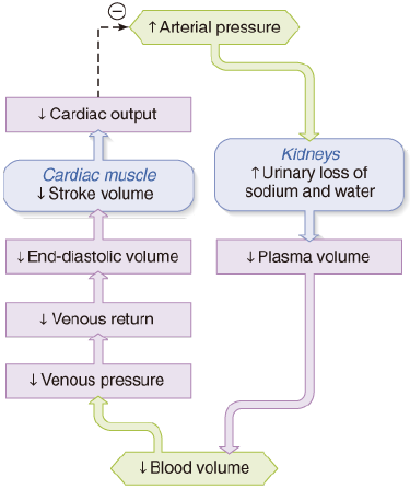
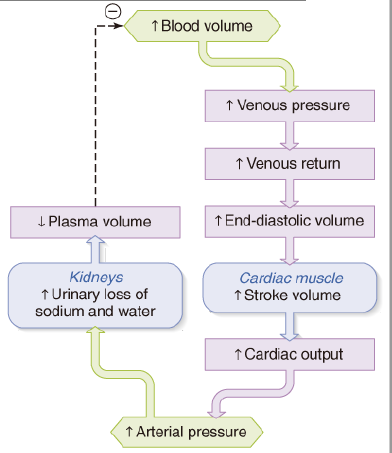
Arterial pressure influences blood volume but blood volume also influences arterial pressure, blood pressure can stabilize, in the long run, only at a value at which blood volume is also stable. Consequently, changes in steady-state blood volume are the single most important long-term determinant of blood pressure. 动脉压影响血容量,但血容量也会影响动脉压,从长远来看,血压只能稳定在血容量也稳定的值。因此,稳态血容量的变化是血压最重要的长期决定因素。
利尿剂:氢氯噻嗪
Chapter 11 Reproduction
Reproduction is not necessary for survival of an individual, but is necessary for survival of the species.
- sperm and ovum develop and grow; 精子和卵子发育生长
- sperm and ovum unite to produce a new and unique combination of genes in a new organism; 精子和卵子结合,在新的有机体中产生新的、独特的基因组合
- sexual maturation (puberty); 性成熟(青春期)
- pregnancy and lactation in women; 女性妊娠期及哺乳期
- gonads hormones, which are controlled by and influence secretion of hormones from hypothalamus and anterior pituitary gland. 性腺激素,受下丘脑和垂体前叶控制并影响激素的分泌
11.1 Male Reproductive Physiology
11.1.1 Structure of Male Reproduction System
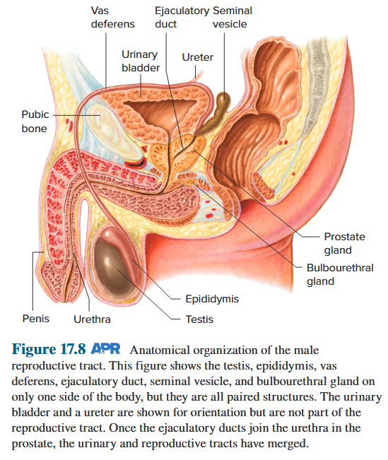
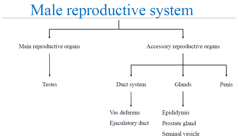
11.1.2 Spermatogenesis and Transport of Sperms 精子发生和精子运输
11.1.2.1 Spermatogenesis
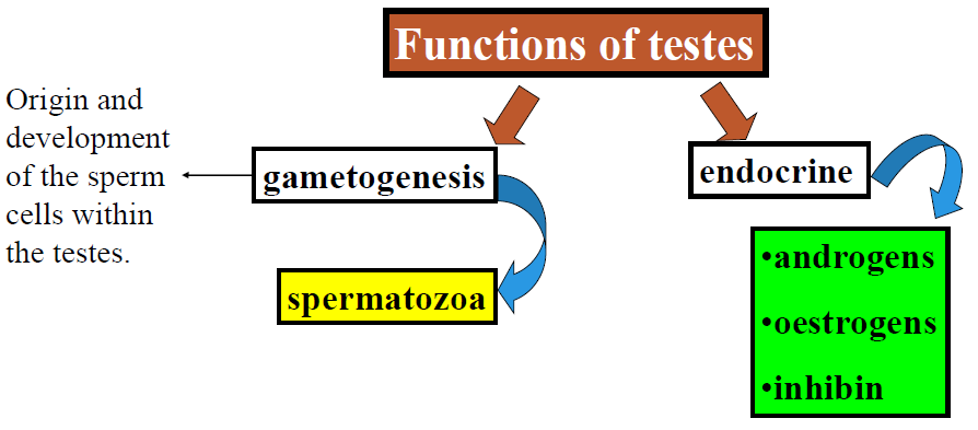
11.1.2.2 Testis
- Rete testis: a network of interconnected tubes from different seminiferous tubules. 睾丸网:由不同的生精小管相互连接的管道网络
- Efferent ductules: leave the rete testis, pierce the fibrous covering of the testis, and empty into epididymis. 传出小管:离开睾丸网,刺穿睾丸的纤维覆盖层,排入附睾
- Epididymis: loosely attaches to the outside of the testis, and draining each testis leads to a vas deferens. 附睾:松散地附着在睾丸外部,每个睾丸的液体都排入输精管
- Vas deferens: a large, thick-walled tube lined with smooth muscle. 输精管:一种大而厚壁的管道,内衬平滑肌
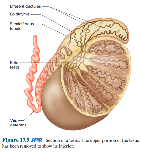
11.1.2.3 Seminiferous tubules
The sites of spermatogenesis in the testes 睾丸中精子发生的部位
Basement membrane 基底膜
Lumen: the center of each tubule, a fluid-filled containing mature sperm cells. 管腔:每个小管的中心,充满液体,内含成熟精子细胞
Developing germ cells 发育中的生殖细胞
Sertoli cells: supporting cells, produce sperm 塞托利细胞:支持细胞,产生精子
Leydig cells: interstitial cells, lie in small, connective-tissue spaces between the tubules. synthesize and release testosterone. 莱迪希细胞:间质细胞,位于小管之间的小结缔组织空间中。合成并释放睾酮
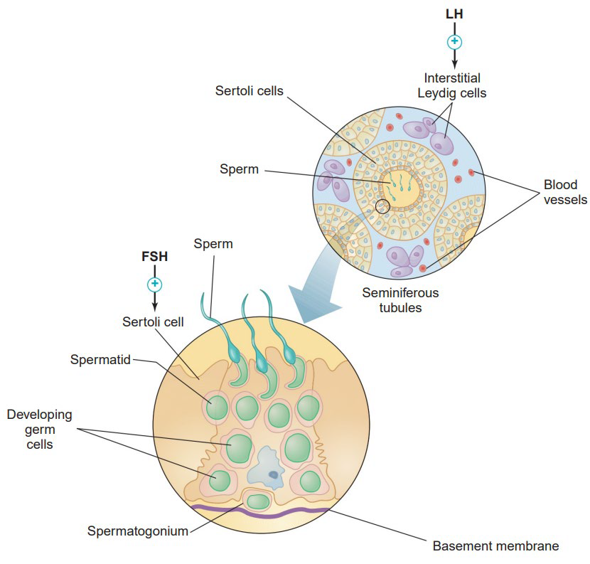
11.1.2.4 Sertoli cells
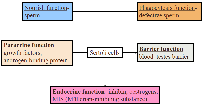
11.1.2.5 Spermatogenesis
Spermatogonia: undifferentiated germ cells, found only in the basal compartment. After several mitotic cycles, each primary spermatocytes crosses a tight junction, enlarges, and divides into two secondary spermatocytes, then divide into spermatids. The final phase of spermatogenesis is the differentiation of the spermatids into sperm. This process involves extensive cell remodeling, including elongation, but no further cell divisions. 精原细胞:未分化的生殖细胞,仅存在于基底区。经过几个有丝分裂周期后,每个初级精母细胞穿过紧密连接,扩大并分裂成两个次级精母细胞,然后分裂成精子细胞。精子发生的最后阶段是精子细胞分化成精子。该过程涉及广泛的细胞重塑,包括伸长,但没有进一步的细胞分裂。
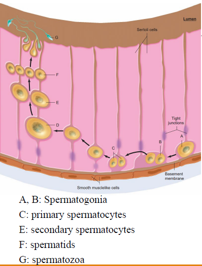
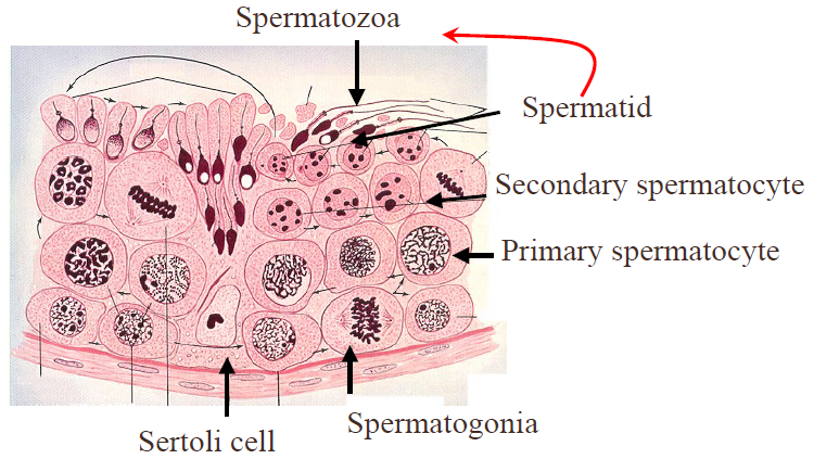
Each spermatogonium yields, by mitosis, a clone of spermatogonia; for simplicity, the figure shows only two such cycles, with a third mitotic cycle generating two primary spermatocytes. The arrow from one of the spermatogonia back to a stem cell spermatogonium denotes the fact that one cell of the clone does not go on to generate primary spermatocytes but reverts to an undifferentiated spermatogonium that gives rise to a new clone. Each primary spermatocyte produces four spermatozoa. 每个精原细胞通过有丝分裂产生一个精原细胞克隆;为简单起见,图中仅显示两个这样的周期,第三个有丝分裂周期产生两个初级精母细胞。从一个精原细胞返回到干细胞精原细胞的箭头表示克隆中的一个细胞不会继续产生初级精母细胞,而是恢复为未分化的精原细胞,从而产生新的克隆。每个初级精母细胞产生四个精子。
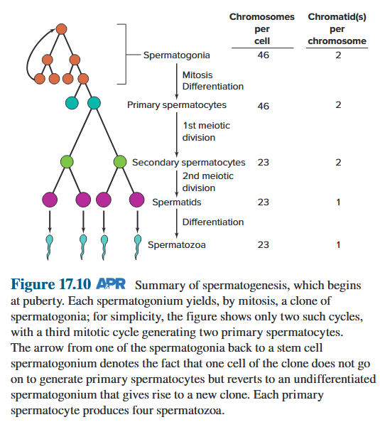
11.1.2.6 Requirements for spermatogenesis
- optimal temperature: The testes are suspended outside the abdomen in the scrotum, which is an outpouching of the abdominal wall and is divided internally into two sacs, one for each testis. During early fetal development, the testes are located in the abdomen; but during later gestation, they usually descend into the scrotum. This descent is essential for normal sperm production during adulthood, because sperm formation requires a temperature approximately 2 ℃lower than normal internal body temperature. 最佳温度:睾丸悬于腹腔外的阴囊中,阴囊是腹壁的外袋,内部分为两个囊,每个睾丸一个。在胎儿早期发育期间,睾丸位于腹部;但在妊娠后期,睾丸通常会下降到阴囊中。这种下降对于成年期正常的精子生成至关重要,因为精子形成需要的温度比正常体内温度低约 2 ℃。
- action of various hormones 各种激素的作用
11.1.2.7 Testicles develop intra-abdominally
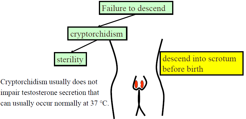
11.1.2.8 Transport of Sperm
- From the seminiferous tubules, the sperm pass into the epididymis, where they are concentrated and become mature. 精子从曲细精管进入附睾,在那里浓缩并成熟
- The epididymis and vas deferens store the sperm, and the seminal vesicles and prostate secrete most of the semen. 附睾和输精管储存精子,精囊和前列腺分泌大部分精液
- Erection of the penis occurs because of vascular engorgement accomplished by relaxation of the small arteries and passive occlusion of the veins. 阴茎勃起是由于小动脉松弛和静脉被动闭塞引起的血管充血
- Ejaculation includes emission—emptying of semen into the urethra—followed by expulsion of the semen from the urethra. 射精包括射精——将精液排入尿道——然后将精液从尿道排出
11.1.3 Endocrine of Testis and Function of Hormones
11.1.3.1 Endocrine of Testis
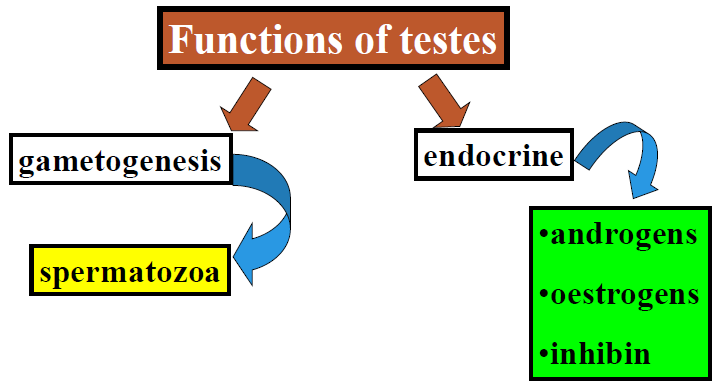
11.1.3.2 Function of Testosterone
- Initiates and maintains spermatogenesis; 启动和维持精子发生
- Decreases GnRH secretion via hypothalamus; 减少下丘脑分泌的 GnRH
- Inhibits LH secretion via anterior pituitary gland; 抑制垂体前叶分泌的 LH
- Induces differentiation of male accessory reproductive organs and maintains their function; 诱导男性附属生殖器官分化并维持其功能
- Induces male secondary sex characteristics; opposes action of estrogen on breast growth; 诱导男性第二性征;对抗雌激素对乳房生长的作用
- Stimulates protein anabolism, bone growth, and cessation of bone growth; 刺激蛋白质合成代谢、骨骼生长和骨骼生长停止
- Improves sex drive and enhances aggressive behavior; 提高性欲并增强攻击性行为
- Stimulates erythropoietin secretion by the kidneys. 刺激肾脏分泌促红细胞生成素
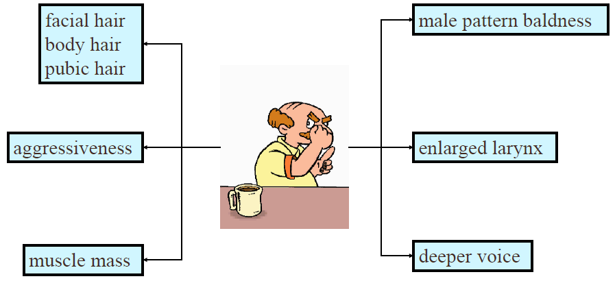
11.1.4 Hormonal Control of Male Reproductive Function
11.1.4.1 Hypothalamus-Pituitary-Gonad Axis
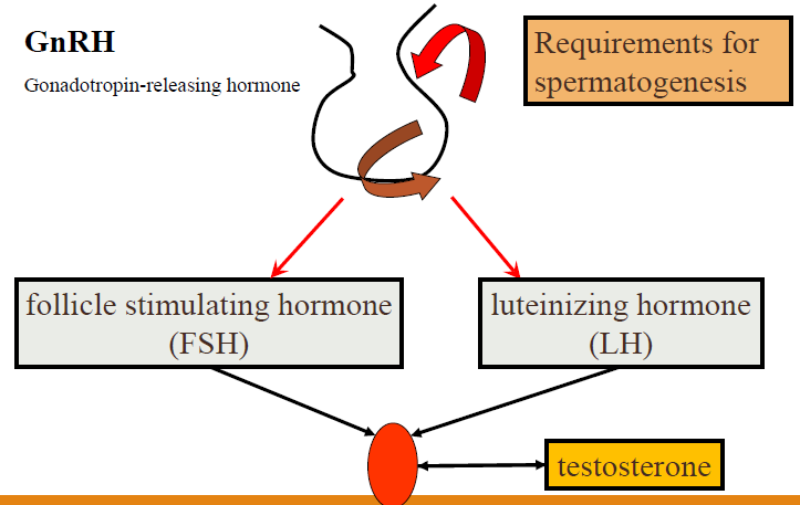
FSH acts only on the Sertoli cells, whereas LH acts primarily on the Leydig cells. The secretion of FSH is inhibited mainly by inhibin, a protein hormone secreted by the Sertoli cells, and the secretion of LH is inhibited mainly by testosterone, the steroid hormone secreted by the Leydig cells. Testosterone, acting locally on Sertoli cells, stimulates spermatogenesis, whereas FSH stimulates inhibin release from Sertoli cells. FSH 仅作用于塞托利细胞(支持细胞),而 LH 主要作用于莱迪希细胞(间质细胞)。FSH 的分泌主要受到塞托利细胞分泌的蛋白质激素抑制素的抑制,而 LH 的分泌主要受到间质细胞分泌的类固醇激素睾酮的抑制。睾酮局部作用于塞托利细胞,刺激精子发生,而 FSH 则刺激塞托利细胞释放抑制素。
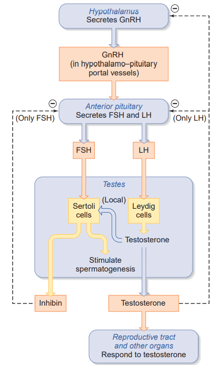
11.1.4.2 Andropause
Changes in the male reproductive system with aging are less drastic than those in women. Testosterone secretion, beginning at about 40 years of age, which apparently reflects slow deterioration of testicular function and failure of the gonads to respond to the pituitary gland gonadotropins. 男性生殖系统随年龄增长的变化不如女性剧烈。睾酮分泌开始于约 40 岁,这显然反映了睾丸功能的缓慢退化和性腺对垂体促性腺激素的反应丧失。
- decreased testosterone concentrations in the blood 血液中睾酮浓度下降
- decreased libido 性欲下降
- decreased sperm motility 精子活力下降
- emotional problems: depression 情绪问题:抑郁
11.2 Female reproductive physiology
11.2.1 Structure of female reproduction system
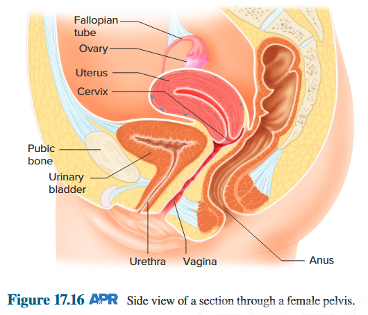
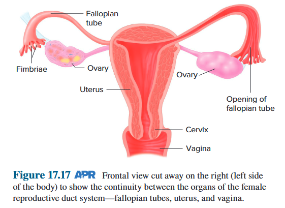
11.2.2 Oogenesis, Follicle Growth, and Formation of Corpus Luteum
11.2.2.1 Function of ovarian
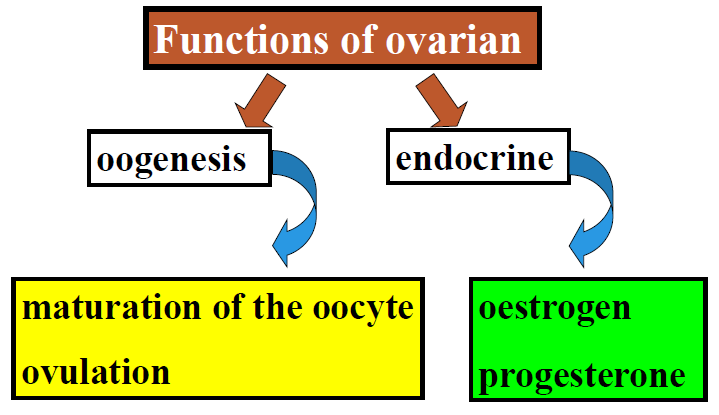
11.2.2.2 Oogenesis
Just before ovulation, primary oocyte started meiotic division, and separated into the secondary oocyte and the first polar body (very small and non-functional). The second meiotic division occurs in a fallopian tube after ovulation, but only if the secondary oocyte is fertilized. Secondary oocyte separated into ovum and second polar body (very small and non-functional). 排卵前,初级卵母细胞开始减数分裂,并分裂成次级卵母细胞和第一极体(非常小且无功能)。第二次减数分裂发生在排卵后的输卵管中,但前提是次级卵母细胞受精。次级卵母细胞分裂成卵子和第二极体(非常小且无功能)。
The secondary oocyte is ovulated and does not complete its meiotic division unless it is penetrated (fertilized) by a sperm. Thus, the oocyte is not termed an egg or ovum until after fertilization occurs. Each primary oocyte yields only one secondary oocyte, which can yield only one ovum. 次级卵母细胞排卵后,除非精子穿透(受精),否则不会完成减数分裂。因此,卵母细胞在受精后才被称为卵子或卵子。每个初级卵母细胞只能产生一个次级卵母细胞,而次级卵母细胞只能产生一个卵子。
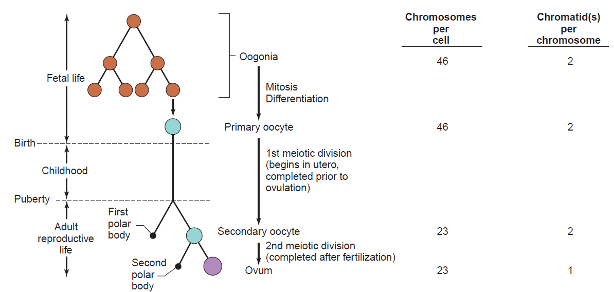
Oogenesis is the differentiation of the ovum (egg cell) into a cell competent to further develop when fertilized. 卵子发生是指卵子(卵细胞)在受精后分化为能够进一步发育的细胞
- Occurs during intrauterine development in female foetus
- At birth: 2-4 million primary oocytes -arrested in meiosis
- By puberty: around 400
- Number decreases throughout life
11.2.2.3 Oocyte development
Primordial follicles: consist of one primary oocyte surrounded by a single layer of granulosa cells. The granulosa cells secrete estrogen, small amounts of progesterone, and inhibin. 原始卵泡:由一个初级卵母细胞组成,周围环绕着一层颗粒细胞。颗粒细胞分泌雌激素、少量孕酮和抑制素
Primary and Preantral follicle: The size of the oocyte increases; granulosa cells proliferates into multiple layers; the oocyte separates from the inner granulosa cells by a thick layer of zona pellucida. 初级卵泡和窦前卵泡:卵母细胞的大小增加;颗粒细胞增殖成多层;卵母细胞通过一层厚厚的透明带与内层颗粒细胞分离
Zona pellucida: secreted by surrounding follicular cells, contains glycoproteins that binds a sperm cell to the surface of an egg after ovulation. 透明带:由周围的卵泡细胞分泌,含有糖蛋白,可在排卵后将精子细胞结合到卵子表面
Early antral follicle: a fluid-filled space (antrum) begins to form in the midst of the granulosa cells as a result of fluid they secrete. 早期窦卵泡:由于颗粒细胞分泌的液体,在颗粒细胞中间开始形成一个充满液体的空间(窦)
Theca: function together with the granulosa cells in the synthesis of estrogen. 卵泡膜:与颗粒细胞一起参与雌激素的合成
The progression of some primordial follicles to the preantral and early antral stages occurs throughout infancy and childhood and then during the entire menstrual cycle. 一些原始卵泡会在整个婴儿期和儿童期进展到窦前阶段和早期窦阶段,然后在整个月经周期中
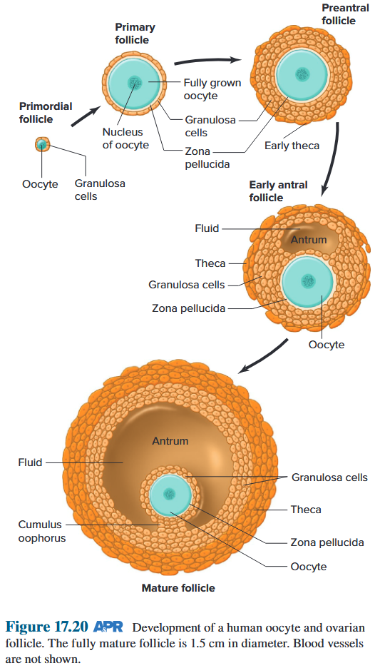
11.2.2.4 Ovulation
- Several follicles start to develop each month 每个月有几个卵泡开始发育
- Usually only one reaches maturity (dominant follicle) - rest undergo atresia 通常只有一个达到成熟(优势卵泡) - 其余的卵泡会闭锁
- In human corpus luteum seems to have a pre-determined life-span and starts to degenerate ~ day 23 if a blastocyst does not implant 人类的黄体似乎有预定的寿命,如果胚泡没有植入,黄体会在约第 23 天开始退化
Atresia: The nondominant follicles began to enlarge undergo a degenerative process and programmed cell death, or apoptosis. The eggs in the degenerating follicles also die. Atresia continues all through prepubertal life so that only 200,000 to 400,000 follicles remain when active reproductive life begins. 闭锁:非优势卵泡开始增大,经历退化过程和程序性细胞死亡或凋亡。退化卵泡中的卵子也会死亡。闭锁会持续整个青春期前期,因此当活跃的生殖生命开始时,只剩下 200,000 到 400,000 个卵泡
11.2.2.5 Corpus luteum
After ovulation, follicle collapses around the antrum and undergoes a rapid transformation. The granulosa cells enlarge greatly, and the entire glandlike structure formed corpus luteum, which secretes estrogen, progesterone, and inhibin. 排卵后,卵泡在窦周围塌陷并经历快速转化。颗粒细胞大大增大,整个腺状结构形成黄体,分泌雌激素、孕激素和抑制素。
If discharged egg (now in a fallopian tube) is not fertilized, the corpus luteum reaches its maximum development within approximately 10 days, and then rapidly degenerates by apoptosis. 如果排出的卵子(现在在输卵管中)未受精,黄体在约 10 天内达到最大发育,然后通过细胞凋亡迅速退化。
The loss of corpus luteum function leads to menstruation and the beginning of a new menstrual cycle: the follicular phase and the luteal phase. 黄体功能丧失导致月经和新月经周期的开始:卵泡期和黄体期。
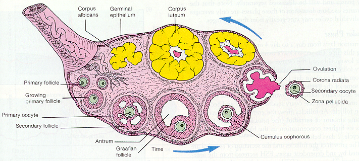
11.2.3 Endocrine of Ovary and Functions of Female Hormones
11.2.3.1 Function of ovarian
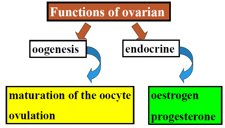
11.2.3.2 Ovarian Hormones
Estrogenis synthesized and released into the blood during the follicular phase mainly by the granulosa cells. After ovulation, estrogen is synthesized and released by the corpus luteum. 雌激素在卵泡期主要由颗粒细胞合成并释放到血液中。排卵后,雌激素由黄体合成并释放。
Progesterone, the other major ovarian steroid hormone, is synthesized and released in very small amounts by the granulosa and theca cells just before ovulation, but its major source is the corpus luteum. 另一种主要的卵巢类固醇激素孕酮在排卵前由颗粒细胞和卵泡膜细胞合成和释放极少量,但其主要来源是黄体。
Inhibin, a peptide hormone, is secreted by both the granulosa cells and the corpus luteum. 抑制素是一种肽类激素,由颗粒细胞和黄体分泌。
11.2.3.3 Types of oestrogens
Estrogen, oroestrogen, is a category of sex hormone responsible for the development and regulation of the female reproduction. 雌激素,又称雌激素,是一类负责女性生殖发育和调节的性激素。
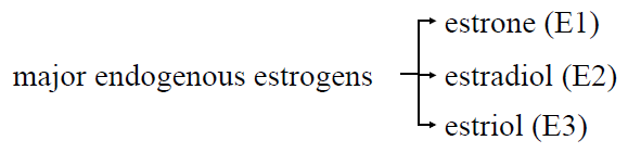
Another estrogen called estetrol (E4) is produced only during pregnancy. 另一种雌激素,称为雌四醇 (E4),仅在怀孕期间产生。
11.2.3.4 Actions of oestrogens
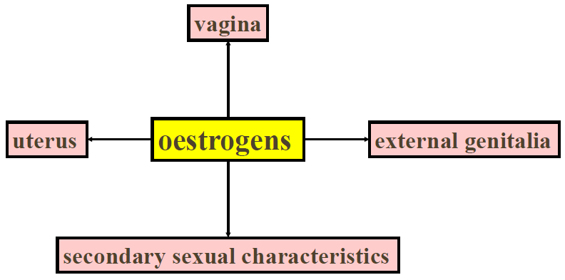
uterus: Increases myometrial contractions and responsiveness to oxytocin. Stimulates secretion of abundant, watery cervical mucus. Prepares endometrium for progesterone’s actions by inducing progesterone receptors. 子宫:增加子宫肌层收缩和对催产素的反应。刺激大量水样宫颈粘液的分泌。通过诱导孕酮受体为子宫内膜做好孕酮作用的准备
vagina: Increases layering of epithelial cells. 阴道:增加上皮细胞的分层
external genitalia: Fallopian tubes: Increases contractions and ciliary activity. 外生殖器:输卵管:增加收缩和纤毛活动
secondary sexual characteristics:
- Stimulates growth of ovary and follicles (local effects). 刺激卵巢和卵泡的生长(局部效应)
- Stimulates breast growth, particularly ducts and fat deposition during puberty. 刺激青春期乳房的生长,特别是乳腺管和脂肪沉积
- Stimulates female body configuration development during puberty: narrow shoulders, broad hips, female fat distribution (deposition on hips and breasts). 刺激青春期女性体型的发育:窄肩、宽臀、女性脂肪分布(臀部和乳房上的沉积)
Each breast contains numerous mammary glands, each with ducts that branch all through the tissue and converge at the nipples. These ducts start in saclike structures called alveoli. The breast alveoli, which are the sites of milk secretion, look like bunches of grapes with stems terminating in the ducts. The alveoli and the ducts immediately adjacent to them are surrounded by specialized contractile cells called myoepithelial cells. 每个乳房都包含许多乳腺,每个乳腺都有导管,这些导管贯穿整个组织并汇聚在乳头。这些导管始于称为肺泡的囊状结构。腺泡是乳汁分泌的部位,看起来像一串葡萄,茎终止于导管。腺泡和紧邻它们的导管被称为肌上皮细胞的特殊收缩细胞包围着。
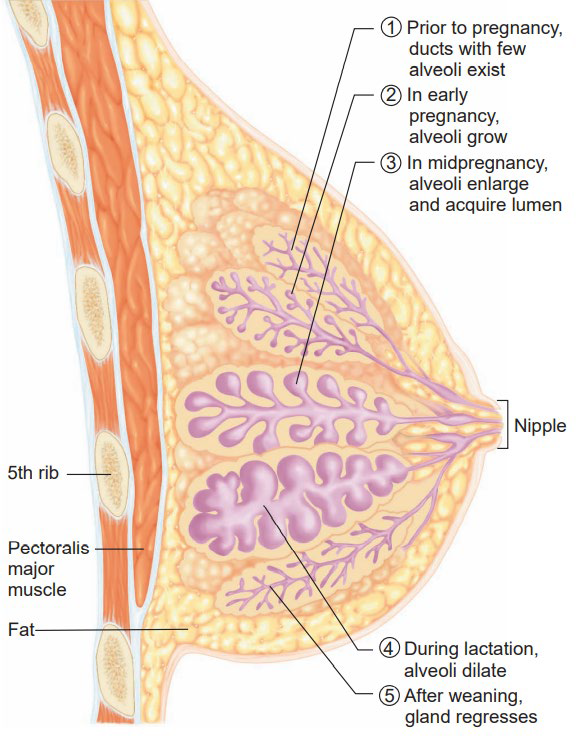
Others:
- Stimulates fluid secretion from lipid (sebum)-producing skin glands (sebaceous glands (皮脂腺)). 刺激产生脂质(皮脂)的皮肤腺(皮脂腺)分泌液体
- Stimulates bone growth and ultimate cessation of bone growth (closure of epiphyseal plates); protects against osteoporosis; does not have an anabolic effect on skeletal muscle. 刺激骨骼生长并最终停止骨骼生长(骨骺板闭合);预防骨质疏松症;对骨骼肌无合成代谢作用
- Vascular effects (deficiency produces “hot flashes”). 对血管有影响(缺乏会产生“潮热”)
- Has feedback effects on hypothalamus and anterior pituitary gland. 对下丘脑和垂体前叶有反馈作用
- Stimulates prolactin secretion but inhibits prolactin’s milk-inducing action on the breasts. 刺激催乳素分泌,但抑制催乳素对乳房的催乳作用
- Protects against atherosclerosis by effects on plasma cholesterol, blood vessels, and blood clotting. 通过对血浆胆固醇、血管和血液凝固的影响预防动脉粥样硬化
11.2.3.5 Actions of progesterone
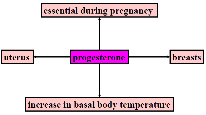
- Converts the estrogen-primed endometrium to an activity secreting tissue suitable for implantation of an embryo. 将雌激素引发的子宫内膜转化为适合胚胎植入的活性分泌组织
- Induces thick, sticky cervical mucus. 诱导粘稠的宫颈粘液
- Decreases contractions of fallopian tubes and myometrium. 减少输卵管和子宫肌层的收缩
- Decreases proliferation of vaginal epithelial cells. 减少阴道上皮细胞的增殖
- Stimulates breast growth, particularly glandular tissue. 刺激乳房生长,特别是腺体组织
- Inhibits milk-inducing effects of prolactin. 抑制催乳素的催乳作用
- Has feedback effects on hypothalamus and adenohypophysis. 对下丘脑和腺垂体有反馈作用
- Increases body temperature. 升高体温
11.2.4 Hormonal Control of Ovarian Function 卵巢功能的激素控制
11.2.4.1 Systemic plasma hormone concentrations during menstrual cycle
Estrogen: remaining fairly low and stable for the first week, and increases rapidly during the second week as the dominant ovarian follicle grows and secretes more estrogen. Then starts decreasing shortly before LH has peaked. This is followed by a second increase due to secretion by the corpus luteum and, finally, a rapid decrease during the last days of the cycle. 雌激素:第一周保持相当低且稳定,第二周随着优势卵泡生长并分泌更多雌激素而迅速增加。然后在 LH 达到峰值前不久开始下降。随后由于黄体分泌而再次增加,最后在周期的最后几天迅速下降。
Progesterone: Very small amounts during the follicular phase until just before ovulation. Very soon after ovulation, the developing corpus luteum begins to release large amounts of progesterone; from this point, the progesterone pattern is similar to that for estrogen. 孕酮:在卵泡期直到排卵前,孕酮含量非常少。排卵后不久,发育中的黄体开始释放大量孕酮;从此时起,孕酮模式与雌激素相似。
FSH (Follicle stimulating hormone) increases in the early part of the follicular phase and then steadily decreases throughout the remainder of the cycle except for a small midcycle peak. FSH(促卵泡激素)在卵泡期早期增加,然后在整个周期的剩余时间内稳步下降,除了周期中期出现一个小的峰值。
LH (Luteinizing hormone) is constant during most of the follicular phase but then shows a very large midcycle increase—the LH surge—peaking approximately 18 h before ovulation. This is followed by a rapid decrease and then a further slow decline during the luteal phase. LH(促黄体激素)在卵泡期的大部分时间里保持恒定,但随后在排卵中期出现大幅增加——LH 激增——在排卵前约 18 小时达到峰值。随后迅速下降,然后在黄体期进一步缓慢下降。
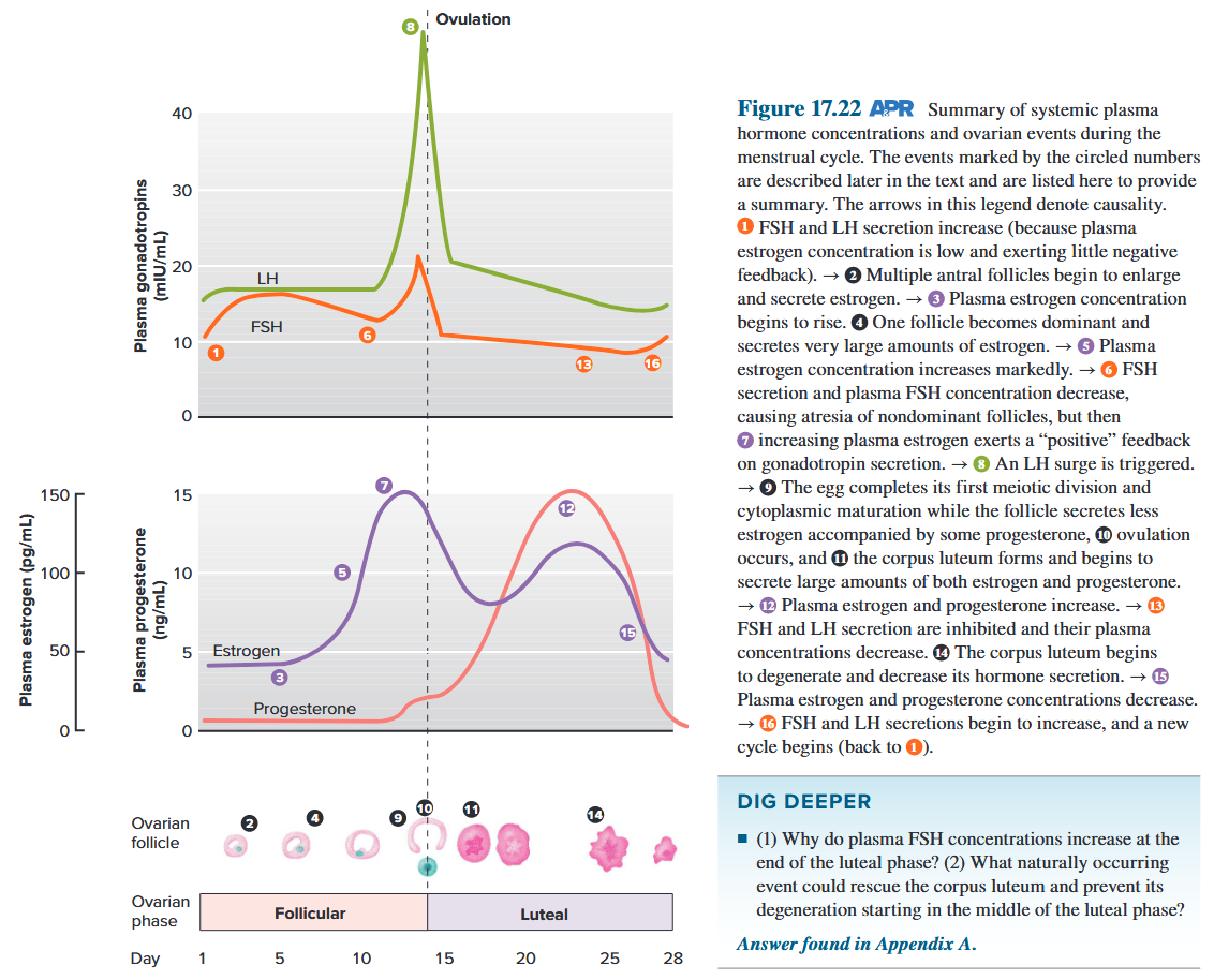
Inhibin increases during the late follicular phase, remains high during the luteal phase, and then decreases as the corpus luteum degenerates. 抑制素在卵泡晚期增加,在黄体期保持较高水平,然后随着黄体退化而降低。
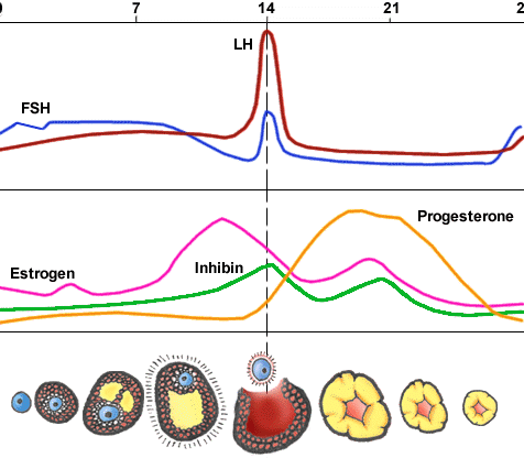
FSH acts on the granulosa cells, and LH acts on the theca cells. Granulosa cells have FSH receptors but no LH receptors and theca cells have just the reverse. FSH stimulates the granulosa cells to multiply and produce estrogen, and it also stimulates enlargement of the antrum. Estrogen also functions as a paracrine or autocrine agent within the follicle, which stimulates the proliferation of granulosa cells, which further increases estrogen production. FSH作用于颗粒细胞,LH作用于卵泡膜细胞。颗粒细胞有FSH受体,没有LH受体,卵泡膜细胞正好相反。FSH刺激颗粒细胞增殖并产生雌激素,同时刺激窦腔扩大。雌激素在卵泡内还起旁分泌或自分泌作用,刺激颗粒细胞增殖,进而增加雌激素的产生。
LH acts upon the theca cells, stimulating them not only to proliferate but also to synthesize androgens. The androgens diffuse into the granulosa cells and are converted to estrogen by aromatase(芳香化酶). By the beginning of the second week, one follicle has become dominant and the other developing follicles degenerate because plasma concentration of FSH begins to decrease and there is no longer enough FSH to prevent atresia. LH作用于卵泡膜细胞,刺激其增殖,并合成雄激素。雄激素扩散到颗粒细胞中,在芳香化酶的作用下转化为雌激素。到第二周初,一个卵泡已占主导地位,其他正在发育的卵泡由于血浆中FSH浓度开始下降,不再有足够的FSH来防止卵泡闭锁而退化。
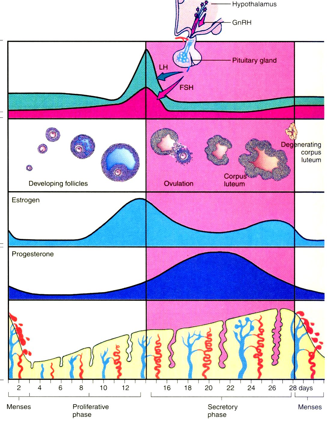
Estrogen negative feedback: when plasma estrogen concentration is relatively low, as during the early and middle follicular phases, estrogen acts on anterior pituitary gland and hypothalamus, negative regulate GnRH (Gonadotropin-releasing hormone), FSH (and LH, to a lesser extent) excretion. 雌激素负反馈:当血浆雌激素浓度较低时,如卵泡早、中期,雌激素作用于垂体前叶和下丘脑,负向调节GnRH(促性腺激素释放激素)、FSH(以及LH,作用较小)的排泄。
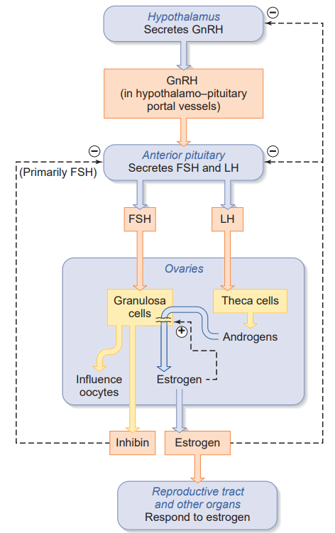
Estrogen positive feedback: increasing plasma concentrations of estrogen for 1 to 2 days, as occurs during the estrogen peak of the late follicular phase, act upon the anterior pituitary gland to enhance the sensitivity of LH-releasing cells to GnRH and may also stimulate GnRH release from the hypothalamus. The rapidly increasing estrogen leads to the LH surge. 雌激素正反馈:在卵泡晚期雌激素高峰期间,血浆雌激素浓度持续增加 1 至 2 天,作用于垂体前叶,增强 LH 释放细胞对 GnRH 的敏感性,也可能刺激下丘脑释放 GnRH。雌激素迅速增加导致 LH 激增。
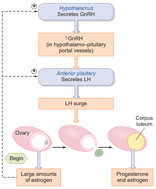
11.2.4.2 Hormonal control
Suppression of FSH and LH during luteal phase. If implantation of a developing conceptus does not occur and hCG(human chorionic gonadotropin) does not appear in the blood, the corpus luteum dies, progesterone and estrogen decrease, menstruation occurs, and the next menstrual cycle begins. 黄体期 FSH 和 LH 受到抑制。如果发育中的胚胎没有着床,并且血液中没有出现 hCG(人绒毛膜促性腺激素),黄体就会死亡,孕酮和雌激素就会减少,月经就会来,下一个月经周期就会开始。
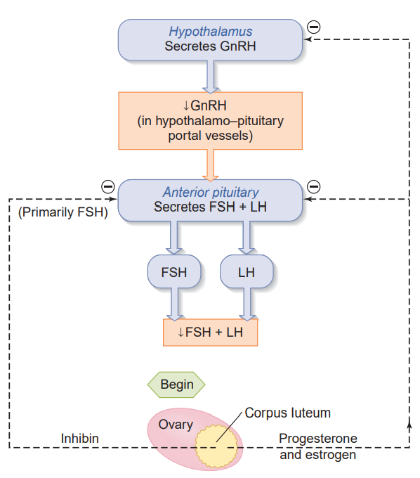
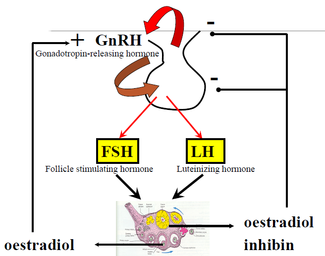
11.2.4.3 Hypothalamus-Pituitary-Gonad Axis
GnRH is secreted in the hypophysial portal bloodstream, and the portal blood carries the GnRH to the anterior pituitary gland. GnRH activates GnRHR, and then stimulate synthesis and secretion of LH and FSH. GnRH activity is very low during childhood, and is activated at puberty or adolescence. During the reproductive years, pulse activity is critical for successful reproductive function as controlled by feedback loops. GnRH 在垂体门脉血流中分泌,门脉血将 GnRH 输送到垂体前叶。GnRH 激活 GnRHR,然后刺激 LH 和 FSH 的合成和分泌。GnRH 活性在儿童时期非常低,在青春期或青少年期被激活。在生育年龄,脉冲活动对于成功的生殖功能至关重要,因为它受反馈回路的控制。
The synthesis and secretion of FSH and LH are controlled by the size and frequency of GnRH pulses, as well as by feedback from androgens and estrogens. Low-frequency GnRH pulses are required for FSH release, whereas high-frequency GnRH pulses stimulate LH pulses. In males, GnRH is secreted in pulses at a constant frequency. In females, the frequency of the pulses varies during the menstrual cycle, and there is a large surge of GnRH just before ovulation. FSH 和 LH 的合成和分泌受 GnRH 脉冲的大小和频率以及雄激素和雌激素的反馈控制。低频 GnRH 脉冲是 FSH 释放所必需的,而高频 GnRH 脉冲会刺激 LH 脉冲。在男性中,GnRH 以恒定频率脉冲分泌。在女性中,脉冲频率在月经周期中会发生变化,并且在排卵前会出现大量的 GnRH 激增。
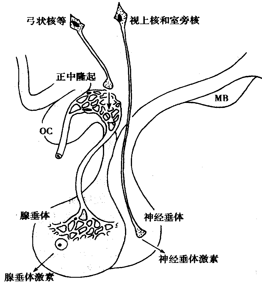
11.2.5 Uterine Changes in the Menstrual Cycle
11.2.5.1 Uterine events
- Influenced by the cyclical production of steroid hormones. 受类固醇激素周期性产生的影响
- Causes changes to the uterus. 导致子宫变化
- Involving the endometrium and cervical mucus. 涉及子宫内膜和宫颈粘液
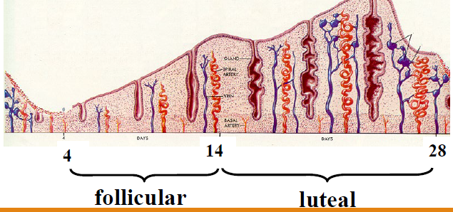
• Endometrium thickens 子宫内膜增厚
• Glands grow 腺体生长
• Blood vessels grow - spiral arteries 血管生长 - 螺旋动脉
• Thickening of endometrium declines 子宫内膜增厚下降
• Glands branch 腺体分支
• Glands secrete 腺体分泌
Proliferative phase 增生期
- Occurs during follicular phase of ovarian cycle 发生在卵巢周期的卵泡期
- Endometrium exposed to oestrogen - regeneration from the last menstrual cycle 子宫内膜暴露于雌激素 - 从最后一次月经周期再生
- Glands in endometrium are straight in this phase, parallel to one another and contain little secretion 子宫内膜中的腺体在此阶段是直的,彼此平行且含有少量分泌物
Secretory phase 分泌期
- After ovulation - progesterone production causes secretory changes in endometrial glands 排卵后 - 孕酮产生导致子宫内膜腺体分泌变化
- Appearance of secretory vacuoles in glandular epithelium below nuclei 腺上皮细胞核下出现分泌空泡
- Secretions in lumen of glands-which become tortuous and develop serrated margins 腺腔内分泌物 - 变得曲折并形成锯齿状边缘
Menstrual phase 月经期
- Regression of corpus luteum-decline in oestrogenand progesterone 黄体退化 - 雌激素和孕酮下降
- Intense spasmodic contraction of spiral section of endometrial arterioles-ischemic necrosis-shedding of superficial layer-bleeding. 子宫内膜小动脉螺旋段强烈痉挛性收缩-缺血性坏死-浅层脱落-出血
- These spasms are associated with prostaglandin, which are also associated with increased uterine contractions during menstrual flow. 这些痉挛与前列腺素有关,前列腺素还与月经期间子宫收缩增强有关
11.2.5.2 Menstrual phase
Constriction of the uterine blood vessels: leads to a diminished supply of oxygen and nutrients to the endometrial cells. The uterine smooth muscle begins to undergo rhythmic contractions. 子宫血管收缩:导致子宫内膜细胞的氧气和营养供应减少。子宫平滑肌开始有节奏地收缩
Dilation of the endometrial arterioles: just after the vascular constriction, which resulting in haemorrhage through the weakened capillary walls. The menstrual flow consists of this blood mixed with endometrial debris. 子宫内膜小动脉扩张:血管收缩后,导致毛细血管壁变弱而出血。月经血由这种血液和子宫内膜碎片混合而成
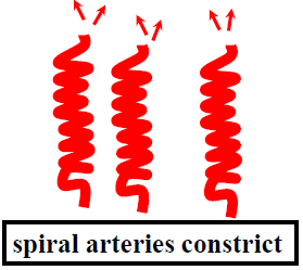
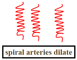
11.2.5.3 The menstrual cycle
The menstrual cycle is the regular natural change that occurs in the female reproductive system(specifically the uterus and ovaries) that makes pregnancy possible. 月经周期是女性生殖系统(特别是子宫和卵巢)发生的正常自然变化,使怀孕成为可能
Average length of human cycle = 28.1 days (26-35 days)
Typical blood loss per menstrual period is about 50 to 150 mL.
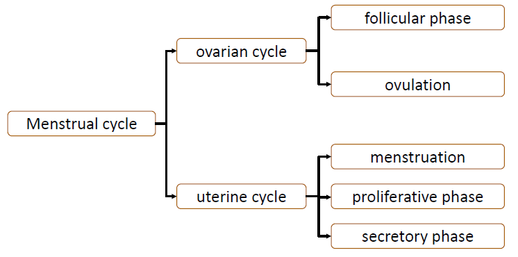
The menstrual cycle may be divided into two phases approximately equal in length and separated by ovulation: 月经周期可分为两个大致相等且以排卵为间隔的阶段
- the follicular phase, during which a mature follicle and secondary oocyte develop; 卵泡期,在此期间成熟卵泡和次级卵母细胞发育
- the luteal phase, beginning after ovulation and lasting until the death of the corpus luteum. 黄体期,从排卵后开始,一直持续到黄体死亡
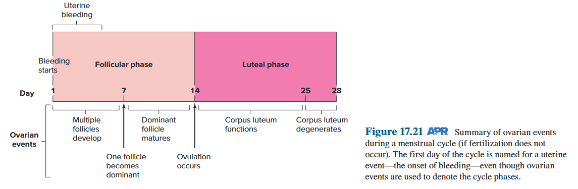
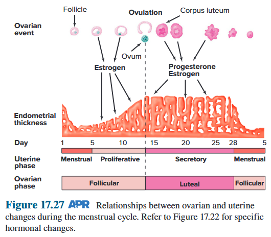
11.2.5.4 Summary of the menstrual cycle
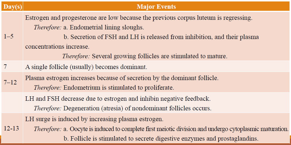
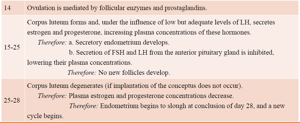
11.3 Pregnancy
11.4 Parturition and lactation
Chapter 8 Urine Formation and Excretion
Functions of the kidneys
- Regulation of H~2~O inorganic ion balance, and acid-base balance (in cooperation with the lungs) (most important function) 水盐平衡
- Removal of metabolic waste products from the blood and their excretion in the urine
- Removal of foreign chemicals from the blood and their excretion in the urine
- Gluconeogenesis 糖异生
- Production of hormones / enzymes:
- Erythropoietin, which controls erythrocyte production 促红细胞生成素,控制红细胞的产生
- Renin, an enzyme that controls the formation of angiotensin, which influences blood pressure and sodium balance 肾素,一种控制血管紧张素形成的酶,血管紧张素影响血压和钠平衡
- Conversion of 25-hydroxyvitamin D to 1,25-dihydroxyvitamin D, which influences calcium balance 25-羟基维生素 D 转化为 1,25-二羟基维生素 D,影响钙平衡
肾脏是机体最重要的排泄器官,通过尿的生成和排出,肾脏能够排出机体代谢终产物、进入机体过剩的物质和异物,调节水、电解质和酸碱平衡,调节动脉血压等,从而维持机体内环境的稳态。肾脏也是一个内分泌器官,它能合成和释放多种生物活性物质,如合成和释放肾素,参与动脉血压的调节;合成和释放促红细胞生成素,促进红细胞的生成;肾脏中的1α-羟化酶可使25-羟维生素D~3~转化为1,25-二羟维生素D~3~, 调节钙的吸收和血钙水平;肾脏还能生成激肽和前列腺素,参与局部或全身血管活动的调节。
8.1 Structure of the Kidneys and Urinary System
Each nephron in the kidneys consists of a renal corpuscle and a tubule.
- Each renal corpuscle comprises a glomerulus, and a Bowman’s capsule. 每个肾小体都包含一个肾小球和一个鲍曼氏囊。
- The tubule extends from Bowman’s capsule and is subdivided into the proximal tubule, loop of Henle, distal convoluted tubule, and collecting-duct system. At the level of the collecting ducts, multiple tubules join and empty into the renal pelvis, from which urine flows through the ureters to the bladder. 小管从鲍曼氏囊延伸,分为近端小管、亨利袢、远端曲小管和集合管系统。在集合管的水平,多个小管连接并排入肾盂,尿液从肾盂流经输尿管到达膀胱。
- Each glomerulus is supplied by an afferent arteriole, and an efferent arteriole leaves the glomerulus to branch into peritubular capillaries. 每个肾小球由传入小动脉供血,传出小动脉离开肾小球分支成管周毛细血管。
8.1.1 Renal Nephron
8.1.1.1 Renal System
- Kidneys have an excellent blood supply: 0.5% total body weight but ~20% of CO (Cardiac Output). 肾脏具有良好的血液供应:占总体重的 0.5%,但约占 CO(心输出量)的 20%。
- Kidneys process plasma portion of blood by removing substances from it, and in a few cases, by adding substances to it. 肾脏通过去除血液中的物质来处理血液的血浆部分,在少数情况下,还通过向血液中添加物质来处理。
- Works with cardiovascular system and others in integrated manner. 与心血管系统和其他系统综合工作。
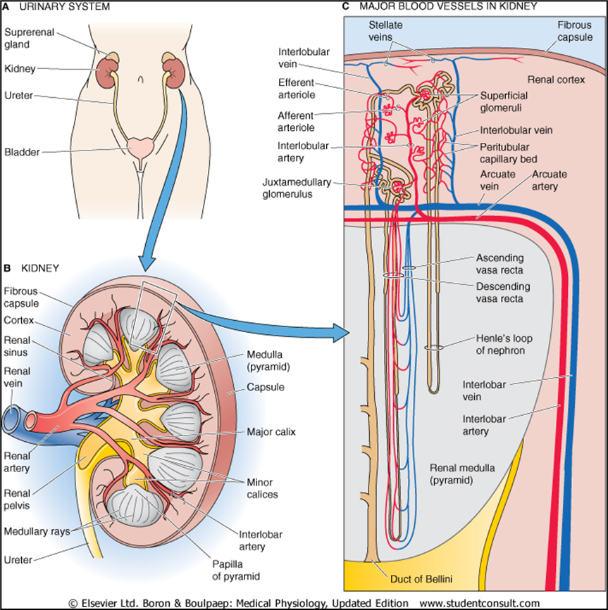
The two kidneys lie at the back of the abdominal wall but not actually in the abdominal cavity. They are retroperitoneal, meaning they are just behind the peritoneum, the lining of this cavity. The urine flows from the kidneys through the ureters into the bladder and then is eliminated via the urethra (Figure 14.1). The major structural components of the kidney are shown in cross section in Figure 14.2. The indented surface of the kidney is called the hilum, through which courses the blood vessels perfusing (renal artery) and draining (renal vein) the kidneys. The nerves that innervate the kidney and the tube that drains urine from the kidney (the ureter) also pass through the hilum. The ureter is formed from the calyces (singular, calyx), which are funnel-shaped structures that drain urine into the renal pelvis, from which the urine enters the ureter. Also notice that the kidney is surrounded by a protective capsule made of connective tissue. The kidney is divided into an outer renal cortex and inner renal medulla, described in more detail later. The connection between the tip of the medulla and the calyx is called the papilla.
两个肾脏位于腹壁后部,但实际上并不在腹腔内。它们是腹膜后的,这意味着它们位于腹膜(该腔的内壁)后面。尿液从肾脏通过输尿管流入膀胱,然后通过尿道排出(图14.1)。肾脏的主要结构部件如图 14.2 中的横截面所示。肾脏的凹进表面称为肾门,灌注肾脏(肾动脉)和引流肾脏(肾静脉)的血管穿过该肾门。支配肾脏的神经和从肾脏排出尿液的管道(输尿管)也穿过肺门。输尿管由肾盏(单数,calyx)形成,肾盏是漏斗状结构,将尿液排入肾盂,尿液从肾盂进入输尿管。另请注意,肾脏被结缔组织制成的保护性胶囊包围。肾脏分为外肾皮质和内肾髓质,稍后将更详细地描述。髓质尖端和花萼之间的连接称为乳头。
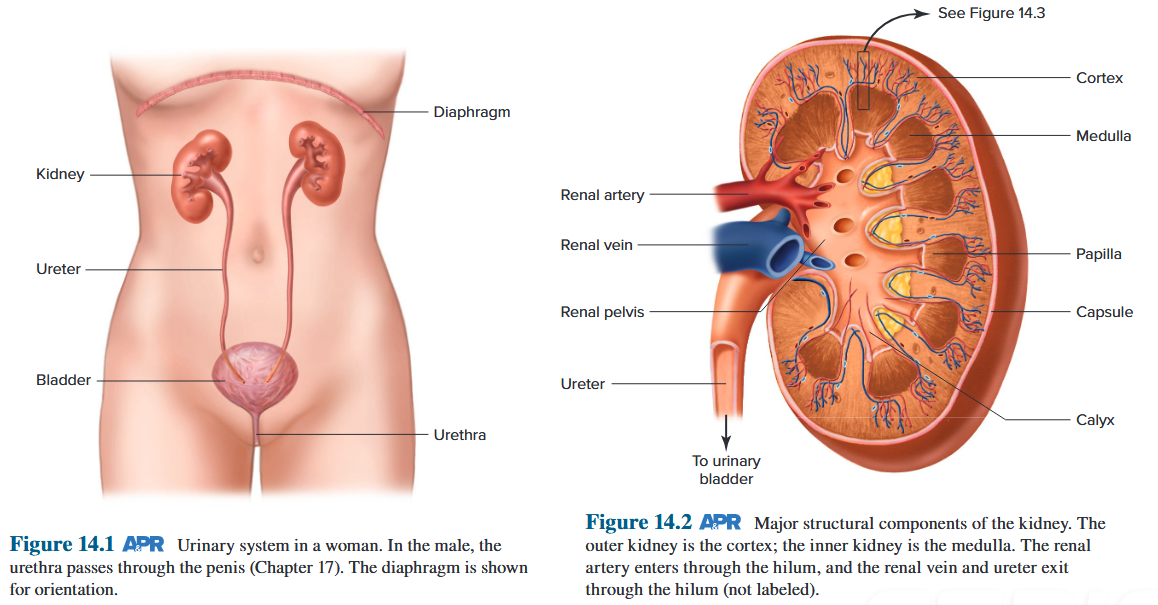
8.1.1.2 Renal Nephron
-
Total of about 2.5 million in the 2 kidneys.
-
Each nephron consists of 2 functional components:
- The tubular component (contains what will eventually become urine) 管状成分(包含最终变成尿液的成分)
- The vascular component (blood supply) 血管成分(血液供应)
-
The mechanisms by which kidneys perform their functions depends upon the relationship between these two components. 肾脏执行其功能的机制取决于这两个组成部分之间的关系。
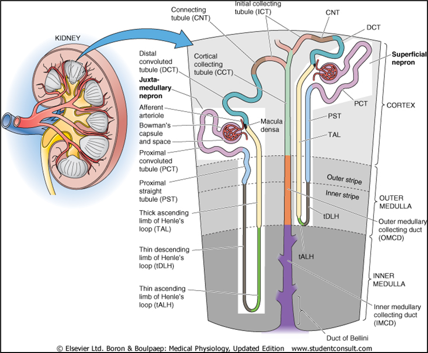
Each nephron consists of:
- an initial filtering component called the renal corpuscle: glomerulus, Bowman’s capsule
- a tubule that extends from the renal corpuscle
Each renal corpuscle contains a compact tuft of interconnected capillary loops called the glomerulus or glomerular capillaries.
Each glomerulus is supplied with blood by an arteriole called an afferent arteriole.
The glomerulus protrudes into a fluid-filled capsule called Bowman’s capsule.
The combination of a glomerulus and a Bowman’s capsule constitutes a renal corpuscle. As blood flows through the glomerulus, about 20% of the plasma filters into Bowman’s capsule. The remaining blood then leaves the glomerulus by the efferent arteriole.
The part of Bowman’s capsule in contact with the glomerulus becomes pushed inward but does not make contact with the opposite side of the capsule. Accordingly, a fluid-filled space called the Bowman’s space exists within the capsule. Fluid essentially free of proteins filters from the glomerulus into this space.
每个肾小体都包含一簇紧密相连的毛细血管袢,称为肾小球或肾小球毛细血管。 每个肾小球由称为传入小动脉的小动脉提供血液。 肾小球突出到一个充满液体的囊中,称为鲍曼囊。 肾小球和鲍曼囊的组合构成肾小体。当血液流经肾小球时,大约 20% 的血浆过滤到鲍曼氏囊中。然后剩余的血液通过传出小动脉离开肾小球。 鲍曼囊与肾小球接触的部分被向内推,但不与囊的另一侧接触。因此,胶囊内存在一个充满液体的空间,称为鲍曼空间。基本上不含蛋白质的液体从肾小球过滤到该空间。
- two sets of capillaries in the kidneys:
- the glomerular capillaries
- the peritubular capillaries
- two sets of arterioles capillaries int he kidneys:
- the afferent arteriole
- the efferent arteriole
- the renal circulation is very unusual in that it includes two sets of arterioles and two sets of capillaries.
8.1.1.3 Consecutive Segments of the Nephron
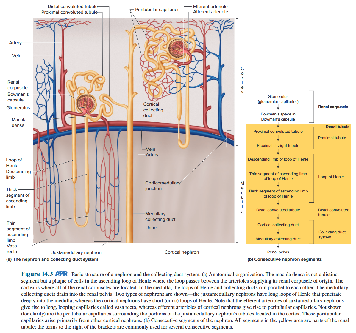
Two types of nephrons are shown - the juxtamedullary nephrons have long loops of Henle that penetrate deeply into the medulla, whereas the cortical nephrons have short (or no) loops of Henle. 近髓肾单位具有深入髓质的长亨利环,而皮质肾单位具有短(或无)亨利环。
致密斑不是一个独特的部分,而是亨利升袢中的细胞斑块,该袢在供应其起源肾小体的小动脉之间穿过。皮质是所有肾小体所在的地方。在髓质中,亨利环和集合管彼此平行。髓质集合管流入肾盂。显示了两种类型的肾单位——近髓肾单位具有深入髓质的长亨利环,而皮质肾单位具有短(或无)亨利环。请注意,近髓肾单位的传出小动脉产生称为直血管的长的环状毛细血管,而皮质肾单位的传出小动脉产生管周毛细血管。未显示(为了清楚起见)围绕位于皮质中的近髓肾单位小管部分的管周毛细血管。这些管周毛细血管主要来自其他皮质肾单位。
8.1.2 Juxtaglomerular Apparatus
8.1.2.1 Glomerulus and Bowman’s capsule
Glomerular filtrate drains into Bowman’s space, and then into proximal convoluted tubule.
Endothelium has pores to allow small molecules through.
Podocytes have negative charge. This and the basement membrane stops proteins getting through into tubular fluid.
Macula densa senses GFR by [Na^+^]
Juxtaglomerular (JG) apparatus also includes JG cells that secrete renin.
JGA helps regulate renal blood flow, GFR and also indirectly, modulates Na^+^ balance and systemic BP.
肾小球滤液流入鲍曼氏腔,然后流入近曲小管。 内皮细胞具有允许小分子通过的孔隙。 足细胞带负电荷。它和基底膜阻止蛋白质进入管状液体。 致密斑通过 [Na^+^] 肾小球旁 (JG) 装置感知 GFR,还包括分泌肾素的 JG 细胞。 JGA 有助于调节肾血流量、GFR,并间接调节 Na^+^ 平衡和全身血压。
One additional anatomical detail involving both the tubule and the arterioles is important. Near its end, the ascending limb of each loop of Henle passes between the afferent and efferent arterioles of that loop’s own nephron (see Figure 14.3). At this point, there is a patch of cells in the wall of the ascending limb as it becomes the distal convoluted tubule called the macula densa, and the wall of the afferent arteriole contains secretory cells known as juxtaglomerular (JG) cells. The combination of macula densa and juxtaglomerular cells is known as the juxtaglomerular apparatus (JGA) (see Figure 14.4a and Figure 14.5). As described later, the JGA has important functions in the regulation of ion and water balance, and the production of factors that control blood pressure. 涉及小管和小动脉的另一项解剖细节很重要。在其末端附近,每个亨利环的升肢在该环自身肾单位的传入和传出小动脉之间穿过(见图 14.3)。此时,升肢的壁上有一片细胞,因为它成为远端曲管,称为致密斑,传入小动脉的壁包含称为肾小球旁(JG)细胞的分泌细胞。致密斑和肾小球旁细胞的组合被称为肾小球旁装置(JGA)(见图 14.4a 和图 14.5)。如下所述,JGA在离子和水平衡的调节以及控制血压的因子的产生方面具有重要的功能。
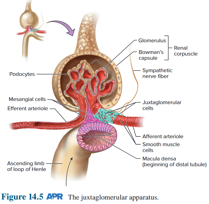
8.2 Basic Renal Processes
The three basic renal processes:
- Glomerular filtration
- Tubular reabsorption
- Tubular secretion
The excretion of a substance is equal to the amount filtered plus the amount secreted minus the amount reabsorbed. 物质的排泄量等于过滤量加上分泌量减去重吸收量。
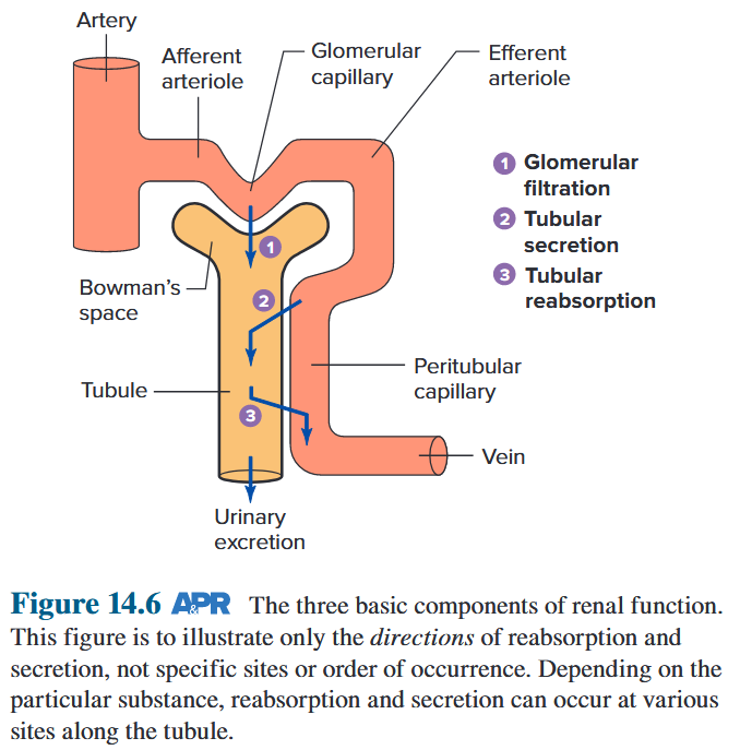 $$
\text{Amount\ excreted} = \text{Amount\ filtered + Amount\ secreted - Amount\ reabsorbed}
$$
$$
\text{Amount\ excreted} = \text{Amount\ filtered + Amount\ secreted - Amount\ reabsorbed}
$$
8.2.1 Glomerular Filtration
8.2.1.1 GFR
The volume of fluid filtered from the glomeruli into Bowman’s space per unit time is known as the glomerular filtration rate (GFR). GFR is determined by:
- the net filtration pressure 净过滤压力
- the permeability of the corpuscular membranes 红细胞膜的通透性
- the surface area available for filtration 可用于过滤的表面积
At any given net filtration pressure, the GFR will be directly proportional to the membrane permeability and the surface area. 在任何给定的净过滤压力下,GFR 将与膜渗透性和表面积成正比。
- GFR controlled by diameters of afferent and efferent arterioles GFR 由传入和传出小动脉的直径控制
- Sympathetic vasoconstrictor nerves 交感血管收缩神经
- ADH and RAAS also have an effect on GFR ADH 和 RAAS 也对 GFR 有影响
- Autoregulation maintains blood supply and so maintains GFR. Also prevents high pressure surges damaging kidneys. 自动调节可维持血液供应,从而维持 GFR,还可以防止高压浪涌损害肾脏
- Remember: high hydrostatic pressure (P~GC~) at glomerular capillaries is due to short, wide afferent arteriole (low R to flow) and the long, narrow efferent arteriole (high R). 肾小球毛细血管的高静水压 (PGC) 是由于短而宽的传入小动脉(低 R 流动)和长而窄的传出小动脉(高 R)造成的。
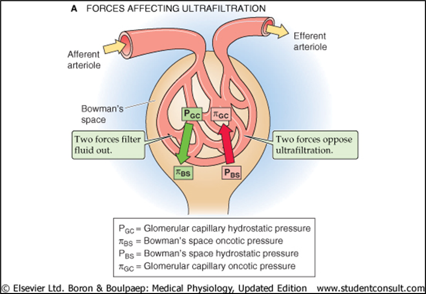
8.2.1.2 GFR depends on diameters of afferent and efferent arterioles
Control of GFR by constriction or dilation of afferent arterioles (AA) or efferent arterioles (EA). (a) Constriction of the afferent arteriole or © dilation of the efferent arteriole reduces PGC, thus decreasing GFR. (b) Constriction of the efferent arteriole or (d) dilation of the afferent arteriole increases PGC, thus increasing GFR. 通过收缩或扩张传入小动脉 (AA) 或传出小动脉 (EA) 来控制 GFR。 (a) 传入小动脉收缩或 © 传出小动脉扩张会降低 PGC,从而降低 GFR。 (b) 传出小动脉收缩或 (d) 传入小动脉扩张会增加 PGC,从而增加 GFR。
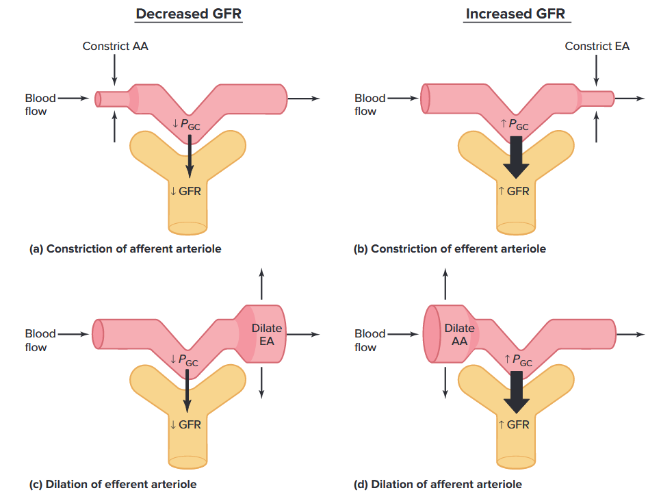
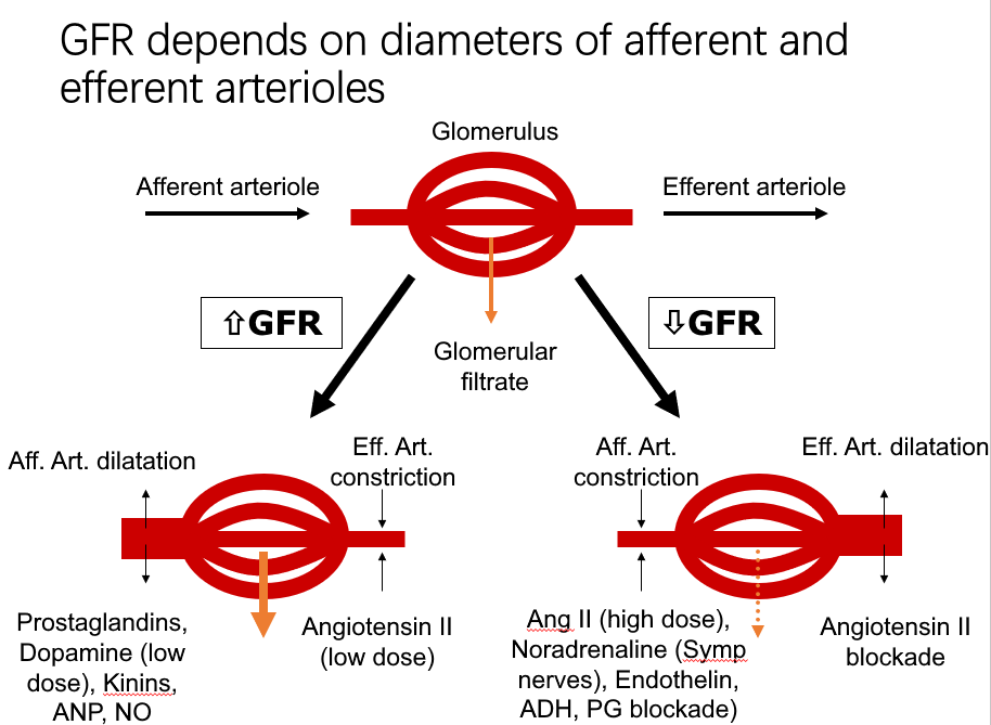
8.2.2 Tubular Reabsorption
GFR (glomerular filtration rate) is very high: ~180L/day. Lots of opportunity to precisely regulate ECF (extracellular fluid) and get rid of unwanted substances.
It is the ECF that is being regulated, NOT the urine.
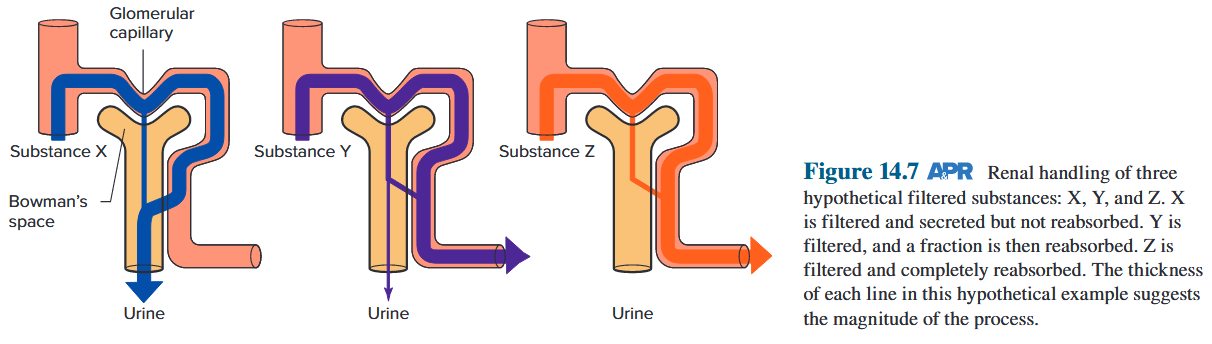
Assume that the tubule can secrete 100% of the peritubular capillary substance X into the tubular lumen but cannot reabsorb X. Therefore, by the combination of filtration and tubular secretion, the plasma that originally entered the renal artery is cleared of all of its substance X, which leaves the body via the urine. Logically, this tends to be the pattern for renal handling of foreign substances that are potentially harmful to the body. 假设肾小管可以将100%的肾小管周围毛细血管物质X分泌到肾小管腔内,但不能重吸收X。因此,通过滤过和肾小管分泌的结合,最初进入肾动脉的血浆中的所有物质X被清除,通过尿液离开身体。从逻辑上讲,这往往是肾脏处理对身体可能有害的异物的模式。
By contrast, assume that the tubule can reabsorb but not secrete Y and Z. The amount of Y reabsorption is moderate so that some of the filtered material is not reabsorbed and escapes from the body. For Z, however, the reabsorptive mechanism is so powerful that all the filtered Z is reabsorbed back into the plasma. Therefore, no Z is lost from the body. Hence, for Z, the processes of filtration and reabsorption have canceled each other out and the net result is as though Z had never entered the kidney. Again, it is logical to assume that substance Y is important to retain but requires maintenance within a homeostatic range; substance Z is presumably very important for health and is therefore completely reabsorbed. 相比之下,假设肾小管可以重吸收但不分泌Y和Z。Y重吸收的量是适中的,因此一些过滤的物质不会被重吸收并从体内逸出。然而,对于 Z 而言,重吸收机制非常强大,以至于所有过滤的 Z 都会被重吸收回血浆中。因此,Z 不会从体内丢失。因此,对于 Z 来说,过滤和重吸收过程相互抵消,最终结果就好像 Z 从未进入肾脏一样。同样,假设物质 Y 很重要,但需要维持在稳态范围内,这是合乎逻辑的; Z 物质可能对健康非常重要,因此会被完全重新吸收。
- The filtered loads are enormous, generally larger than the total amounts of the substances in the body. For example, the body contains about 40 L of water, but the volume of water filtered each day is 180 L. 过滤的负荷是巨大的,通常大于体内物质的总量。例如,人体含有约40升水,但每天过滤的水量为180升。
- Reabsorption of waste products is relatively incomplete (as in the case of urea), so that large fractions of their filtered loads are excreted in the urine. 废物的重吸收相对不完全(如尿素),因此其过滤负荷的大部分通过尿液排出。
- Reabsorption of most useful plasma components, such as water, inorganic ions, and organic nutrients, is relatively complete so that the amounts excreted in the urine are very small fractions of their filtered loads. 大多数有用的血浆成分(例如水、无机离子和有机营养物)的重吸收相对完全,因此尿液中排出的量仅占其过滤负荷的很小一部分。
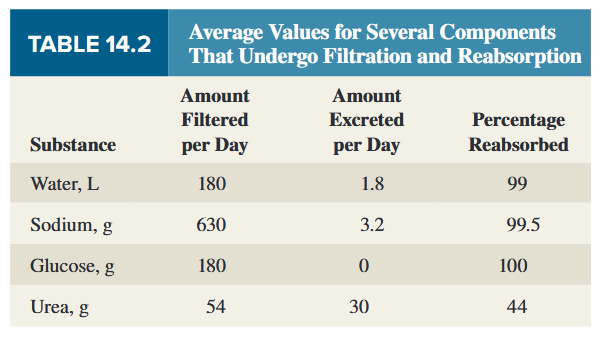
8.2.2.1 Control of reabsorption rate
The reabsorption rates of most organic nutrients, such as glucose, are always very high and are not physiologically regulated. Therefore, the filtered loads of these substances are completely reabsorbed in a healthy kidney, with none appearing in the urine. For these substances, like substance Z in Figure 14.7, it is as though the kidneys do not exist because healthy kidneys do not eliminate these substances from the body at all. Therefore, the kidneys do not regulate the plasma concentrations of these organic nutrients. Rather, the kidneys merely maintain whatever plasma concentrations already exist. 大多数有机营养素(例如葡萄糖)的重吸收率总是非常高并且不受生理调节。因此,这些物质的过滤负荷完全被健康肾脏重新吸收,尿液中不会出现任何物质。对于这些物质,如图 14.7 中的 Z 物质,就好像肾脏不存在一样,因为健康的肾脏根本不会将这些物质从体内消除。因此,肾脏不调节这些有机营养素的血浆浓度。相反,肾脏只是维持已经存在的血浆浓度。
The reabsorptive rates for water and many ions, although also very high, are under physiological control. For example, if water intake is decreased, the kidneys can increase water reabsorption to minimize water loss. 水和许多离子的重吸收率虽然也很高,但受到生理控制。例如,如果水摄入量减少,肾脏可以增加水的重吸收,以最大限度地减少水的流失。
In contrast to glomerular filtration, the crucial steps in tubular reabsorption do not occur by bulk flow because there are inadequate pressure differences across the tubule and limited permeability of the tubular membranes. Instead, two other processes are involved: 肾小管上的压差不足并且肾小管膜的渗透性有限
- The reabsorption of some substances from the tubular lumen is by diffusion, often across the tight junctions connecting the tubular epithelial cells (Figure 14.10). 一些物质从肾小管腔的重吸收是通过扩散进行的,通常穿过连接肾小管上皮细胞的紧密连接。
- The reabsorption of all other substances involves mediated transport, which requires the participation of transport proteins in the plasma membranes of tubular cells. 所有其他物质的重吸收都涉及介导的转运,这需要肾小管细胞质膜中转运蛋白的参与。
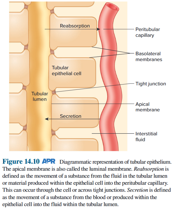
8.2.2.2 Reabsorption by Diffusion
The reabsorption of urea by the proximal tubule provides an example of passive reabsorption by diffusion. 近曲小管对尿素的重吸收提供了通过扩散被动重吸收的例子。
- Some urea is reabsorbed from the proximal tubule in a process that facilitates water reabsorption farther down the nephron. 一些尿素从近端肾小管重吸收,这一过程有利于肾单位更深处的水重吸收。
- Because the corpuscular membranes are freely filterable to urea, the urea concentration in the fluid within Bowman’s space is the same as that in the peritubular capillary plasma and the interstitial fluid surrounding the tubule. 由于红细胞膜可自由过滤尿素,鲍曼间隙内液体中的尿素浓度与肾小管周围毛细血管血浆和肾小管周围间质液中的尿素浓度相同。
- Then, as the filtered fluid flows through the proximal tubule, water reabsorption occurs (by mechanisms to be described later). This removal of water increases the concentration of urea in the tubular fluid so it is higher than in the interstitial fluid and peritubular capillaries. 然后,当过滤后的液体流过近端小管时,会发生水的重吸收(通过稍后描述的机制)。这种水的去除增加了肾小管液中尿素的浓度,因此其浓度高于间质液和肾小管周围毛细血管中的尿素浓度。
- Therefore, urea diffuses down this concentration gradient from tubular lumen to peritubular capillary. 因此,尿素沿着该浓度梯度从肾小管腔扩散到肾小管周围毛细血管。
- Urea reabsorption is thus dependent upon the reabsorption of water. 因此,尿素重吸收取决于水的重吸收。
8.2.2.3 Reabsorption by Mediated Transport
Many solutes are reabsorbed by primary or secondary active transport. These substances must first cross the apical membrane (also called the luminal membrane) that separates the tubular lumen from the cell interior. Then, the substance diffuses through the cytosol of the cell and, finally, crosses the basolateral membrane, which begins at the tight junctions and constitutes the plasma membrane of the sides and base of the cell. The movement by this route is termed transcellular epithelial transport.
A substance does not need to be actively transported across both the apical and basolateral membranes in order to be actively transported across the overall epithelium, moving from lumen to interstitial fluid against its electrochemical gradient.
For example, Na^+^ moves “downhill” (passively) into the cell across the apical membrane through specific channels or transporters and then is actively transported “uphill” out of the cell across the basolateral membrane via Na^+^/K^+^-ATPases in this membrane.
The reabsorption of many substances is coupled to the reabsorption of Na^+^. The cotransported substance moves uphill into the cell via a secondary active cotransporter as Na^+^ moves downhill into the cell via this same cotransporter. This is precisely how glucose, many amino acids, and other organic substances undergo tubular reabsorption. The reabsorption of several inorganic ions is also coupled in a variety of ways to the reabsorption of Na^+^.
许多溶质通过初级或次级主动转运被重吸收。这些物质必须首先穿过将管状腔与细胞内部分开的顶膜(也称为腔膜)。然后,物质扩散通过细胞的细胞质,最后穿过基底外侧膜,基底外侧膜从紧密连接处开始,构成细胞侧面和基底的质膜。通过该途径的运动称为跨细胞上皮运输。物质不需要主动转运穿过顶膜和基底外侧膜,以便主动转运穿过整个上皮,逆其电化学梯度从管腔移动到间质液。例如,Na^+^通过特定通道或转运蛋白穿过顶膜“下坡”(被动)进入细胞,然后通过基底外侧膜通过该膜中的Na^+^/K^+^-ATP酶主动“上坡”运输出细胞。许多物质的重吸收与Na^+^的重吸收相关。当 Na^+^ 通过相同的协同转运蛋白下坡进入细胞时,协同转运物质通过次级活性协同转运蛋白上坡进入细胞。这正是葡萄糖、许多氨基酸和其他有机物质经历肾小管重吸收的方式。几种无机离子的重吸收也以多种方式与Na^+^的重吸收耦合。
Many of the mediated-transport-reabsorptive systems in the renal tubule have a limit to the amounts of material they can transport per unit time known as the transport maximum ($T_m$). This is because the binding sites on the membrane transport proteins become saturated when the concentration of the transported substance increases to a certain level. 肾小管中的许多介导转运重吸收系统对单位时间内可转运的物质量有限制,称为最大转运量。这是因为当转运物质的浓度增加到一定水平时,膜转运蛋白上的结合位点变得饱和。
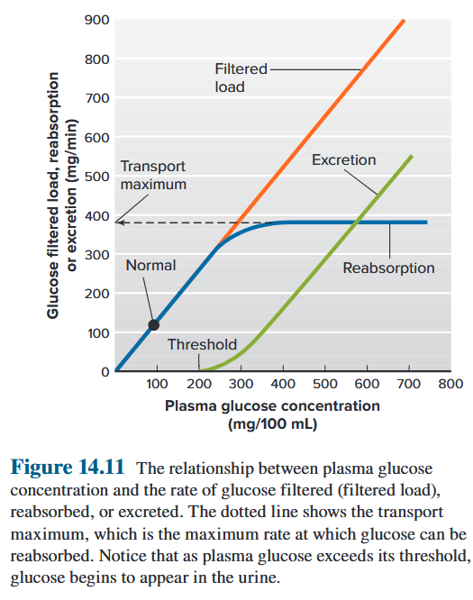
Secondary active-transport proteins for glucose, located in the proximal tubule.
Glucosuria
When the filtered load of glucose exceeds the glucose transport maximum for a significant number of nephrons (typically during hyperglycaemia), glucose starts to appear in urine. *in people with sustained hyperglycaemia (for example, in poorly controlled diabetes mellitus), 当葡萄糖的过滤负荷超过大量肾单位的葡萄糖转运最大值时(通常在高血糖期间),葡萄糖开始出现在尿液中。持续高血糖的人(例如,糖尿病控制不佳)
8.2.2.4 “Division of Labour” in the Tubules
The primary role of the proximal tubule is to reabsorb most of this filtered water and these solutes. 近端小管的主要作用是重新吸收大部分过滤水和这些溶质。此外,除了 K^+^ 以外,近曲小管是溶质分泌的主要部位。
Furthermore, with K^+^ as a major exception, the proximal tubule is the major site of solute secretion. Henle’s loop also reabsorbs relatively large quantities of the major ions and, to a lesser extent, water. 亨利环还重新吸收相对大量的主要离子,并在较小程度上重新吸收水。
Extensive reabsorption by the proximal tubule and Henle’s loop ensures that the masses of solutes and the volume of water entering the tubular segments beyond Henle’s loop are relatively small. 近端小管和亨利袢的广泛重吸收确保了进入亨利袢之外的管状段的溶质质量和水量相对较小。
These distal segments then do the fine-tuning for most low-molecular-weight substances, determining the final amounts excreted in the urine by adjusting their rates of reabsorption and, in a few cases, secretion. 然后,这些远端部分对大多数低分子量物质进行微调,通过调整其重吸收率以及在少数情况下的分泌率来确定尿液中排泄的最终量。
Most homeostatic controls act upon the more distal segments of the tubule. 大多数稳态控制作用于肾小管的更远端部分。
8.3 Renal Clearance
A useful way of quantifying renal function is in terms of clearance. The renal clearance of any substance is the volume of plasma from which that substance is completely removed (“cleared”) by the kidneys per unit time. Every substance has its own distinct clearance value, but the units are always in volume of plasma per unit of time. The basic clearance formula for any substance S is 量化肾功能的一个有用方法是清除率。任何物质的肾脏清除率是单位时间内肾脏完全清除(“清除”)该物质的血浆体积。 每种物质都有其独特的清除率值,但单位始终为每单位时间的血浆体积。任何物质 S 的基本清除公式为
$$
\text{Clearance\ of\ S} = \frac{\text{Mass\ of\ S\ excreted\ per\ unit\ time}}{\text{plasma\ concentration\ of\ S}}
$$
The renal clearance of any substance is the volume of plasma from which that substance is completely removed (“cleared”) by the kidneys per unit time. 任何物质的肾清除率是单位时间内肾脏完全清除(“清除”)该物质的血浆体积。
Special Cases:
-
glucose (no excretion): clearance=0
-
inulin, creatitine (no absorption, no secretion): GFR=clearance
-
PAH (no absorption): clearance = renal plasma flow
8.4 Basic Renal Processes
glomerular filtration
tubular reabsorption
tubular secretion
The excretion of a substance is equal to the amount filtered plus the amount secreted minus the amount reabsorbed. 物质的排泄量等于过滤量加上分泌量减去再吸收量。
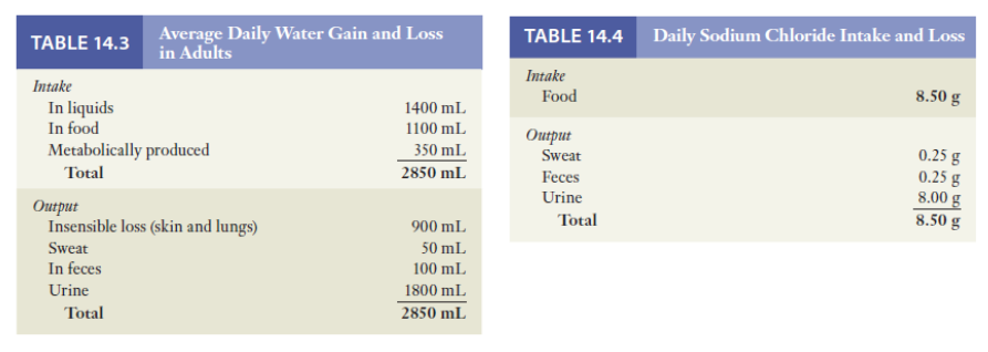
8.4.1 Primary Active Na^+^ Reabsorption
The bulk of Na and water reabsorption (about two-thirds) occurs in the proximal tubule, but the major hormonal control of reabsorption is exerted on the distal convoluted tubules and collecting ducts. 大部分钠和水的重吸收(约三分之二)发生在近曲小管,但重吸收的主要激素控制作用于远曲小管和集合管。
The mechanisms of Na^+^ and water reabsorption can be summarized in two generalizations:
- Na^+^ reabsorption is an active process occurring in all tubular segments except the descending limb of the loop of Henle.
- Water reabsorption is by osmosis (passive) and is dependent upon Na^+^ reabsorption.
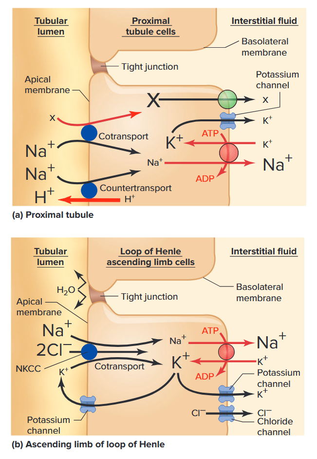
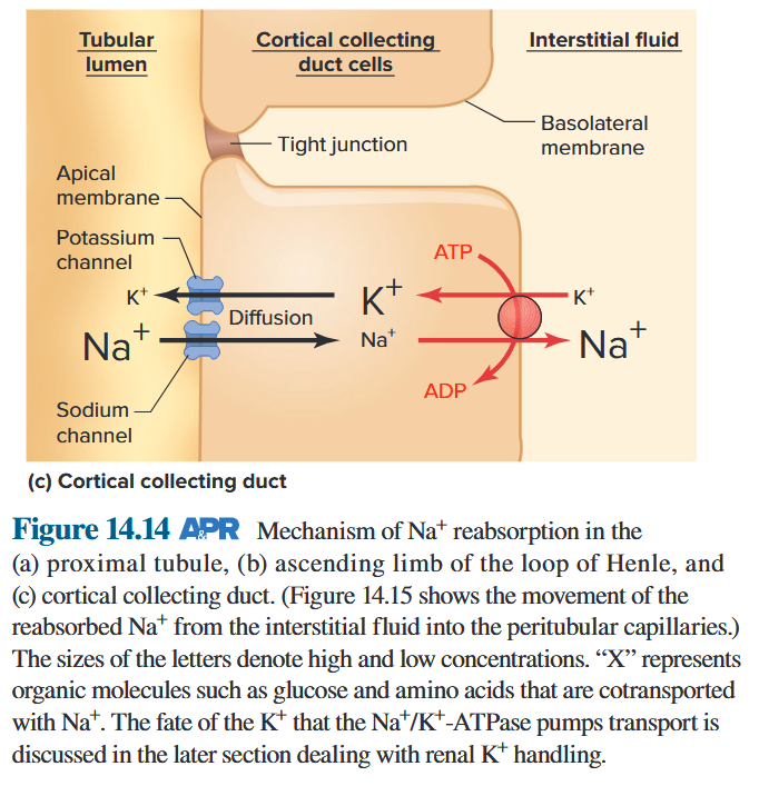
8.4.2 Coupling of Water Reabsorption to Na^+^ Reabsorption
- Na^+^ is transported from the tubular lumen to the interstitial fluid across the epithelial cells. Other solutes, such as glucose, amino acids, and HCO~3~^−^, whose reabsorption depends on Na^+^ transport, also contribute to osmosis. Na^+^ 从肾小管腔穿过上皮细胞转运至间质液。其他溶质,如葡萄糖、氨基酸和 HCO~3~^−^,其重吸收依赖于 Na^+^ 转运,也有助于渗透。
- The removal of solutes from the tubular lumen decreases the local osmolarity of the tubular fluid adjacent to the cell (i.e., the local water concentration increases). At the same time, the appearance of solute in the interstitial fluid just outside the cell increases the local osmolarity (i.e., the local water concentration decreases). 从管状腔中去除溶质会降低细胞附近管状液体的局部渗透压(即局部水浓度增加)。同时,细胞外间质液中溶质的出现增加了局部渗透压(即局部水浓度降低)。
- The difference in water concentration between lumen and interstitial fluid causes net diffusion of water from the lumen across the tubular cells’ plasma membranes and/or tight junctions into the interstitial fluid. 管腔和间质液之间的水浓度差异导致水从管腔穿过肾小管细胞的质膜和/或紧密连接进入间质液的净扩散。
- From there, water, Na^+^, and everything else dissolved in the interstitial fluid move together by bulk flow into peritubular capillaries as the final step in reabsorption. 从那里,水、Na^+^ 和溶解在间质液中的其他物质一起大量流入肾小管周围毛细血管,作为重吸收的最后一步。
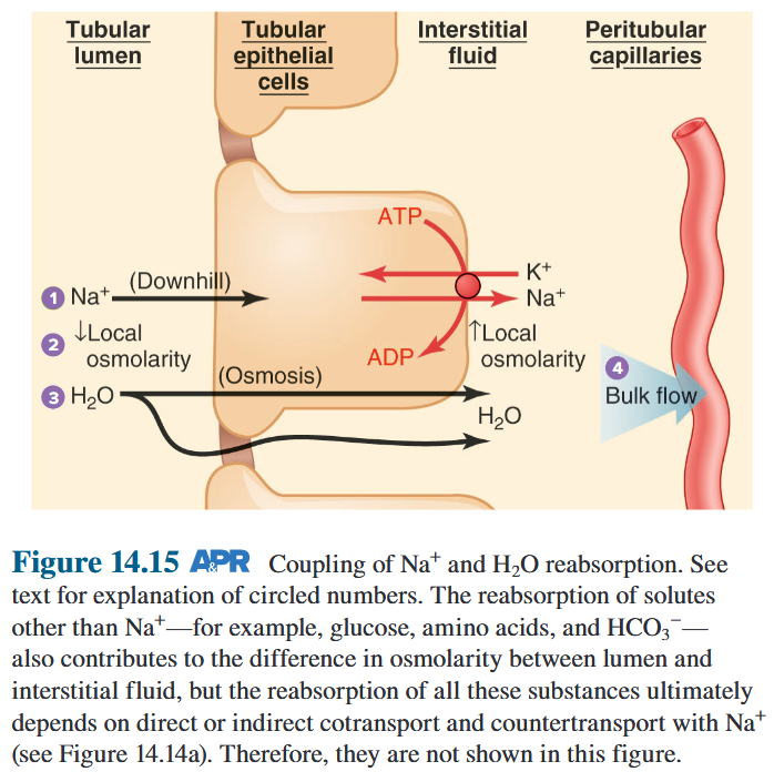
8.4.3 AQPs
8.4.3.1 AQPs
Water permeability varies from tubular segment to segment and depends largely on the presence of water channels, called aquaporins, in the plasma membranes. 不同管段的水渗透性各不相同,很大程度上取决于质膜中水通道(称为水通道蛋白)的存在。
The proximal tubule reabsorbs large amounts of Na and water in the same proportions. 近端小管以相同比例重吸收大量的钠和水。
Crucial point: the water permeability of the cortical and medullary collecting ducts, can vary greatly due to physiological control. These are the only tubular segments in which water permeability is under such control. 关键点:皮质和髓质集合管的透水性会因生理控制而发生很大变化。这些是唯一水渗透率受到这种控制的管状部分。
8.4.3.2 ADH
The major determinant of this controlled permeability and, therefore, of passive water reabsorption in the collecting ducts is a peptide hormone secreted from the posterior pituitary and known as vasopressin, or antidiuretic hormone (ADH; see Chapter 11). Vasopressin stimulates the insertion into the apical membrane of a particular aquaporin water channel made by the collecting-duct cells. More than 10 different aquaporins have been identified throughout the body, and they are identified as AQP1, AQP2, and so on. Figure 14.16 shows the function of the aquaporin water channels in the cells of the collecting ducts. 这种受控渗透性以及集合管中被动水重吸收的主要决定因素是垂体后叶分泌的肽激素,称为加压素或抗利尿激素(ADH;参见第 11 章)。加压素刺激由集合管细胞形成的特定水通道蛋白水通道插入顶膜。全身各处已鉴定出10多种不同的水通道蛋白,它们被鉴定为AQP1、AQP2等。图 14.16 显示了集合管细胞中水通道蛋白水通道的功能。
diuresis: large urine flow
- water diuresis (low ADH)
- osmotic diuresis (increase in solute secretion)
diabetes insipidus: ADH disorder
- central diabetes insipidus (failure of ADH release)
- nephrogenic diabetes insipidus (inability of kidney to respond to ADH)
- any loss of solute in the urine must be accompanied by water loss (osmotic diuresis)
- but water diuresis is not necessarily accompanied by equivalent solute loss
8.4.4 Urine Concentration: The Countercurrent Multiplier System 逆流倍增系统
8.4.4.1 Countercurrent Multiplier System
- hypoosmotic, isoosmotic, hyperosmotic
- obligatory water loss 这个尿液量被称为每日强制性失水量。当饮水量非常低时,这种最小量尿液的流失会导致脱水。
Urinary concentration takes place as tubular fluid flows through the medullary collecting ducts. The interstitial fluid surrounding these ducts is very hyperosmotic. In the presence of vasopressin, water diffuses out of the ducts into the interstitial fluid of the medulla and then enters the blood vessels of the medulla to be carried away. 当管状液体流经髓质集合管时,尿液就会浓缩。这些导管周围的间质液渗透压非常高。在加压素存在的情况下,水从导管扩散到髓质的间质液中,然后进入髓质的血管被带走。
The key question is, How does the medullary interstitial fluid become hyperosmotic? The answer involves several interrelated factors: 髓质间质液如何变得高渗
- the countercurrent anatomy of the loop of Henle of juxtamedullary nephrons 近髓肾单位亨利环的逆流解剖
- reabsorption of NaCl in the ascending limbs of those loops of Henle 亨利袢的上升支对 NaCl 的重吸收
- impermeability to water of those ascending limbs 上升支的不透水性
- trapping of urea in the medulla 尿素滞留在髓质中
- hairpin loops of vasa recta to minimize washout of the hyperosmotic medulla 直肠血管的发夹环可最大限度地减少高渗髓质的冲刷
Along the entire length of the ascending limb, Na and Cl are reabsorbed from the lumen into the medullary interstitial fluid (impermeable to H~2~O). 沿着升肢的整个长度,Na 和 Cl 从管腔被重吸收到髓质间质液(H~2~O 不可渗透)中。
Interstitial fluid of the medulla becomes hyperosmotic. 髓质间质液变得高渗。
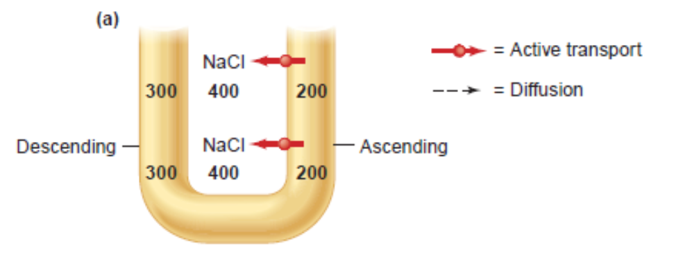
The descending limb does not reabsorb sodium chloride and is highly permeable to water. 下降支不重吸收氯化钠并且对水具有高度渗透性。
Passive reabsorption of H~2~O in descending limb. The osmolarities of the descending limb and interstitial fluid become equal, and both are higher than that of the ascending limb. 降肢对 H~2~O 的被动重吸收。降肢和间质液的渗透压变得相等,并且均高于升肢。
This is the essence of the system: The loop countercurrent multiplier causes the interstitial fluid of the medulla to become concentrated. It is this hyperosmolarity that will draw water out of the collecting ducts and concentrate the urine. 这就是该系统的本质:环路逆流倍增器使髓质的间质液浓缩。正是这种高渗透压将水从集合管中吸出并浓缩尿液。
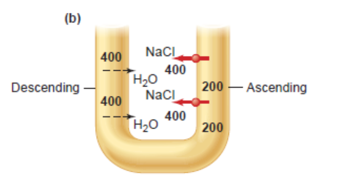
The osmolarity that exists at each horizontal level is “multiplied” as the fluid goes deeper into the medulla. By the time the fluid reaches the bend in the loop, the osmolarity of the tubular fluid and interstitium has been multiplied to a very high osmolarity that can be as high as 1400 mOsmol/L. 随着液体深入髓质,每个水平层存在的渗透压会“成倍增加”。当流体到达环路中的弯曲部时,管状流体和间质的渗透压已倍增至非常高的渗透压,可高达 1400 mOsmol/L。
Keep in mind that the active NaCl transport mechanism in the ascending limb (coupled with low water permeability in this segment) is the essential component of the system. Without it, the countercurrent flow would have no effect on loop and medullary interstitial osmolarity, which would simply remain 300 mOsmol/L throughout. 请记住,升肢中的主动 NaCl 转运机制(加上该部分的低透水性)是该系统的重要组成部分。如果没有它,逆流将对袢和髓质间质渗透压没有影响,整个渗透压将保持在 300 mOsmol/L。
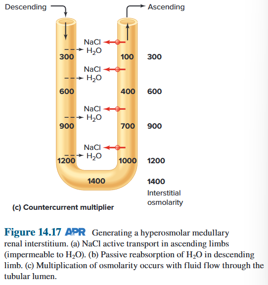
Simplified depiction of the generation of an interstitial fluid osmolarity gradient by the renal countercurrent multiplier system and its role in the formation of hyperosmotic urine in the presence of vasopressin. Notice that the hyperosmotic medulla depends on NaCl reabsorption and urea trapping 简化描述了肾逆流倍增系统产生的间质液渗透压梯度及其在加压素存在下形成高渗尿中的作用。请注意,高渗髓质依赖于 NaCl 重吸收和尿素捕获 ▼
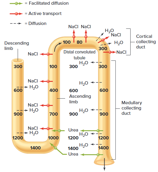
- ADH increases tubular permeability to water in both the cortical and medullary collecting ducts. ADH 增加皮质和髓质集合管中水的管状渗透性。
- In contrast, ADH does not directly influence water reabsorption in the parts of the tubule prior to the collecting ducts. 相反,ADH 不会直接影响集合管之前的肾小管部分的水重吸收。
- Thus, regardless of the plasma concentration of this hormone, the fluid entering the cortical collecting duct is hypoosmotic. 因此,无论这种激素的血浆浓度如何,进入皮质集合管的液体都是低渗的。
- From there on, however, ADH is crucial. 然而,从那时起,ADH 就变得至关重要。
- In the presence of high concentrations of vasopressin, water reabsorption occurs by diffusion from the hypoosmotic fluid in the cortical collecting duct until the fluid in this segment becomes isoosmotic to the interstitial fluid and peritubular plasma of the cortex. 在存在高浓度加压素的情况下,水通过从皮质集合管中的低渗液扩散而发生重吸收,直到该段中的液体与皮质间质液和管周血浆等渗。
- The isoosmotic tubular fluid then enters and flows through the medullary collecting ducts. 然后等渗管状流体进入并流经髓质集合管。
- In the presence of high plasma concentrations of ADH, water diffuses out of the ducts into the medullary interstitial fluid as a result of the high osmolarity that the loop countercurrent multiplier system and urea trapping establish there. 在存在高血浆 ADH 浓度的情况下,由于回路逆流倍增器系统和尿素捕集在那里建立的高渗透压,水从导管扩散到髓质间质液中。
- Water reabsorption occurs all along the lengths of the medullary collecting ducts so that, in the presence of ADH, the fluid at the end of these ducts has essentially the same osmolarity as the interstitial fluid surrounding the bend in the loops—that is, at the bottom of the medulla. 水的重吸收沿着髓质集合管的整个长度发生,因此,在 ADH 存在的情况下,这些导管末端的液体与环路弯曲处周围的间质液具有基本相同的渗透压,即在髓质底部。
- By this means, the final urine is hyperosmotic. By retaining as much water as possible, the kidneys minimize the rate at which dehydration occurs during water deprivation. 通过这种方式,最终的尿液是高渗的。通过保留尽可能多的水分,肾脏可以最大限度地减少缺水期间发生脱水的速度。
8.5.4.2 The Medullary Circulation
Function of the vasa recta to maintain the hypertonic interstitial renal medulla. All movements of water and solutes are by diffusion. 直肠血管维持肾髓质间质高渗的功能。水和溶质的所有运动都是通过扩散进行的。

Why doesn’t the blood flowing through medullary capillaries eliminate the countercurrent gradient set up by the loops of Henle?
- The blood vessels in the medulla (vasa recta) form hairpin loops that run parallel to the loops of Henle and medullary collecting ducts.
- As the blood flows down the loop deeper and deeper into the medulla, Na and Cl do indeed diffuse into—and water out of—the vessel.
- However, after the bend in the loop is reached, the blood then flows up the ascending vessel loop, where the process is almost completely reversed.
- Because of NaCl and water reabsorbed from the loop of Henle and collecting ducts, the amount of blood flow leaving the vasa recta is at least twofold higher than the blood flow entering the vasa recta.
髓质(直肠血管)中的血管形成发夹环,与亨利氏环和髓质集合管的环平行。 当血液沿着循环越来越深入髓质时,Na 和 Cl 确实会扩散到血管中,而水则会从血管中扩散出来。 然而,在到达环路的弯曲处后,血液然后向上流动到升血管环路,该过程几乎完全相反。 由于从亨利袢和集合管中重吸收的氯化钠和水,离开直肠血管的血流量至少比进入直血管的血流量高两倍。
8.5.4.3 Role of Urea in Concentrating Urine
- Urea very useful in concentrating urine 尿素对于浓缩尿液非常有用
- High protein diet = more urea = more concentrated urine 高蛋白饮食 = 更多尿素 = 更浓缩的尿液
- Kidneys filter, reabsorb and secrete urea 肾脏过滤、重吸收和分泌尿素
- Urea excretion rises with increasing urinary flow 尿素排泄随着尿流量的增加而增加
8.5.4.4 The Recycling of Urea Helps to Establish a Hypertonic Medullary Interstitium
- Urea toxic at high levels, but can be useful in small amounts 尿素在高浓度时有毒,但少量即可有用
- Urea recycling causes build up of high [urea] in inner medulla 尿素回收导致髓质内积聚高浓度的尿素
- This helps create the osmotic gradient at loop of Henle so H~2~O can be reabsorbed 这有助于在亨利环处形成渗透梯度,以便水可以被重新吸收
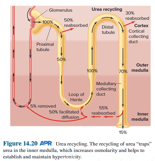
8.5.4.5 Summary of Vasopressin Control of Urine Volume and Osmolarity
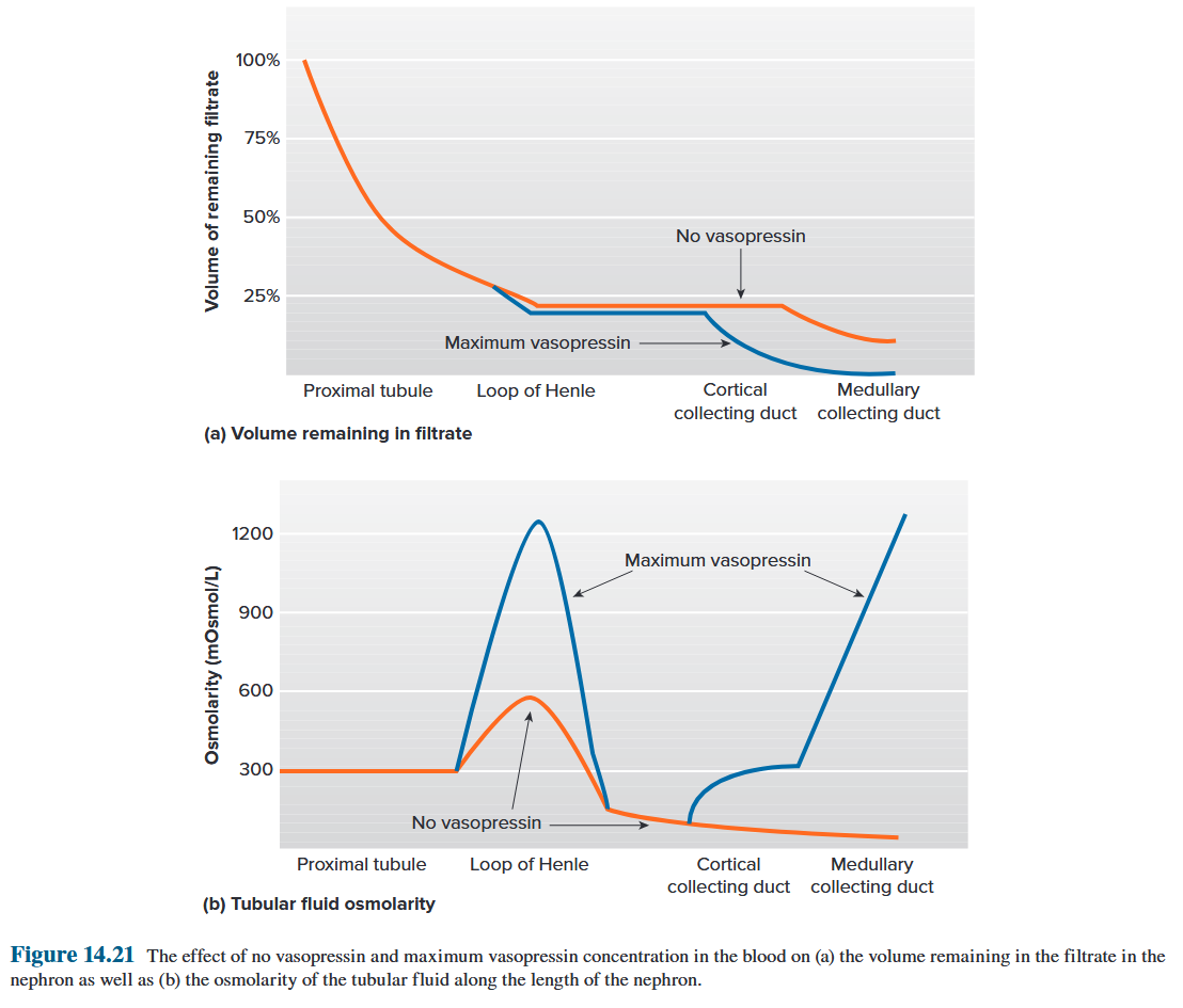
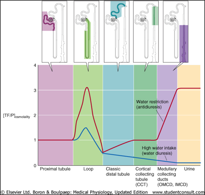
Red = water restriction
Blue = high water intake
Initial concentration of tubular fluid at loop of Henle, then finally at collecting ducts.
8.5.4.6 ADH
ADH also needed to concentrate urine: how does it work?
Antidiuretic Hormone (ADH)
Increases permeability of collecting ducts to H~2~O by inserting H~2~O channels (Aquaporins). 通过插入 H~2~O 通道(水通道蛋白)来增加集合管对 H~2~O 的渗透性。
Helps you make small amount of concentrated urine. 帮助您产生少量浓缩尿液。
Reabsorption of H~2~O increase urea conc. in tubule, increasing its recycling effect.
ADH allows rapid, graded control of urine conc.
ADH released in response to plasma osmolality and ECF(extracellular fluid) volume – osmoreceptors and baroreceptors.
H~2~O 的重吸收增加尿素浓度。在小管中,增加其循环效果。 ADH 可以快速、分级地控制尿液浓度。 ADH 响应血浆渗透压和 ECF(细胞外液)体积而释放 - 渗透压感受器和压力感受器。
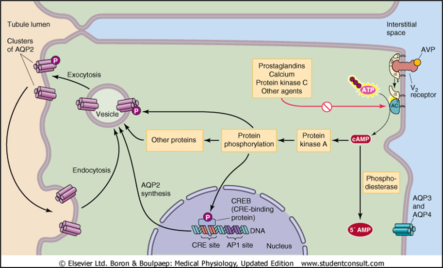
- Increased plasma osmolality stimulates osmoreceptors in the hypothalamus that trigger the release of ADH, which inhibits water excretion.
- Increased osmolality stimulates a second group of osmoreceptors that trigger thirst, which promotes water intake.
- Other factors also trigger ADH release e.g. decreased effective circulating volume, decreased BP, pregnancy, pain, morphine, nausea, congestive heart failure (CHF).
- Hyperaldosteronism = hypernatremia. Due to chronic volume expansion, where osmoreceptors become less sensitive to ADH, reducing ADH inappropriately.
血浆渗透压升高会刺激下丘脑的渗透压感受器,从而引发 ADH 的释放,从而抑制水的排泄。 渗透压的增加会刺激第二组渗透压感受器,从而引发口渴,从而促进水的摄入。 其他因素也会触发 ADH 释放,例如有效循环量减少、血压降低、妊娠、疼痛、吗啡、恶心、充血性心力衰竭 (CHF)。 醛固酮增多症=高钠血症。由于慢性容量扩张,渗透压感受器对抗利尿激素 (ADH) 的敏感性降低,从而不适当地减少了抗利尿激素 (ADH)。
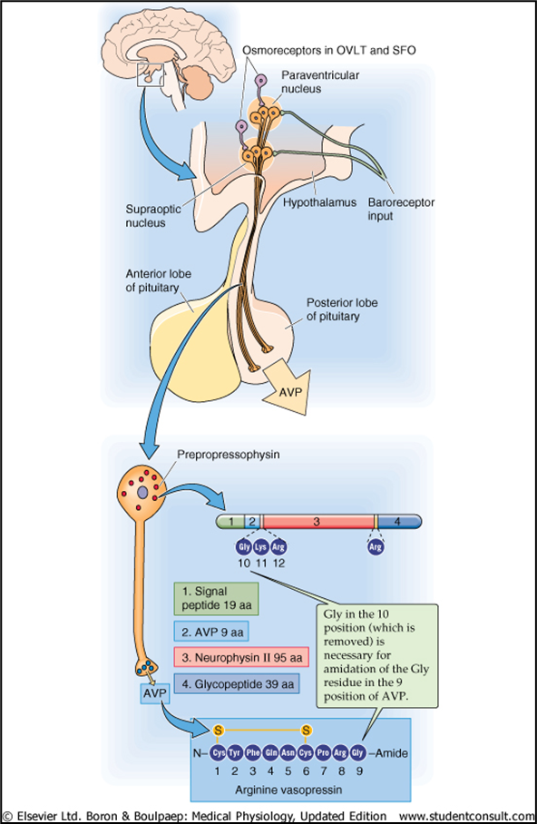
8.5.4.7 RAAS: Renin-Angiotensin-Aldosterone Axis
- Principal factor controlling Ang II levels is renin release.
- Decreased circulating volume stimulates rein release via:
- Decreased BP (symp effects on JGA)
- Decreased [NaCl] at macula densa (“NaCl sensor”)
- Decreased renal perfusion pressure (“renal” baroreceptor)
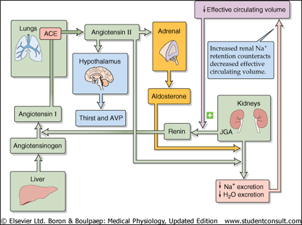
8.5.5 Renal Sodium Regulation
Na excreted = Na filtered + Na reabsorbed
Regulation of cardiovascular pressures by baroreceptors also simultaneously achieves regulation of total-body sodium.
Low total-body sodium leads to low plasma volume, which leads to a decrease in cardiovascular pressures. These lower pressures, via baroreceptors initiate reflexes that influence the renal arterioles and tubules so as to decrease GFR and increase Na reabsorption. These latter events decrease Na excretion, thereby retaining Na in the body and preventing further decreases in plasma volume and cardiovascular pressures.
To summarize, the amount of Na in the body determines the extracellular fluid volume, the plasma volume component of which helps determine cardiovascular pressures, which initiate the responses that control Na excretion.
压力感受器对心血管压力的调节也同时实现了对全身钠的调节。 体内钠含量低会导致血浆容量低,从而导致心血管压力降低。这些较低的压力通过压力感受器启动影响肾小动脉和肾小管的反射,从而降低 GFR 并增加 Na 的重吸收。后面这些事件会减少钠排泄,从而将钠保留在体内并防止血浆容量和心血管压力进一步下降。 总而言之,体内钠的含量决定了细胞外液的体积,其中的血浆体积部分有助于确定心血管压力,从而启动控制钠排泄的反应。
8.5.5.1 Control of GFR
Increased Na loss elicits a decrease in GFR
The main direct cause of the decreased GFR is a decreased net glomerular filtration pressure. This occurs both as a consequence of a decreased arterial pressure in the kidneys and, more importantly, as a result of reflexes acting on the renal arterioles. Note that these reflexes are the basic baroreceptor reflexes - a decrease in cardiovascular pressures causes neurally mediated reflex vasoconstriction in many areas of the body. As we will see, the hormones angiotensin II and vasopressin also participate in this renal vasoconstrictor response. GFR降低的主要原因是肾小球净滤过压降低。这种情况的发生既是肾脏动脉压降低的结果,更重要的是,是作用于肾小动脉的反射的结果。请注意,这些反射是基本压力感受器反射——心血管压力的降低会导致身体许多部位出现神经介导的反射性血管收缩。正如我们将看到的,血管紧张素 II 和加压素激素也参与这种肾血管收缩反应。
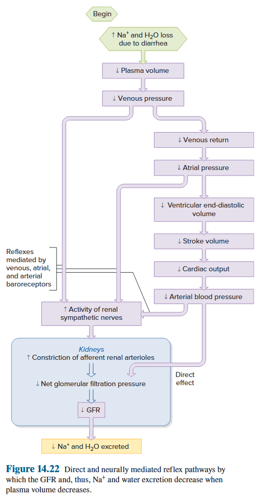
Direct and neurally mediated reflex pathways by which the GFR, Na and water excretion decrease when plasma volume decreases 当血浆容量减少时,GFR、Na 和水排泄减少的直接和神经介导的反射途径
8.5.5.2 Control of Na^+^ Reabsorption
8.5.5.2.1 Aldosterone and the Renin–Angiotensin System (RAAS)
Summary of the renin–angiotensin system and the stimulation of aldosterone secretion by angiotensin II.
Angiotensin-converting enzyme (ACE) is located on the surface of capillary endothelial cells. The plasma concentration of renin is the rate-limiting factor in the renin–angiotensin system.
肾素-血管紧张素系统和血管紧张素 II 刺激醛固酮分泌的总结。 血管紧张素转换酶(ACE)位于毛细血管内皮细胞的表面。肾素的血浆浓度是肾素-血管紧张素系统的限速因素。
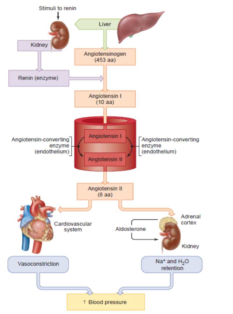
Aldosterone
Aldosterone stimulates Na^+^ reabsorption and K^+^ excretion by the renal tubule.
Aldosterone exerts indirect negative feedback on RAAS by increasing ECV and by lowering plasma [K^+^]
Really important in conserving Na^+^ and water, but also really good at preventing massive swings in K^+^ levels.
醛固酮刺激肾小管的 Na^+^ 重吸收和 K^+^ 排泄。 醛固酮通过增加 ECV 和降低血浆 [K^+^] 对 RAAS 施加间接负反馈。这对于保存 Na^+^ 和水非常重要,而且也非常擅长防止 K^+^ 水平的大幅波动。
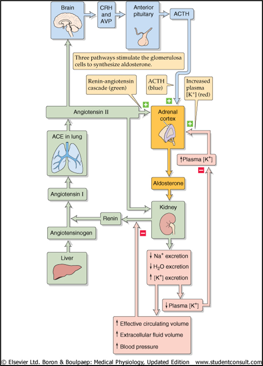
Aldosterone II
- Stimulation of aldosterone release from adrenal cortex.
- Vasoconstriction of renal and other systemic vessels.
- Enhanced tubuloglomerular feedback - makes macula densa more sensitive.
- Enhance Na-H exchanger and Na channel function to promote Na reabsorption.
- Renal hypertrophy.
- Stimulates thirst and ADH release by acting upon hypothalamus.
刺激肾上腺皮质释放醛固酮。 肾血管和其他全身血管的血管收缩。 增强的肾小球反馈 - 使致密黄斑更加敏感。 增强Na-H交换器和Na通道功能,促进Na重吸收。 肾肥大。 通过作用于下丘脑刺激口渴和 ADH 释放。
Pathways by which decreased plasma volume leads, via the renin–angiotensin system and aldosterone, to increased Na reabsorption by the cortical collecting ducts and hence to decreased Na excretion. 血浆容量减少通过肾素-血管紧张素系统和醛固酮导致皮质集合管对钠的重吸收增加,从而减少钠的排泄。
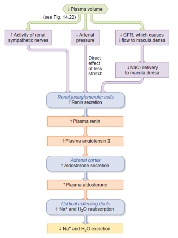
8.5.5.2.2 Atrial Natriuretic Peptide (ANP)
- ANP promotes natriuresis (loss of sodium)
- Atrial myocytes synthesise, store and release ANP in response to stretch (low P volume sensor)
- Major effect is renal vasodilatation. Increase blood flow = increased GFR
- Thus, more Na^+^ reaches macula densa.
- More Na^+^ excreted.
ANP 促进尿钠排泄(钠流失) 心房肌细胞响应拉伸(低 P 容量传感器)合成、储存和释放 ANP 主要作用是肾血管舒张。增加血流量 = 增加 GFR 因此,更多的 Na^+^ 到达致密斑。 排出更多的 Na^+^ 。
May inhibit actions of renin, and generally opposes effects of angiotensin II. 可能抑制肾素的作用,通常对抗血管紧张素 II 的作用。
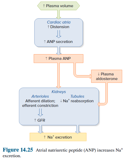
Feedback systems involved in osmolality control
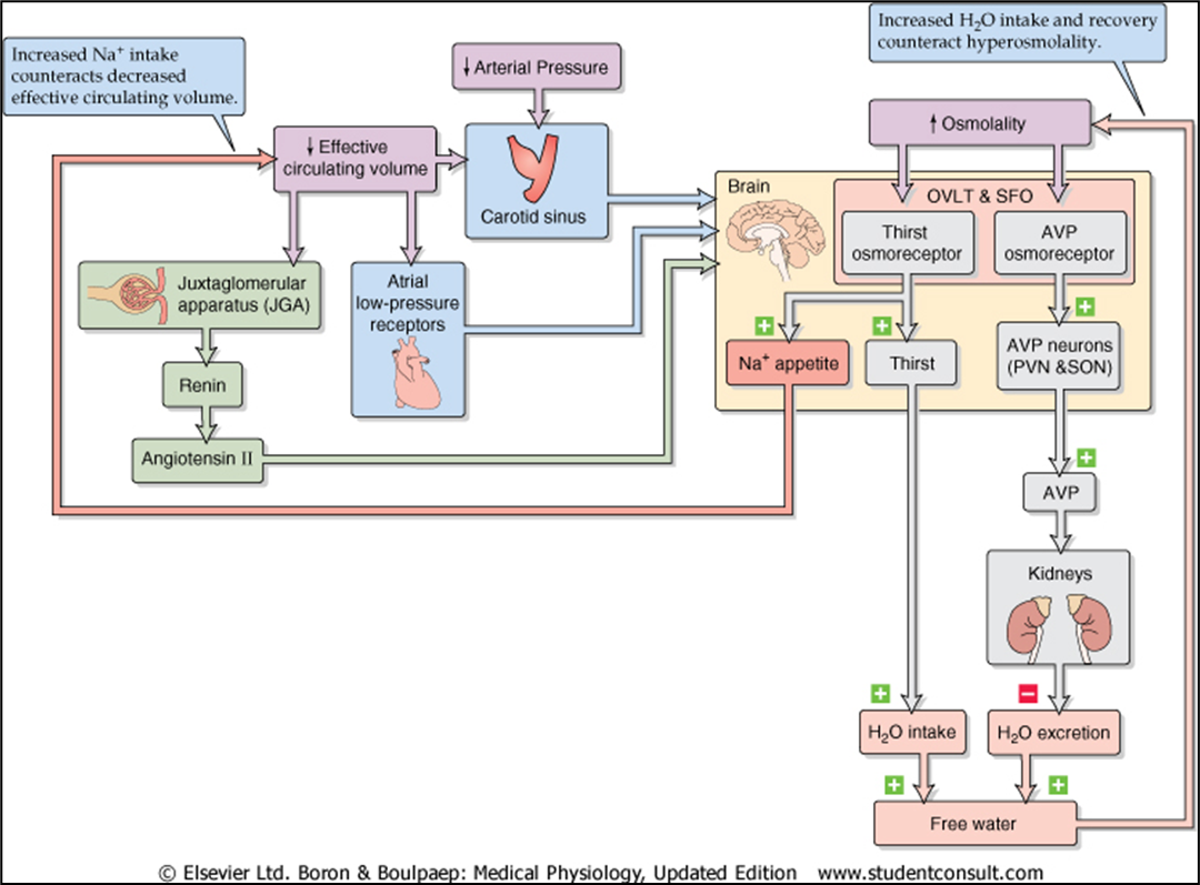
Comparison of systems controlling effective circulating volume and osmolality
Control of effective circulating volume
- Feedback control of effective circulating volume. 有效循环量反馈控制。
- A low effective circulating volume triggers 4 parallel effector pathways that act on the kidney. 低有效循环量会触发作用于肾脏的 4 条平行效应器通路。
- Either changes haemodynamics or changes Na^+^ transport by renal tubule cells. 要么改变血流动力学,要么改变肾小管细胞的 Na^+^ 转运。
ECF volume receptors
“Central” vascular sensors
-
Low pressure (very important)
- Cardiac atria
- Pulmonary vasculature
-
High pressure (less important)
- Carotid sinus
- Aortic arch
- Juxtaglomerular apparatus (renal afferent arteriole)
Sensors in the CNS (less important)
Sensors in the liver (less important)
N.B. Regulation of ECF volume = Regulation of body Na^+^. Thus, regulation of Na^+^ also dependent upon baroreceptors.
8.5.6 Renal Water Regulation
8.5.6.1 Osmoreceptor Control of Vasopressin Secretion 渗透压感受器控制加压素分泌
甲状旁腺激素渗透感受器途径,当摄入过多的水时,会减少加压素的分泌并增加水的排泄。当渗透压增加时,如缺水期间,会发生相反的事件(加压素分泌增加)。
8.5.6.2 Baroreceptor Control of Vasopressin Secretion 加压素分泌的压力感受器控制
当血浆容量减少时,加压素分泌增加的压力感受器途径。当血浆容量增加时,会发生相反的事件(最终导致加压素分泌减少)。
8.5.6.3 Other Stimuli to Vasopressin Secretion
8.5.7 Potassium Regulation
- K^+^ is the most abundant intracellular ion.
- Although only 2% of total-body potassium is in the extracellular fluid, the K^+^ concentration in this fluid is extremely important for the function of excitable tissues, notably, nerve and muscle.
- Resting membrane potentials of these tissues are directly related to the relative intracellular and extracellular K^+^ concentrations.
- Consequently, either increases (hyperkalemia) or decreases (hypokalemia) in extracellular K^+^ concentration can cause abnormal rhythms of the heart (arrhythmias) and abnormalities of skeletal muscle contraction and neuronal action potential conduction.
K^+^ 是最丰富的细胞内离子。 虽然细胞外液中的钾含量仅占全身钾的 2%,但细胞外液中的 K^+^ 浓度对于可兴奋组织(尤其是神经和肌肉)的功能极为重要。这些组织的静息膜电位与相对的细胞内和细胞外 K^+^ 浓度直接相关。 因此,细胞外 K^+^ 浓度的增加(高钾血症)或减少(低钾血症)均可导致心律异常(心律失常)以及骨骼肌收缩和神经元动作电位传导异常。
8.5.7.1 Renal Regulation of K^+^
The cortical collecting ducts can secrete K and changes in K excretion are due mainly to changes in K secretion by this tubular segment.
In cortical collecting ducts, the K pumped into the cell across the basolateral membrane by Na/K-ATPases diffuses into the tubular lumen through K channels in the luminal membrane. Thus, the secretion of K by the cortical collecting duct is associated with the reabsorption of Na.
K secretion does not occur in other Na-reabsorbing tubular segments because there are few K channels in the luminal membranes of their cells. Rather, in these segments, the K pumped into the cell by Na/K-ATPase simply diffuses back across the basolateral membrane through K channels located there.
皮质集合管可以分泌钾,钾排泄的变化主要是由于该管状部分钾分泌的变化。 在皮质集合管中,Na/K-ATP 酶通过基底外侧膜泵入细胞的 K,通过管腔膜中的 K 通道扩散到管腔中。因此,皮质集合管分泌 K 与 Na 的重吸收有关。 其他钠重吸收管段不发生钾分泌,因为其细胞的管腔膜中钾通道很少。相反,在这些片段中,由 Na/K-ATP 酶泵入细胞的 K 简单地通过位于基底外侧膜的 K 通道扩散回细胞。
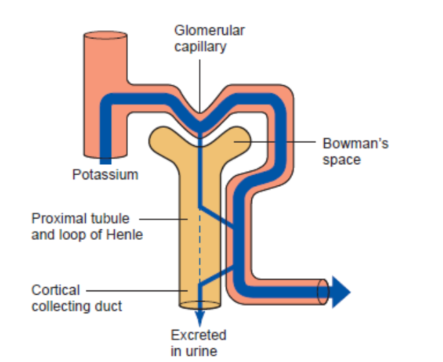
What factors influence K^+^ secretion by the cortical collecting ducts to achieve homeostasis of bodily potassium? 哪些因素影响皮质集合管分泌 K^+^ 以实现体内钾的稳态?
- The single most important factor is as follows. When a highpotassium diet is ingested (Figure 14.31), plasma K^+^ concentration increases, though very slightly, and this directly drives enhanced basolateral uptake via the Na^+^/K^+^-ATPase pumps. Thus, there is an enhanced K^+^ secretion. 最重要的一个因素如下。当摄入高钾饮食时(图 14.31),血浆 K^+^ 浓度会增加,尽管非常轻微,这直接驱动通过 Na^+^/K^+^-ATP 酶泵增强基底外侧摄取。 因此,K^+^ 分泌增强。
- A second important factor linking K^+^ secretion to potassium balance is the hormone aldosterone (see Figure 14.31). Besides stimulating tubular Na^+^ reabsorption by the cortical collecting ducts, aldosterone simultaneously enhances K^+^ secretion by this tubular segment. 将 K^+^ 分泌与钾平衡联系起来的第二个重要因素是醛固酮激素(见图 14.31)。除了刺激皮质集合管对肾小管 Na^+^ 的重吸收外,醛固酮还同时增强该管状管段的 K^+^ 分泌。
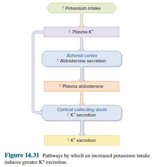
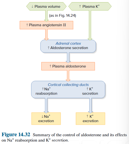
8.6 Summary: Division of Labor
- H~2~O rapidly regulated by vasopressin H~2~O 受加压素快速调节
- vasopressin secretion regulated by osmotic pressure and blood volume 加压素的分泌受渗透压和血容量调节
- Na^+^ and K^+^ regulated by aldosterone and ANP Na^+^ 和 K^+^ 受醛固酮和 ANP 调节
- aldosterone secretion regulated by the renin-angiotensin system and by ANP 醛固酮分泌受肾素-血管紧张素系统和 ANP 调节
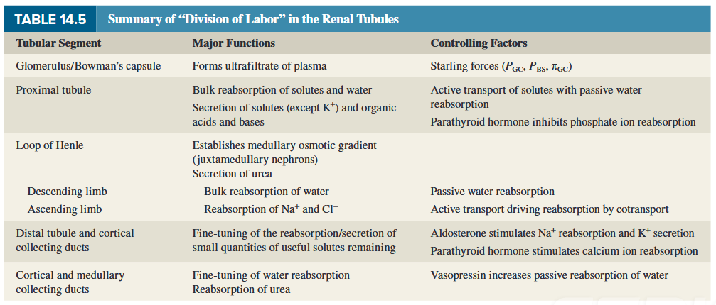
8.7 The Kidneys: Long Term Control (or Loss of Control)
- Long-term blood pressure control – by controlling blood volume
- Reduction in renal pressure - intrarenal redistribution of pressure and increased absorption of salt and water
- Decreased pressure in renal arterioles and sympathetic activity – renin production – angiotensin II production
长期血压控制 - 通过控制血容量 肾压降低 - 肾内压力重新分布以及盐和水的吸收增加 肾小动脉压力和交感神经活动降低 - 肾素生成 - 血管紧张素 II 生成
- Angiotensin II:
- Causes direct constriction of renal arterioles
- Stimulation of aldosterone synthesis – sodium absorption and increase in intravascular blood volume
血管紧张素 II:导致肾直接收缩小动脉刺激醛固酮合成 - 钠吸收和血管内血容量增加
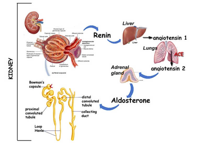
8.7.1 Glomerulus and Bowman’s Capsule (RAAS)
RAAS primary importance in blood pressure
- Activated in response to reduced blood flow
- Renal disease
- Renovascular disease
- Any decrease in kidney blood flow (GFR)
- Angiotensin II
- Aldosterone
- Inappropriate activation of RAAS
8.7.2 Cellular mechanisms controlling smooth muscle contraction and relaxation
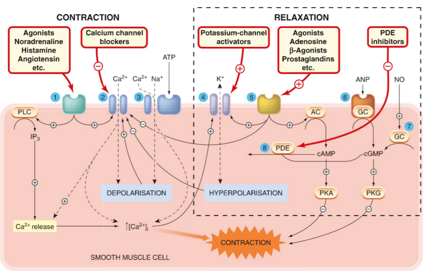
8.7.3 Pharmacologic Treatment of Hypertension
Diuretics
- Sympatholytics - opposes the downstream effects of postganglionic nerve firing in effector organs innervated by the sympathetic nervous system (SNS). They are indicated for various functions; for example, they may be used as anti-hypertensives.
- Act on renin angiotensin aldosterone system / axis (RAAS)
- Calcium channel blockers
- Direct-acting vasodilators
利尿剂 交感神经阻滞剂 - 对抗交感神经系统 (SNS) 支配的效应器器官中节后神经放电的下游效应。它们具有不同的功能;例如,它们可用作抗高血压药。 作用于肾素血管紧张素醛固酮系统/轴 (RAAS) 钙通道阻滞剂 直接作用血管扩张剂
8.7.4 Diuretics
Classification of Diuretics
- All diuretics indirectly prevent the re-absorption of water in the kidneys, most of them by preventing the reabsorption of sodium 所有利尿剂都间接阻止肾脏对水的重吸收,其中大多数是通过阻止钠的重吸收
- Loop diuretics - furosemide supplemented with spironolactone or amiloride 袢利尿剂 - 呋塞米 辅以螺内酯或阿米洛利
- Thiazides - Bendroflumethiazide 噻嗪类 - 苯氟噻嗪
- Potassium sparing diuretics - Not acting directly on sodium channels 保钾利尿剂 - 不直接作用于钠通道
Diuretics - sight of action
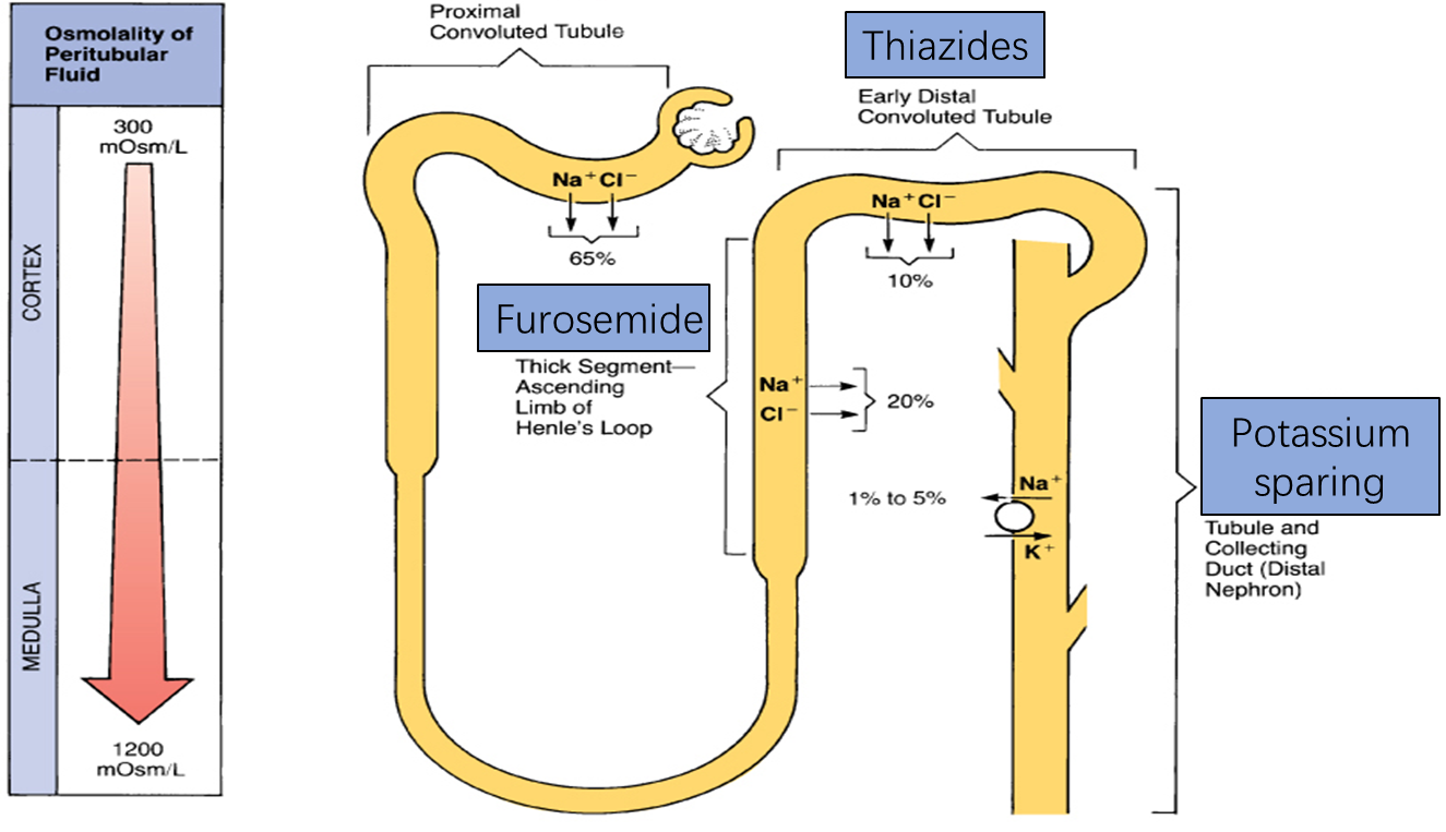
Diuretics mechanisms of action
- Loop diuretics act principally by inhibiting the Na^+^/K^+^/2Cl−co-transporter.
- Thiazide diuretics, which are the most commonly used diuretic, inhibit the sodium-chloride transporter in the distal tubule. 噻嗪类利尿剂是最常用的利尿剂,可抑制远端小管中的氯化钠转运蛋白。
- Because this transporter normally only reabsorbs about 5% of filtered sodium, these diuretics are less efficacious than loop diuretics in producing diuresis and natriuresis. 由于这种转运蛋白通常仅重吸收约 5% 的滤过钠,因此这些利尿剂在产生利尿和尿钠排泄方面不如袢利尿剂有效。
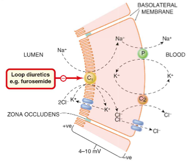
- Potassium sparing diuretics 保钾利尿剂
- Distal tubule Na^+^-channel inhibitor 远端小管 Na^+^ 通道抑制剂
- Aldosterone receptor antagonist 醛固酮受体拮抗剂
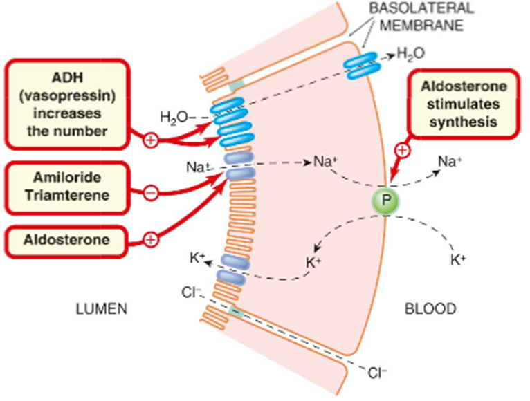
8.7.5 Drugs Acting on RAAS
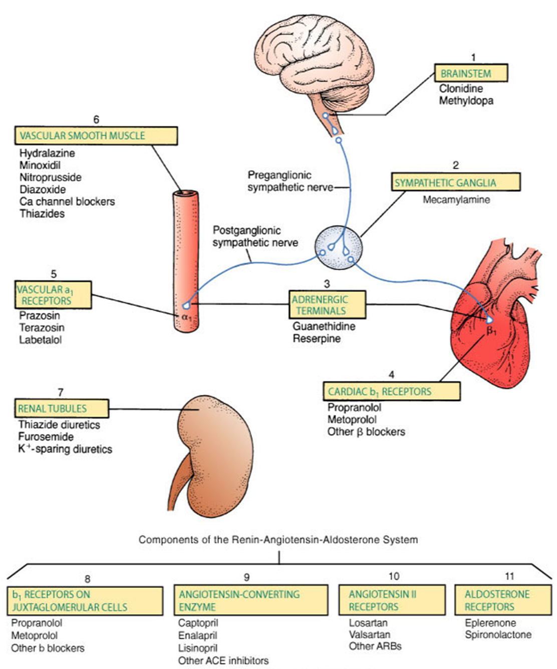
Renin Inhibition
- Aliskiren
- first in class direct renin inhibitor
- used for primary hypertention
- B1 antagonists
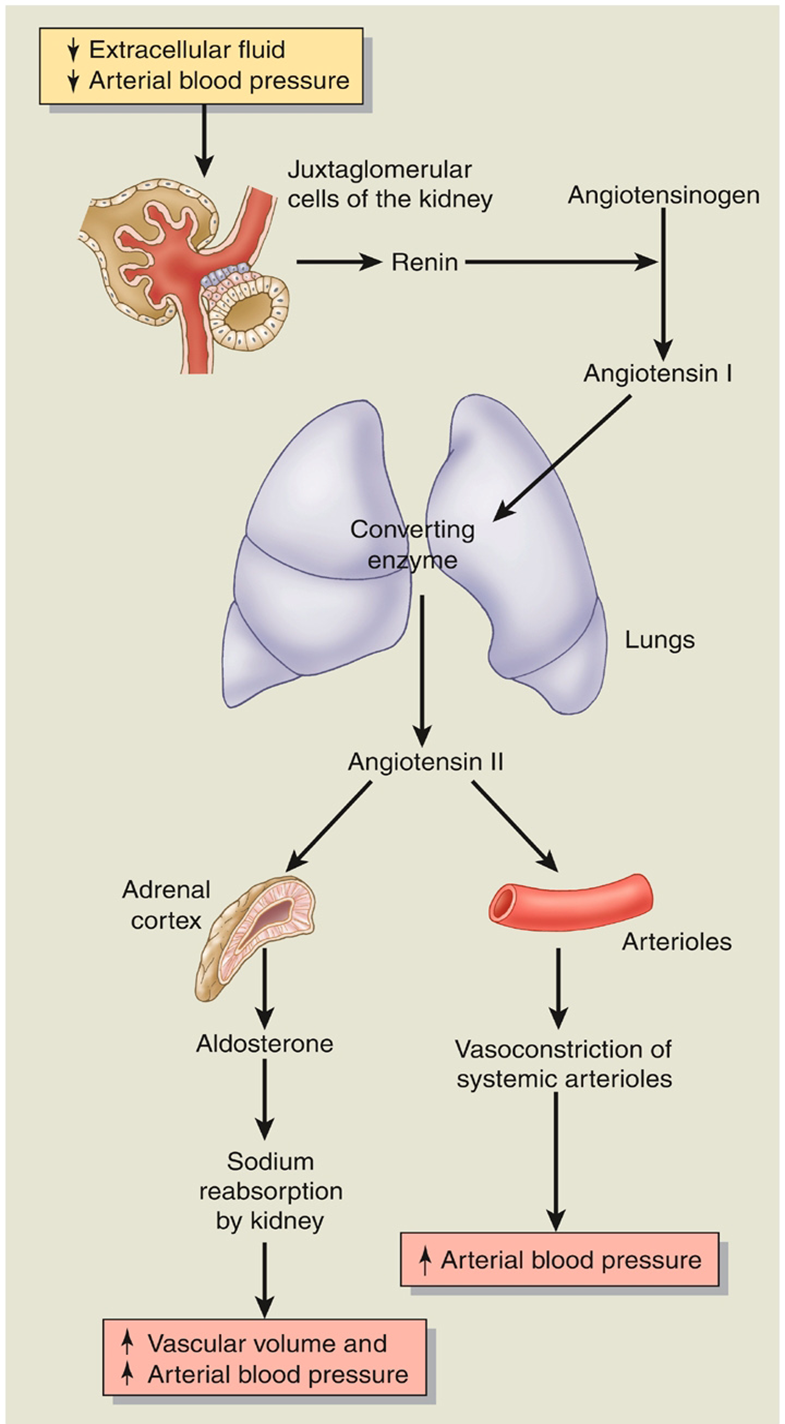
ACE Inhibitors (ACEI)
- Angiotensin converting enzyme inhibitors
- Work primarily in the lung
- Captopril, lisinopril, enalapril, and others - All end in -pril
- ACE inhibitors block the action of the ACE, to prevent the conversion of angiotensin I to angiotensin II.
- A vasoconstrictor and stimulates aldosterone secretion.
- Blocking its action reduces peripheral vascular resistance (afterload), which lowers blood pressure.
- Dilates the efferent glomerular arteriole, which reduces intra-glomerular pressure.
- Reducing the aldosterone level promotes sodium and water excretion. This can help to reduce venous return (preload), which has a beneficial effect in heart failure.
血管紧张素转换酶抑制剂 主要作用于肺部 卡托普利、赖诺普利、依那普利等 - 所有 ACE 抑制剂均能阻断 ACE 的作用,从而阻止血管紧张素 I 转化为血管紧张素 II。 血管收缩剂并刺激醛固酮分泌。 阻断其作用可降低外周血管阻力(后负荷),从而降低血压。 扩张传出肾小球小动脉,从而降低肾小球内压力。 降低醛固酮水平可促进钠和水的排泄。这有助于减少静脉回流(前负荷),这对心力衰竭有有益的作用。
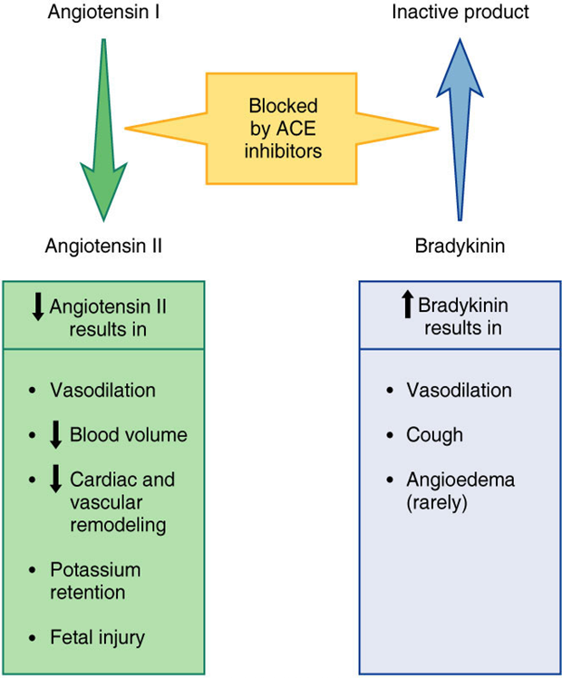
Angiotensin II Receptor Blockers (ARBs)
- Losartan, valsartan, candesartan, and others (sartan)
- ARBs have similar effects to ACE inhibitors, but instead of inhibiting the conversion of angiotensin I to angiotensin II, ARBs block the action of angiotensin II on the AT1 receptor
氯沙坦、缬沙坦、坎地沙坦和其他(沙坦)ARB 与 ACE 抑制剂具有相似的作用,但 ARB 不是抑制血管紧张素 I 向血管紧张素 II 的转化,而是阻断血管紧张素 II 对 AT1 受体的作用
Aldosterone antagonist
- Spironolactone
- Eplerenone
- Potassium-sparing diuretics
- competitively binding to the aldosterone receptor
- Promote Na^+^ and H~2~O excretion in the collecting tubule and duct
螺内酯 依普利酮 保钾利尿剂 竞争性结合醛固酮受体 促进集合管和导管中 Na^+^ 和 H~2~O 的排泄
8.6 Final review of renal integration with respiratory and cardiovascular systems
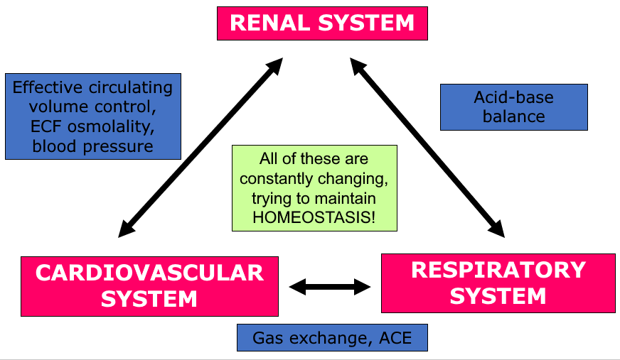
Problems in one system are often only noticed by appearance of problems in another.
This is because of the integrated nature of these systems – they don’t operate in isolation.
Means we can sometimes compensate for problems in another system via reflexes, but also means that when disease progresses, lots of problems in several systems can begin to appear.
If we understand links between systems, we have more targets for drugs and other therapies to correct problems.
May also target actual cause of problem, rather than just worrying about the symptoms. Scientists think laterally – medics have a tendency just to stay inside their little window of expertise.
These three systems are probably the locations of most of the health-related problems you might encounter in whatever career you follow – regardless of whether you are a sports scientist or a physiologist.
Chapter 10 Endocrine Physiology
- Endocrine diseases very common (especially diabetes mellitus, thyroid disease) 内分泌疾病很常见(特别是糖尿病、甲状腺疾病)
- Drugs affecting endocrine system very commonly used (e.g. corticosteroids; oral contraceptives) 非常常用的影响内分泌系统的药物(例如皮质类固醇;口服避孕药)
- especially important for pharmacists, clinical biochemists and all who may work in hospital service or pharmaceutical industry 对于药剂师、临床生物化学家以及所有可能在医院服务或制药行业工作的人员尤其重要
Key points:
- General Characteristics of endocrine system
- Hypothalamus-Pituitary system and their hormones
- Physiological Functions and regulation of secretion of the following hormones:
- Growth hormone
Thyroid hormones
Adrenocortical Hormones
Insulin
Hormones in charge of Calcium and Phosphate metabolism
- Growth hormone
Endocrine system, composed of endocrine gland (classical) and cells (non-classical), independently or along with nervous system and immune system, coordinate complex body functions, such as metabolism, growth and development, reproduction etc. 内分泌系统由内分泌腺(经典)和细胞(非经典)组成,独立或与神经系统、免疫系统一起协调新陈代谢、生长发育、生殖等复杂的机体功能。
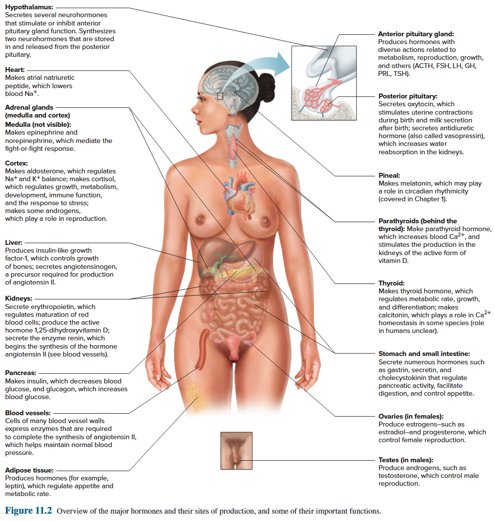
Endocrine system and nervous system
In common:
adjusts and correlates the activities of various body systems, making them appropriate to the demands of external and internal environment changes. 调整和关联身体各个系统的活动,使它们适应外部和内部环境变化的需求。
Differences:
nervous system: signal conduction by action potentials along the nerve fibers is very rapid and precise but of short duration 神经系统:动作电位沿着神经纤维传导信号非常快速和精确,但持续时间短
endocrine system: the signaling mediated by chemical messages via body fluid is relatively slow, diffuse but with a longer duration 内分泌系统:化学信息通过体液介导的信号传导相对缓慢、弥散,但持续时间较长
10.1 General characteristics of hormones
10.1.1 Major Classes of Hormones
10.1.1.1 Definition of Hormone
Hormone is bioactive chemical messenger released from endocrine cells, and carried by extracellular fluid or blood to target cells throughout the body. By binding to its receptor, hormone initiates multiple reactions. More reactions would occur when several different hormones cooperate. 激素是内分泌细胞释放的生物活性化学信使,由细胞外液或血液携带至全身的靶细胞。通过与其受体结合,激素引发多种反应。当几种不同的激素协同作用时,会发生更多的反应。
10.1.1.2 Characteristics of hormone
- Specificity
- Signaling
- Highly effective
- Cooperation
10.1.1.3 Classes of hormones
- Proteins and polypeptides (hormones secreted by pituitary; insulin, glucagon etc.) 蛋白质和多肽(垂体分泌的激素;胰岛素、胰高血糖素等)
- Derivatives of the amino acid (thyroid, adrenal medullae) 氨基酸衍生物(甲状腺、肾上腺髓质)
- Steroids 甾体类 (cortisol, oestrogen etc.)
Peptide hormones
- Rapid onset of action
- Rapid offset of action
- Secreted in “bursts” when required
Steroid type hormones
- Slow onset of action
- Slow offset of action
- Secreted to maintain “constant” plasma hormone concentration
Explain differences between peptide hormones and steroid-type hormones in terms of their: speed of onset / speed of offset
Clue – depends on their two broadly different mechanisms of action!
10.1.1.4 Many Hormones Reach Their Target Cells by Transport in the Bloodstream
- Most amino acid-derived and polypeptide hormones dissolve readily in the plasma, and thus no special mechanisms are required for their transport. 大多数氨基酸衍生的激素和多肽激素很容易溶解在血浆中,因此不需要特殊的机制来运输它们。
- Steroid and thyroid hormones are relatively insoluble in plasma. 90% or more of steroid and thyroid hormones in the blood are bound to plasma proteins. Some of the plasma proteins that bind hormones are specialized. 类固醇和甲状腺激素在血浆中相对难溶。血液中 90% 或更多的类固醇和甲状腺激素与血浆蛋白结合。一些结合激素的血浆蛋白是专门的。
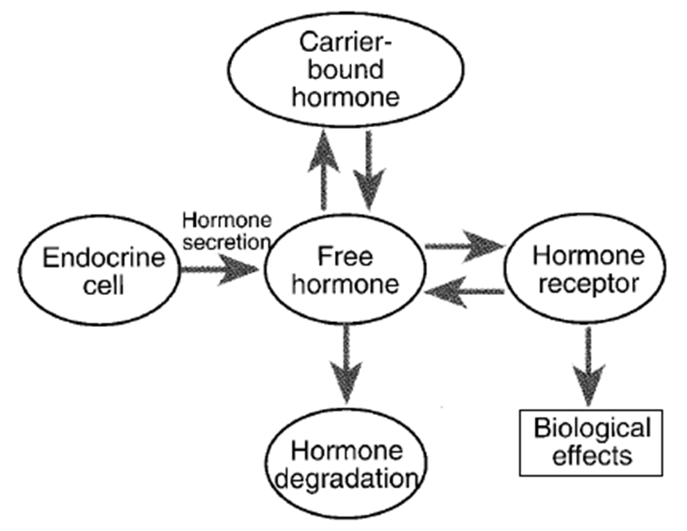
The relationship between hormone secretion, carrier protein binding, and hormone degradation. This relationship determines the amount of free hormone available for receptor binding and the production of biological effects. 激素分泌、载体蛋白结合和激素降解之间的关系。这种关系决定了可用于受体结合和生物效应产生的游离激素的量。
10.1.2 Mechanisms of Hormones Action
-
Binding to receptors (receptors located in membrane, cytoplasm or nucleus) Binding to receptors (receptors located in membrane, cytoplasm or nucleus)
-
Intracellular signalling cascade: The event occurred following the forming of hormone-receptor complex depends on the type of hormone receptor. 细胞内信号级联:激素受体复合物形成后发生的事件取决于激素受体的类型。
- Ion channel-linked receptor
- G protein-linked receptor
- Enzyme-linked receptor
-
Final physiological response:
- Metabolism altered (increased or decreased)
- Genes Activated and protein expressed
e.g. Growth hormone can increase cell metabolism and induce body to grow.
10.1.3 Control of Hormones Secretion
Hormone secretion is mainly under control of three types of inputs to endocrine cells: 激素分泌主要受内分泌细胞的三种输入控制:
- Ions or Nutrients
- Neurotransmitters
- Hormones
10.1.3.1 Negative Feedback
The action of the hormone tend to suppress its further release, thus a hormone can inhibit its own production: negative feedback
- Long-loop negative feedback: The “target gland” hormone exerts negative feedback on the secretion of the hypothalamic and/or anterior pituitary gland hormone in the pathway. 长环负反馈:“靶腺”激素对通路中下丘脑和/或垂体前叶激素的分泌施加负反馈。
- Short-loop negative feedback: A given anterior pituitary hormone inhibits the hypophysiotropic hormone(s) that control its secretion. 短环负反馈:特定的垂体前叶激素会抑制控制其分泌的促垂体激素。
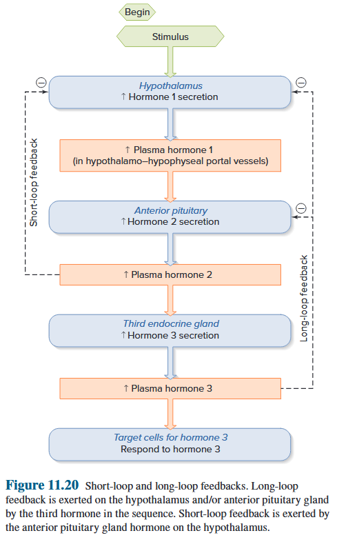
Regulation of secretion of hormones concerned with short-term regulation (Peptide hormones)
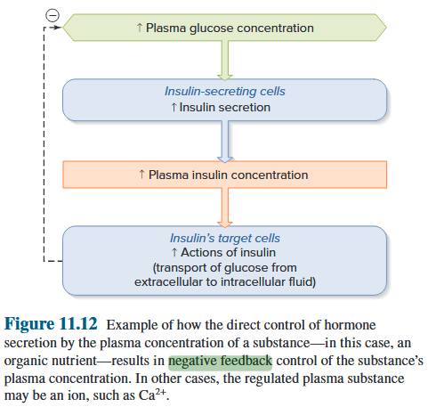
Regulation of secretion of hormones concerned with long-term regulation (Steroid-like hormones)
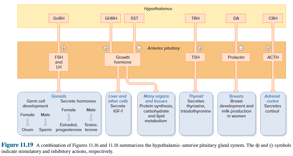
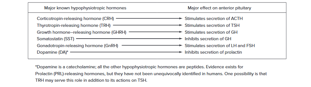
System maintain the concentration of hormones within physiological limits through negative feedback mechanisms 系统通过负反馈机制将激素浓度维持在生理限度内
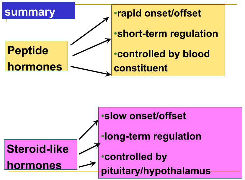
10.2 Endocrine of the hypothalamus and pituitary gland
10.2.1 Location and Structure of Pituitary
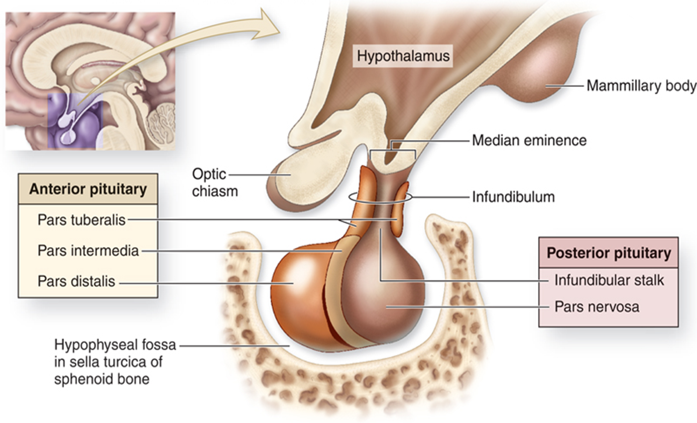
Nucleus in Hypothalamus: Specific areas that produce and release multiple hormones 下丘脑核:产生和释放多种激素的特定区域
10.2.2 Relations between Pituitary and Hypothalamus
- VP and Oxytocin are synthesized in Hypothalamus but released in posterior pituitary (green). VP 和催产素在下丘脑合成,但在垂体后叶(绿色)释放。
- Hormones produced in Hypothalamus are carried via Hypothalamic-Hypophysial portal system to anterior pituitary and regulate the endocrine cells activity in the anterior pituitary (red). 下丘脑产生的激素通过下丘脑-垂体门脉系统输送至垂体前叶,并调节垂体前叶(红色)的内分泌细胞活动。
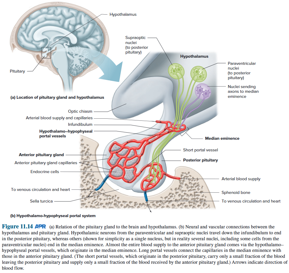
Hypothalamic-Hypophsial Portal System
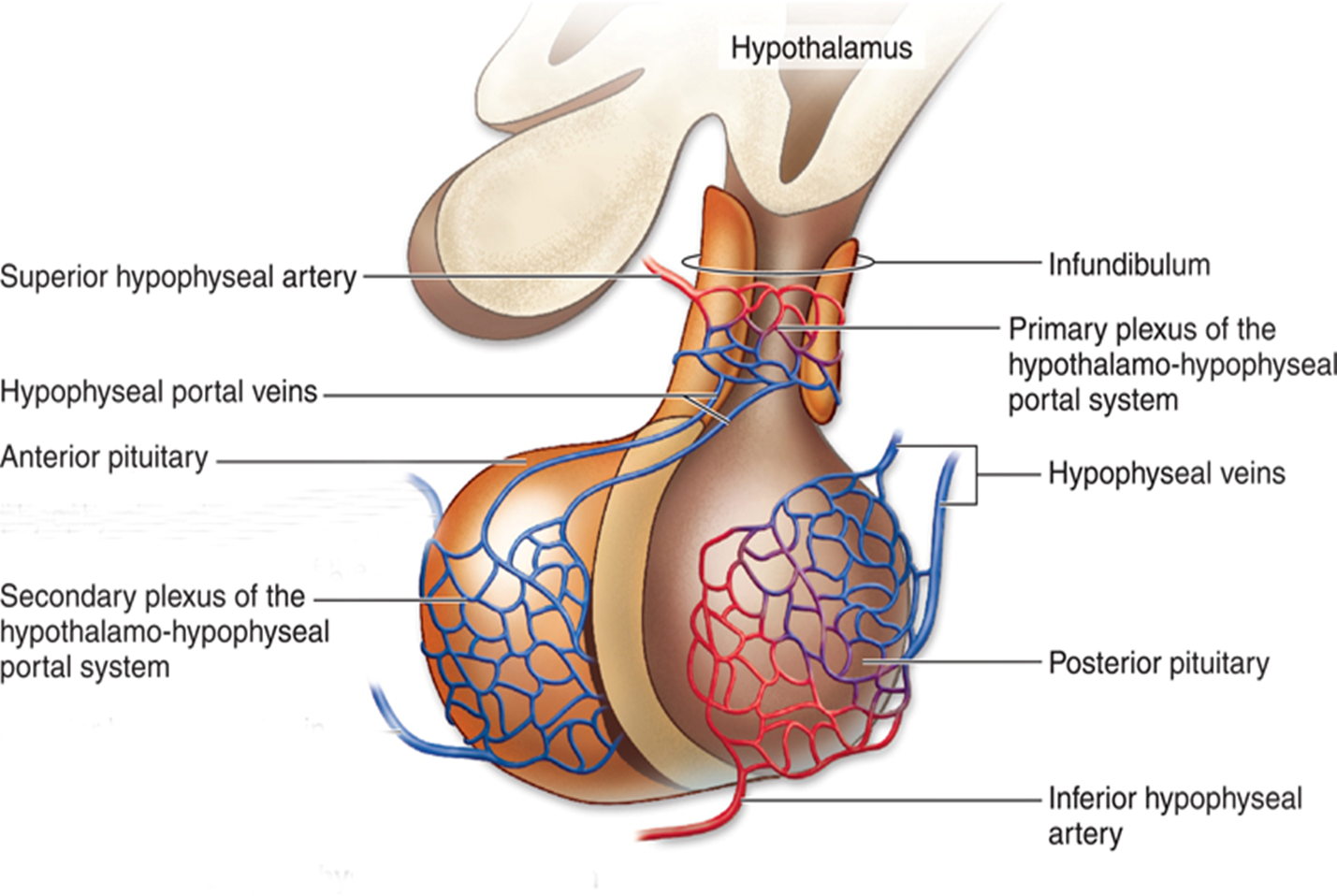
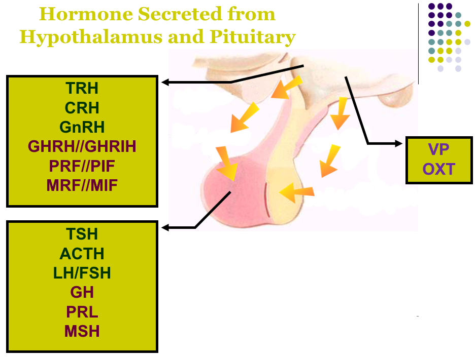
OXT & PRL
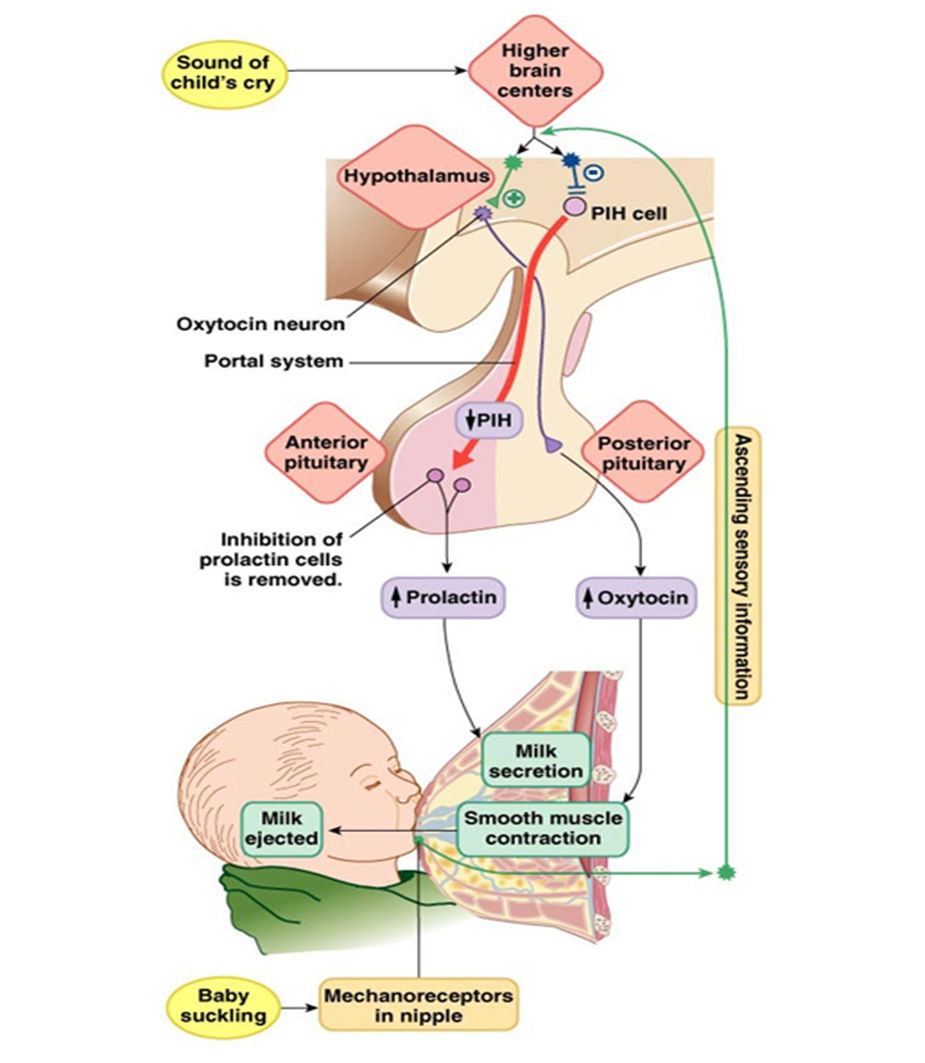
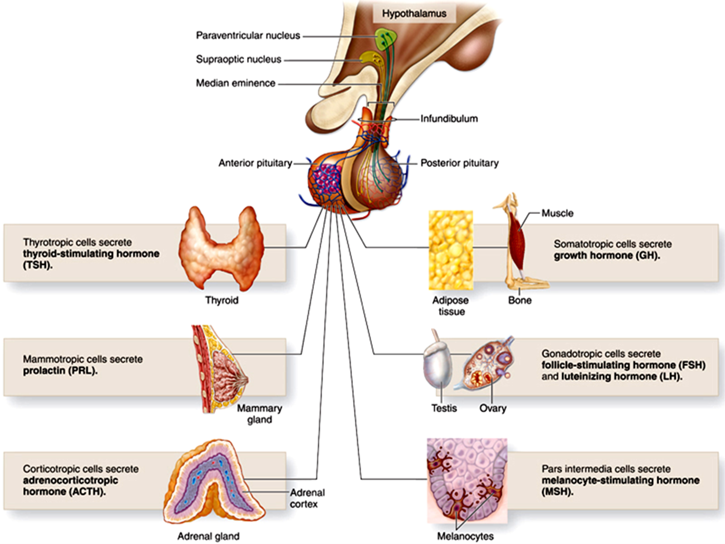
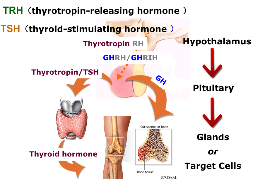
10.2.3 Neural control of hypothalamic releasing and inhibitory hormones
Three – hormone sequence:
(1) A hypophysiotropic hormone controls the secretion of (2) an anterior pituitary hormone, which controls the secretion of (3) a hormone from some other endocrine gland. (1) 促垂体激素控制 (2) 垂体前叶激素的分泌,垂体前叶激素控制 (3) 来自其他内分泌腺的激素的分泌。
e.g. CRH (Corticotropin releasing hormone) - ACTH (adrenocorticotropin) - adrenal cortex - secretes cortisol.
Central Neural system influence the hormones secretion
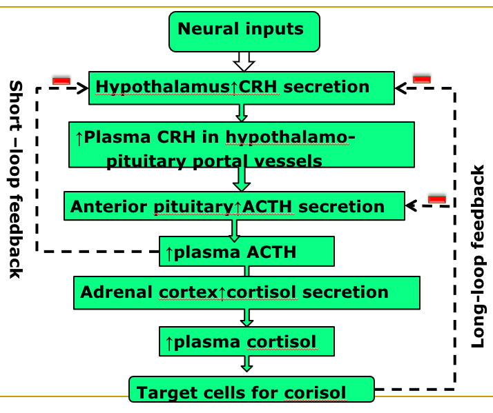
10.3 Endocrine of the Growth Hormone
Hormonal control of growth and development: Insulin, Growth hormone, Thyroid hormones, Sex hormones
10.3.1 Introduction of GH
Effects on almost all tissues of the body, such as bone, muscle etc.
The single most important hormone for postnatal growth
However, most of GH functions are dependent on IGF-1 (Somatomedin), which is produced in liver.
Furthermore, insulin and carbohy-drate are necessary for the growth-promoting action of GH
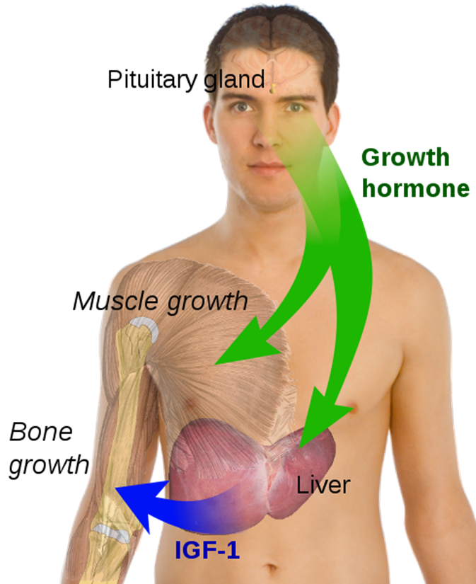
Growth hormone (Somatotrophin)
- Growth
- Ca^2+^ & phosphate retention
- Increased protein synthesis
- Increased blood glucose
- Increased circulating FFA
10.3.2 Physiological functions of GH
Effects on metabolism
(1) Enhance protein synthesis and reducing breakdown of protein;
(2) Enhance fat utilization for energy (adipose tissue);
(3) Increase blood glucose concentration
— Excess GH induces hyperglycemia and subsequently Diabetes (Diabetogenic)
(1) 增强蛋白质合成,减少蛋白质分解; (2) 增强脂肪对能量的利用(脂肪组织); (3) 增加血糖浓度——过量的GH会诱发高血糖,进而引发糖尿病(糖尿病)
GH promotes protein deposition in tissues
- Enhancement of amino acid transport through the cell membrane.
- Enhancement of RNA translation to cause protein synthesis by ribosomes.
- Increased nuclear transcription of DNA to form RNA.
- Decreased catabolism of protein and amino acids.
增强氨基酸通过细胞膜的转运。 增强 RNA 翻译,导致核糖体合成蛋白质。 DNA 的核转录增加,形成 RNA。 蛋白质和氨基酸的分解代谢减少。
GH enhances fat utilization for energy
-
Causing release of fatty acids from adipose tissue
-
Increasing the concentration of fatty acids in the body fluids.
-
Ketogenic effect
Excessive GH cause so much fat mobilization from adipose tissue that larger quantities of acetoacetic acid are formed by the liver and released into the body fluids, thus causing ketosis.
导致脂肪组织释放脂肪酸增加体液中脂肪酸的浓度。 生酮效应 过量的 GH 会导致脂肪组织中大量脂肪动员,导致肝脏形成大量乙酰乙酸并释放到体液中,从而引起酮症。
GH decreases carbohydrate utilization
- Decreased glucose uptake in tissue such as skeletal muscle and fat
- Increased glucose production by the liver
- Increased insulin secretion
- GH induced insulin-resistance
骨骼肌和脂肪等组织中葡萄糖的摄取减少 肝脏产生的葡萄糖增加 胰岛素分泌增加 GH 诱导胰岛素抵抗
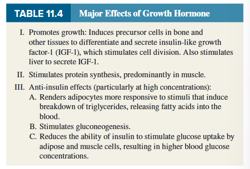
GH stimulates cartilage and bone growth
- Increased deposition of protein by the chondrocytic and osteogenic cells that cause bone growth;
- Increased rate of reproduction of these cells;
- A specific effect of converting specific deposition of new bone.
软骨细胞和成骨细胞的蛋白质沉积增加,导致骨骼生长; 这些细胞的繁殖率提高; 转化新骨的特定沉积的特定效果。
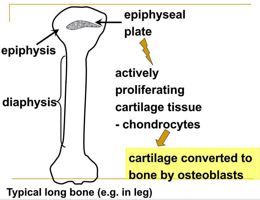
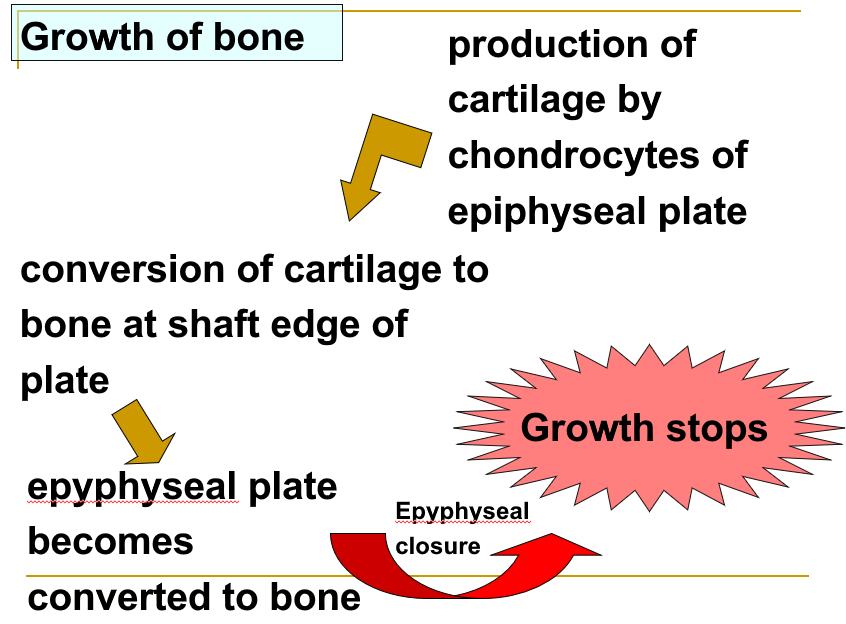
GH increases blood glucose
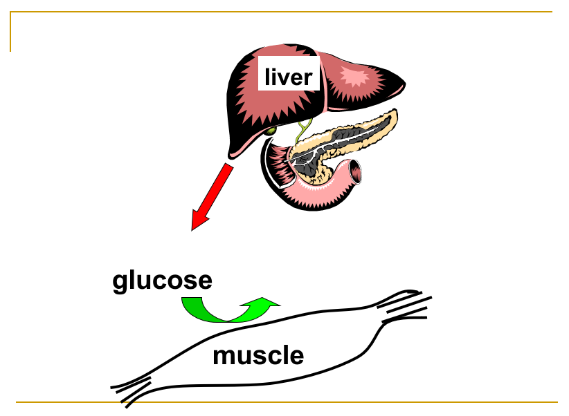
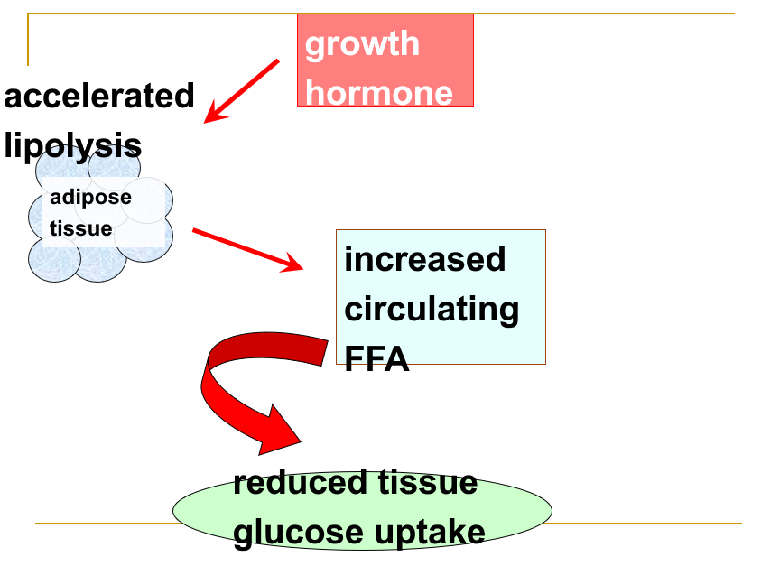
Growth hormone acts indirectly
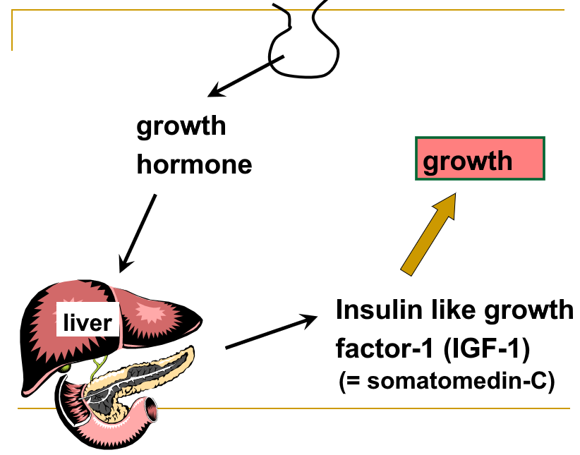
10.3.3 Regulation of GH Secretion
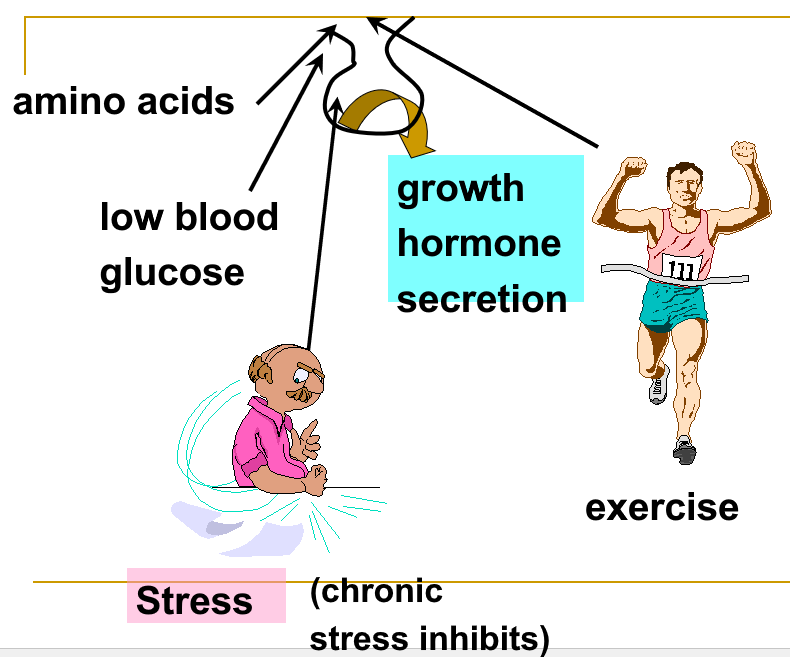
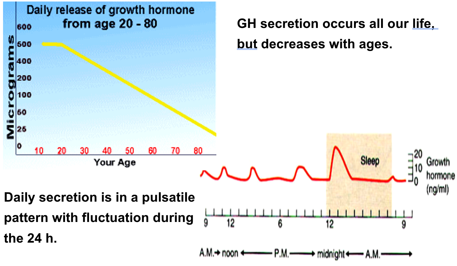
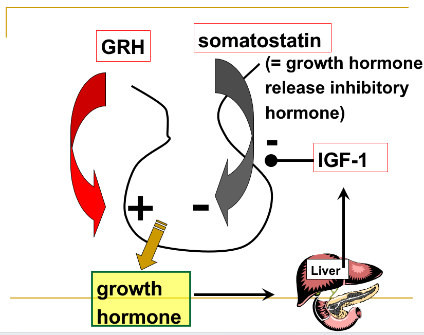
Other factors influencing GH secretion
| Stimulatory factors | Inhibitory factors |
|---|---|
| Low blood glucose, free fatty acids | High blood glucose, free fatty acids |
| Protein deficiency | Aging, Obesity etc |
| Excitement, stress, Deep sleep etc | GH & IGF |
| GHRH | GHIH |
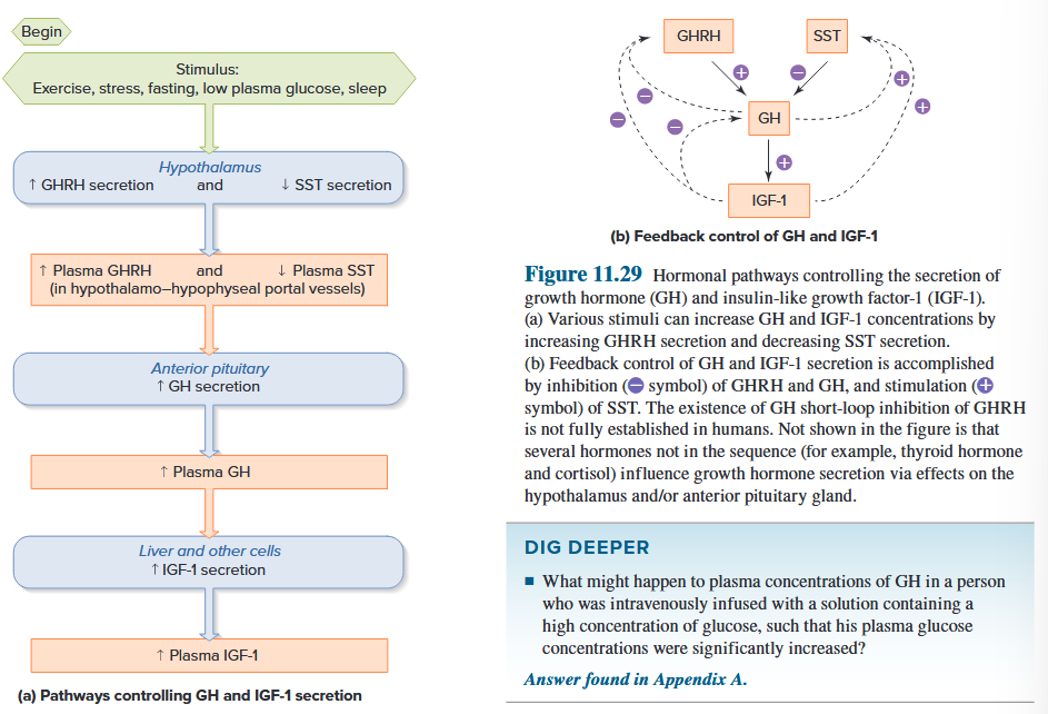
10.3.4 Consequences of growth hormone abnormalities
Dwarfism
- Growth hormone deficiency before epiphyseal closure 骨骺闭合前生长激素缺乏
- IGF-1 deficiency
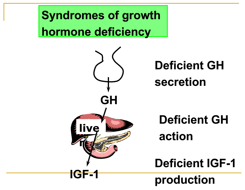
Gigantism and acromegaly
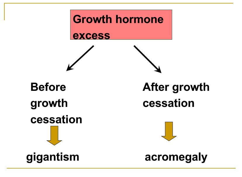
10.4 Endocrine of the thyroid gland
thyroxine (T4) and triiodothyronine (T3)
derivatives of tyrosine 酪氨酸衍生物
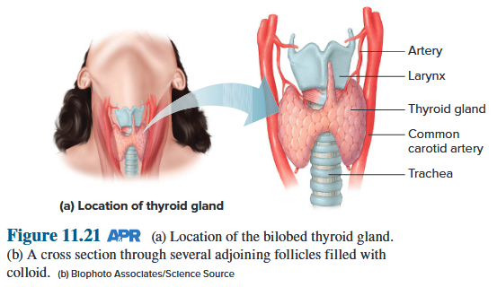
10.3.1 Biosynthesis of thyroid hormones
Step 1: circulating iodide is actively co-transported with sodium ions across the basolateral membranes of the epithelial cells; 循环中的碘化物与钠离子主动共同转运穿过上皮细胞的基底外侧膜;
Step 2: The iodide ions diffuse to the apical membrane of the follicular epithelial cells and are transported into the colloid by a mechanism that is believed to require an integral membrane protein called pendrin; 碘离子扩散到滤泡上皮细胞的顶膜,并通过一种机制转运到胶体中,这种机制被认为需要一种称为pendrin的整合膜蛋白;
Step 3: iodide is rapidly oxidized at the luminal surface of the follicular epithelial cells to iodine, which is then attached to the phenolic rings of tyrosine residues within thyroglobulin; 碘化物在滤泡上皮细胞的管腔表面迅速氧化成碘,然后碘附着在甲状腺球蛋白内酪氨酸残基的酚环上;
Thyroid peroxidase - MIT/DIT
Step 4: The phenolic ring of a molecule of MIT or DIT is removed from the remainder of its tyrosine and coupled to another DIT on the thyroglobulin molecule; MIT或DIT分子的酚环从其剩余的酪氨酸中除去,并与甲状腺球蛋白分子上的另一个DIT偶联;
Step 5: For thyroid hormone to be secreted into the blood, extensions of the colloid-facing membranes of follicular epithelial cells engulf portions of the colloid (with its iodinated thyroglobulin) by endocytosis; 为了将甲状腺激素分泌到血液中,滤泡上皮细胞面向胶体的膜的延伸通过内吞作用吞噬部分胶体(及其碘化甲状腺球蛋白);
Step 6: The thyroglobulin, with its coupled MITs and DITs, is brought into contact with lysosomes in the cell interior; 甲状腺球蛋白及其偶联的 MIT 和 DIT 与细胞内部的溶酶体接触;
Step 7: Proteolysis of thyroglobulin releases T3 and T4 , which then diffuse out of the follicular epithelial cell into the interstitial fluid and from there to the blood. 甲状腺球蛋白的蛋白水解释放 T3 和 T4 ,然后从滤泡上皮细胞扩散到间质液中,并从那里进入血液。
There is sufficient iodinated thyroglobulin stored within the thyroid to provide thyroid hormone for several weeks even in the absence of dietary iodine. This storage capacity makes the thyroid gland unique among endocrine glands but is an essential adaptation considering the unpredictable intake of iodine in the diets of most animals. 甲状腺内储存有足够的碘化甲状腺球蛋白,即使在饮食中缺乏碘的情况下,也可以提供几周的甲状腺激素。这种储存能力使甲状腺在内分泌腺中独一无二,但考虑到大多数动物饮食中碘的摄入量是不可预测的,这是一种重要的适应。
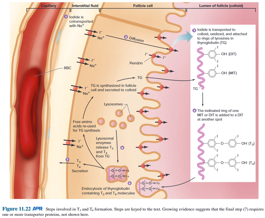
10.3.2 Actions of thyroid hormones
Influence activity of almost every body system: Required for synthesis of various key enzymes involved in cellular functions
- Cardiac sarcoplasmic Ca^2+^-ATPase
- Na-K ATPase
- Glyceraldehyde 3-phosphate dehydrogenase
- β-adrenoceptors
- Enzymes involved in fatty acid synthesis
Na-K ATPase
One of the by-products of this process is heat. Thus, as ATP is consumed in cells by Na/K –ATPases at a high rate due to T3 stimulation, the cellular stores of ATP must be maintained by increased metabolism of fuels. This calorigenic action of T3 represents a very significant fraction of the total heat produced each day in a typical person. Without thyroid hormone, heat production would decrease and body temperature would be compromised. 该过程的副产品之一是热量。因此,由于 T3 刺激,细胞中 ATP 被 Na/K-ATP 酶以高速率消耗,因此必须通过增加燃料代谢来维持 ATP 的细胞储存。 T3 的这种生热作用占典型人每天产生的总热量的很大一部分。如果没有甲状腺激素,产热就会减少,体温就会下降。
Growth and Development
T3 is required for normal production of GH from the pituitary gland. Therefore, in the absence of T3, growth in children is decreased.
T3 is among the most important developmental hormones for the nervous system. During fetal life, T3 exerts many effects on central nervous system development. Absence of T3 during fetal life results in the syndrome called congenital hypothyroidism. This syndrome is characterized by a poorly developed nervous system and severely compromised intellectual function (mental retardation). The most common cause of congenital hypothyroidism is dietary iodine deficiency in the mother. The availability of iodized salt products has eliminated congenital hypothyroidism in many countries.
T3 是垂体正常产生 GH 所必需的。因此,如果缺乏T3,儿童的生长就会减慢。 T3 是神经系统最重要的发育激素之一。在胎儿时期,T3 对中枢神经系统发育产生多种影响。胎儿时期缺乏 T3 会导致称为先天性甲状腺功能减退症的综合征。该综合征的特点是神经系统发育不良和智力功能严重受损(精神发育迟滞)。先天性甲状腺功能减退症最常见的原因是母亲饮食中缺碘。在许多国家,碘盐产品的出现已经消除了先天性甲状腺功能减退症。
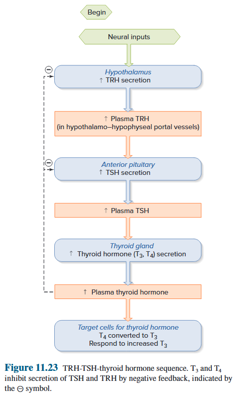
10.3.3 Consequences of thyroid hormone abnormalities
10.3.3.1 Consequences of thyroid hormone deficiency: hypothyroidism
-
Marked reduction in metabolic rate of tissues
-
Reduced function of most organ systems
-
Thyroid deficiency in newborn: Congenital hypothyroidism (cretinism)
- Impaired growth and development
- Mental retardation
-
Adult with congenital hypothyroidism
- Severe mental retardation
- dwarfism
- Impaired sexual development
In iodine deficiency, the synthesis of T3 is compromised, leading to a decrease in the plasma concentration of this hormone.
This, in turn, releases negative feedback on the hypothalamus and anterior pituitary gland, and TRH concentrations become chronically increased in the portal circulation that leads to the anterior pituitary gland. Plasma TSH concentration is increased due to the increased TRH and loss of thyroid hormone negative feedback on the anterior pituitary gland.
The resulting overstimulation of the thyroid gland can produce goiters that can achieve astounding sizes if untreated.
an increased sensitivity to cold (cold intolerance) a tendency toward weight gain.
Both of these symptoms are related to the decreased calorigenic actions normally produced by thyroid hormone.
Many of the other symptoms, such as fatigue and changes in skin tone, hair, appetite, gastrointestinal function, and neurological function (for example, depression).
缺碘时,T3 的合成受到损害,导致该激素的血浆浓度降低。 这反过来会释放下丘脑和垂体前叶的负反馈,并且通向垂体前叶的门脉循环中 TRH 浓度会长期增加。 由于 TRH 增加和垂体前叶甲状腺激素负反馈消失,血浆 TSH 浓度增加。 由此产生的甲状腺过度刺激会产生甲状腺肿,如果不及时治疗,甲状腺肿会达到惊人的大小。 对寒冷的敏感性增加(寒冷不耐受) 体重增加的趋势。 这两种症状都与甲状腺激素通常产生的生热作用减少有关。 许多其他症状,例如疲劳以及肤色、头发、食欲、胃肠功能和神经功能(例如抑郁)的变化。
10.3.3.2 Consequences of thyroid hormone excess: Hyperthyroidism
- Marked increase in metabolic rate of tissues
- Increased function of most organ systems
- Hyperthyroid patients tend to have heat intolerance
- weight loss
- increased appetite
- often show signs of increased sympathetic nervous system activity (anxiety, tremors, jumpiness, increased heart rate).
10.3.4 Summary
Synthesis of Thyroid Hormone
- T3 and T4 are synthesized by sequential iodinations of thyroglobulin in the thyroid follicle lumen, or colloid. Iodinated tyrosines on thyroglobulin are coupled to produce either T3 or T4. T3 is the active hormone.
- The enzyme responsible for T3 and T4 synthesis is thyroid peroxidase.
1.T3和T4是通过甲状腺滤泡腔或胶体中的甲状腺球蛋白连续碘化合成的。甲状腺球蛋白上的碘化酪氨酸偶联产生 T3 或 T4。 T3是活性激素。 2.负责T3和T4合成的酶是甲状腺过氧化物酶。
Control of Thyroid Function
- All of the synthetic steps involved in T3 synthesis are stimulated by TSH. TSH also stimulates uptake of iodide, where it is trapped in the follicle.
- TSH causes growth (hypertrophy) of thyroid tissue. Excessive exposure of the thyroid gland to TSH can cause goiter.
1.T3 合成中涉及的所有合成步骤均由 TSH 刺激。 TSH 还刺激碘化物的吸收,碘化物被困在卵泡中。 2.TSH 导致甲状腺组织生长(肥大)。甲状腺过度接触 TSH 可导致甲状腺肿。
Actions of Thyroid Hormone
- T3 increases the metabolic rate and therefore promotes consumption of calories (calorigenic effect). This results in heat production.
- The actions of the sympathetic nervous system are potentiated by T3. This is called the permissive action of T3.
- Thyroid hormone is essential for normal growth and development—particularly of the nervous system—during fetal life and childhood.
1.T3 提高新陈代谢率,从而促进热量消耗(生热效应)。这导致热量产生。 2.T3 增强交感神经系统的作用。这称为 T3 的许可行为。 3.甲状腺激素对于正常生长和发育至关重要,尤其是胎儿期和儿童期神经系统的正常生长和发育。
10.5 The Endocrine of the adrenal gland
What is stress?
A stressful stimulus is one which is, or is perceived to be, a threat to survival. Stress – disruption of homeostasis
Examples of stressful stimuli
- cold
- heat
- bacterial endotoxin
- surgery
- hypoglycaemia
- heavy exercise
Aim of the hormonal response to stress?
- maintenance of cardiovascular system
- maintenance of blood glucose
- maintenance of fuel supply
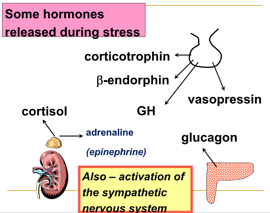
10.5.1 Physiological Functions of Cortisol
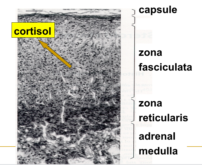
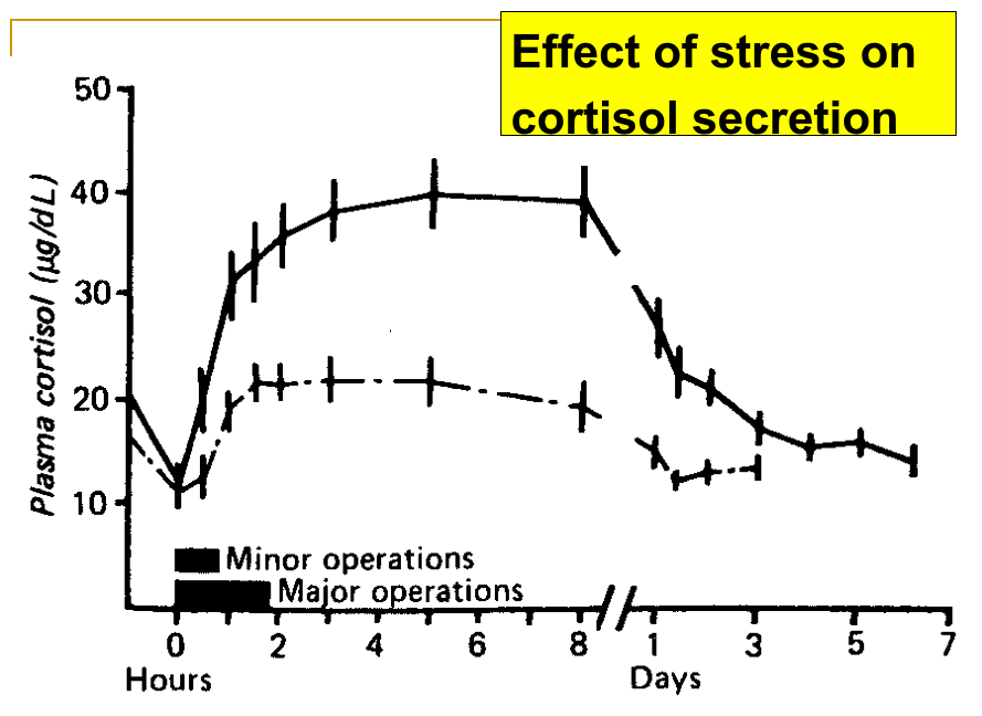
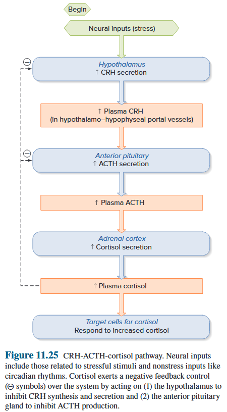
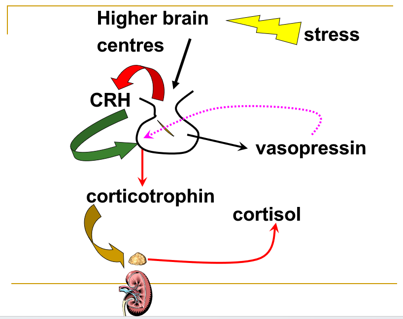
Importance of increased cortisol secretion in response to stress: Failure of cortisol secretion during stress leads to rapid collapse of the circulation and death 增加皮质醇分泌以应对压力的重要性:压力期间皮质醇分泌失败会导致循环迅速崩溃和死亡
10.5.2 Actions of cortisol
Functions of Cortisol in Stress
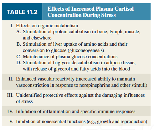
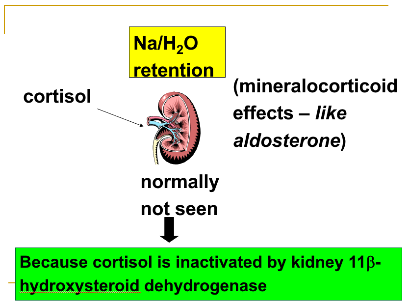
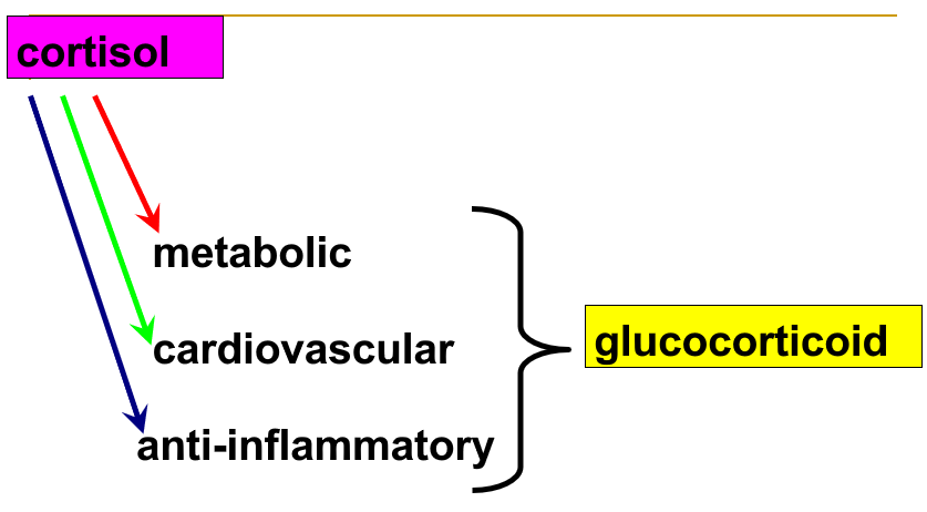
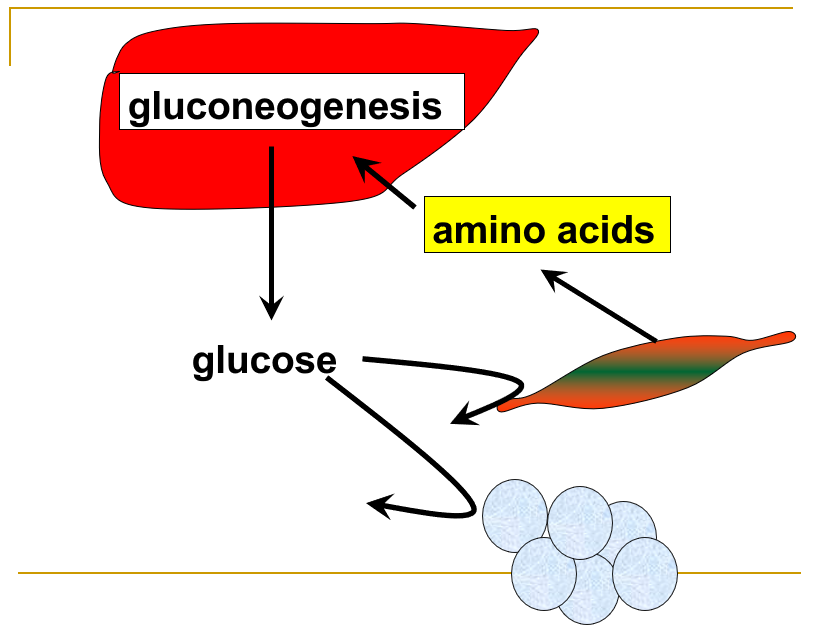
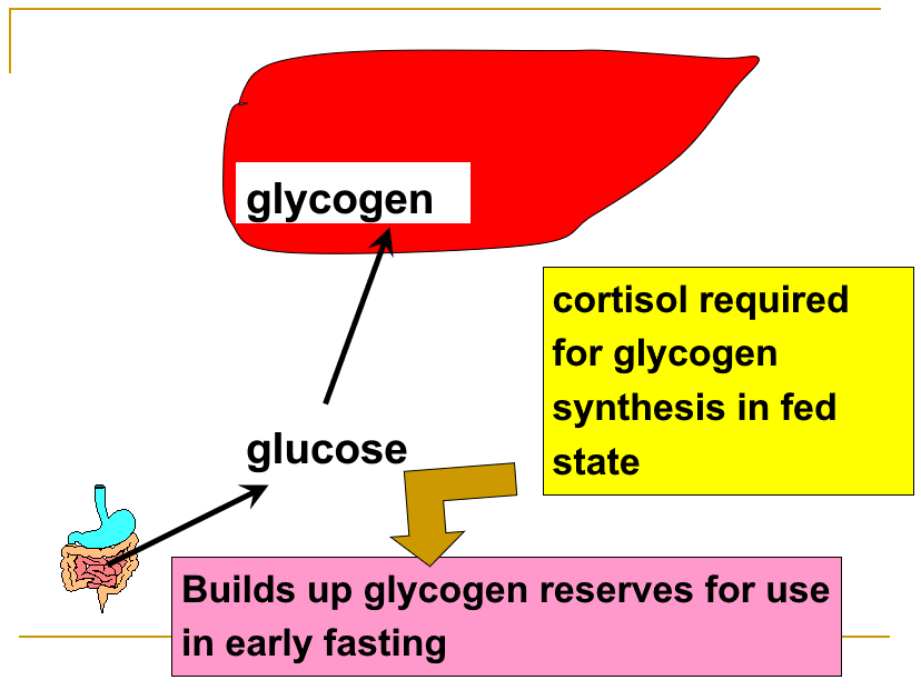
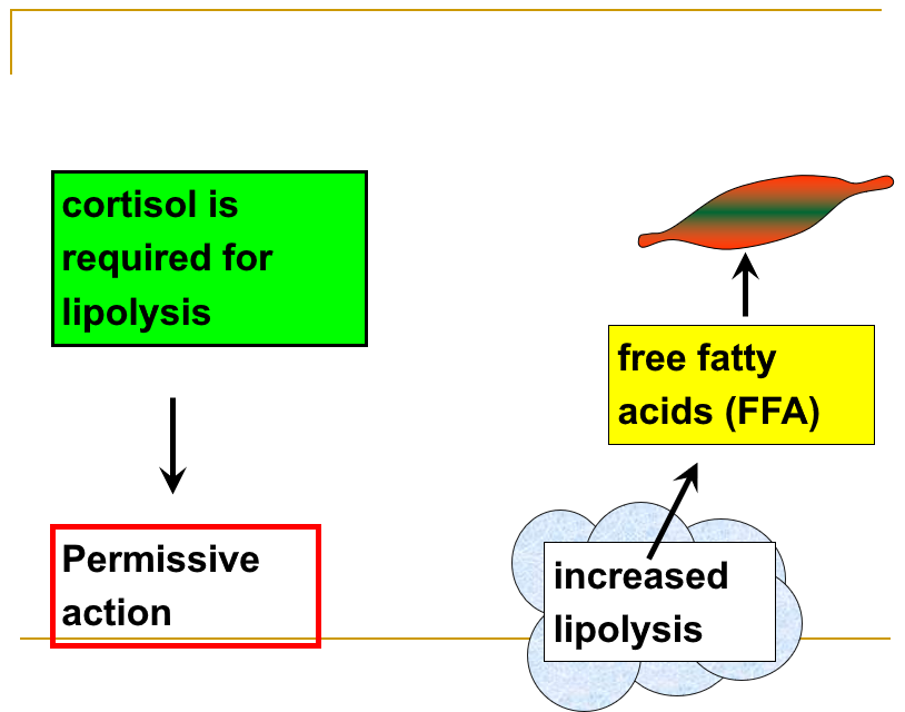
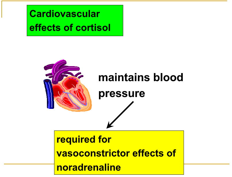
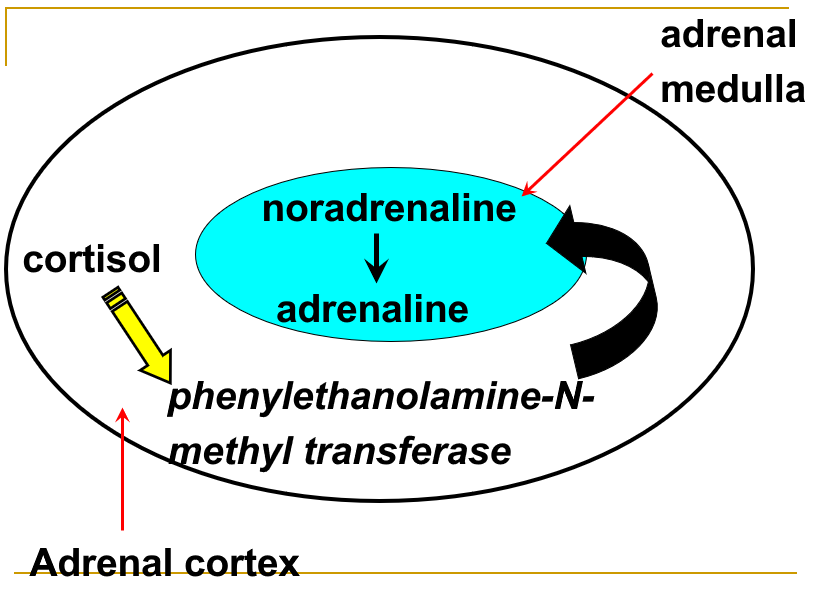
10.5.3 Anti-inflammatory effects of cortisol
Inflammation - the tissue response to injury
Features of inflammation
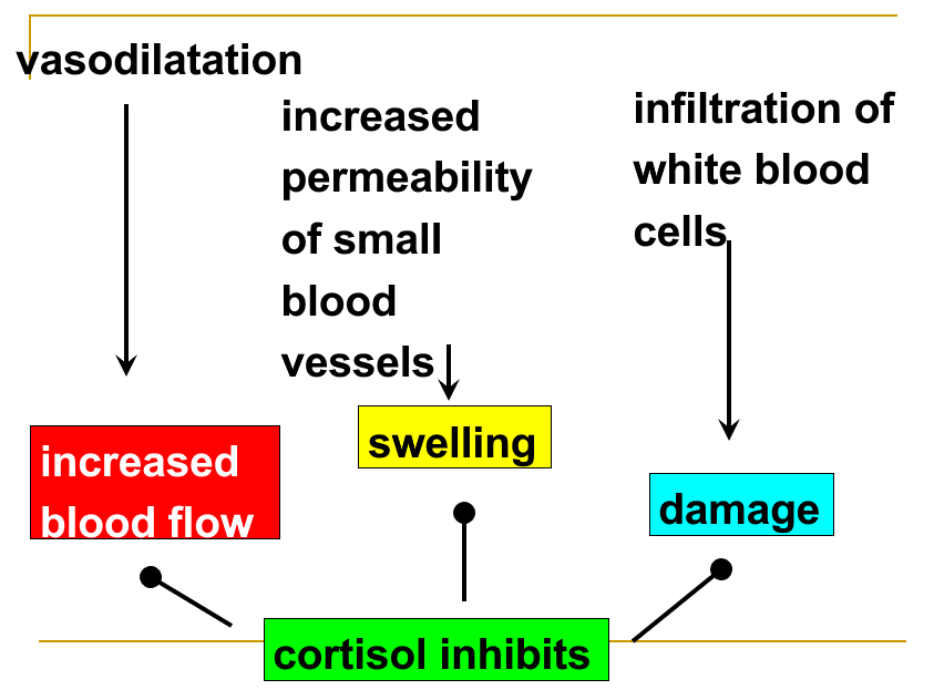
10.6 Endocrine control of the homeostasis of calcium and phosphate
Main aim is to maintain extracellular ionised calcium in range 1.17-1.29 mmol/L
Calcium
- Excitation contraction coupling
- Stimulus-secretion coupling
- Excitability of nerve/muscle cell membranes
- Structural component of bone
- Blood coagulation
兴奋收缩耦合 刺激-分泌耦合 神经/肌肉细胞膜的兴奋性 骨的结构成分 血液凝固
Main hormones regulating calcium
- parathyroid hormone (PTH)
- calcitonin (CT)
- 1,25-dihydroxy vitamin D (1,25(OH)~2~-D~3~)
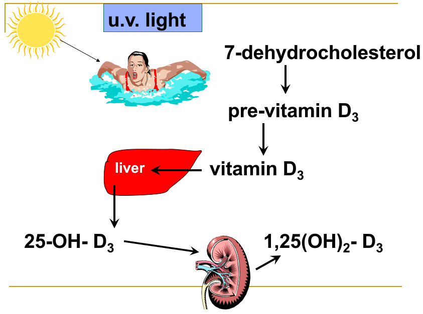
How are calcium and phosphate handled by the body?
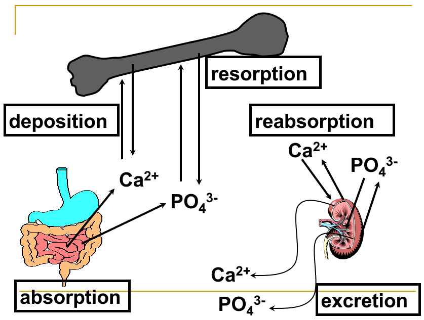
Effects on calcium and phosphate absorption from the small intestine
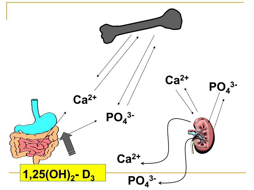
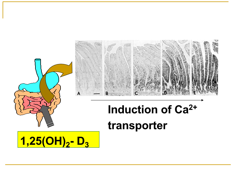
Effects on bone calcium and phosphate
Osteoblasts: Osteoblasts are the bone-forming cells. They secrete collagen to form a surrounding matrix, which then becomes calcified (mineralization). 成骨细胞:成骨细胞是骨形成细胞。它们分泌胶原蛋白形成周围基质,然后钙化(矿化)。
Osteocytes: Once surrounded by calcified matrix, the osteoblasts are called osteocytes. The osteocytes have long cytoplasmic processes that extend throughout the bone and form tight junctions with other osteocytes. 骨细胞:一旦被钙化基质包围,成骨细胞就被称为骨细胞。骨细胞具有长的细胞质突起,延伸到整个骨骼并与其他骨细胞形成紧密连接。
Osteoclasts: Osteoclasts are large, multinucleated cells that break down (resorb) previously formed bone by secreting hydrogen ions, which dissolve the crystals, and hydrolytic enzymes, which digest the osteoid. 破骨细胞:破骨细胞是大型多核细胞,通过分泌氢离子(溶解晶体)和水解酶(消化类骨质)来分解(吸收)先前形成的骨。

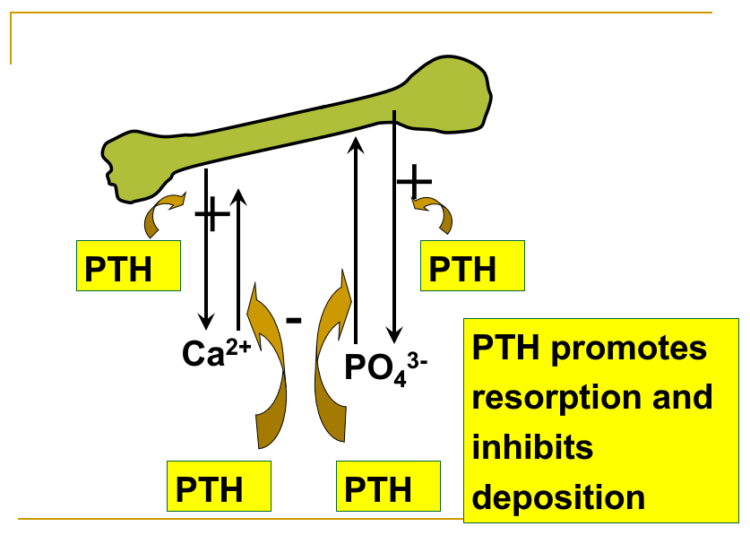
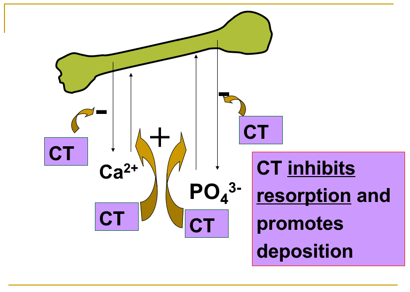
Hormones exert their effects through actions on bone cells
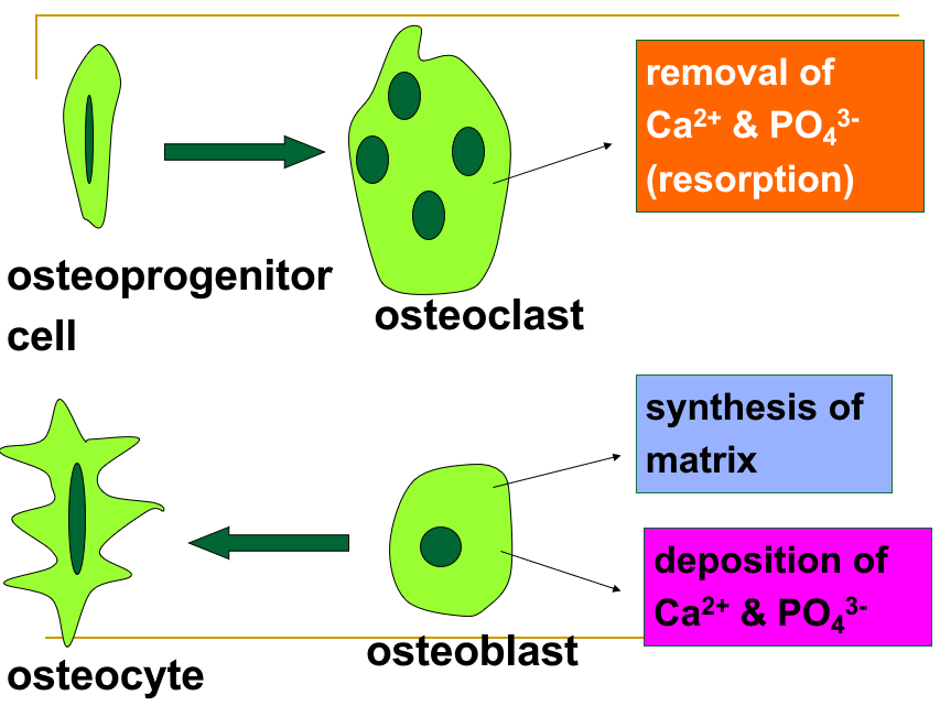
Mechanism of action of PTH on bone cells
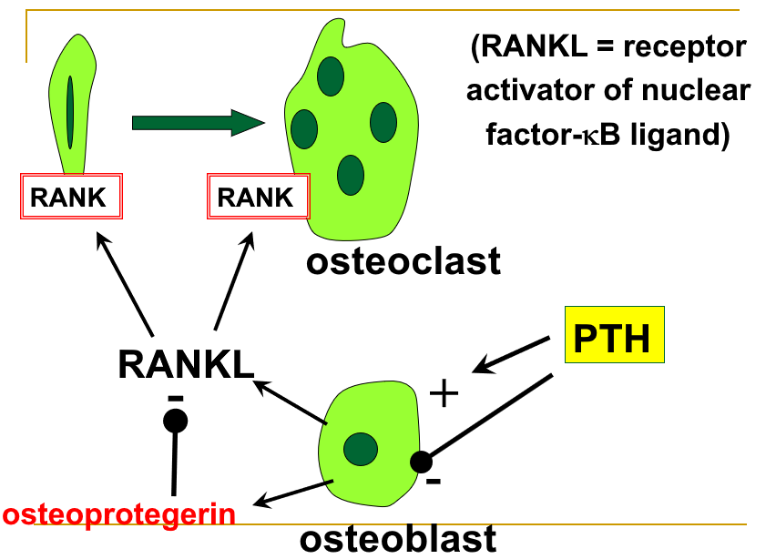
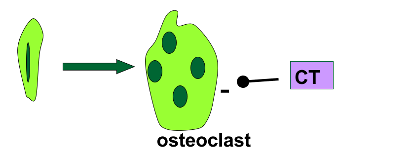
Effects on renal excretion of calcium and phosphate
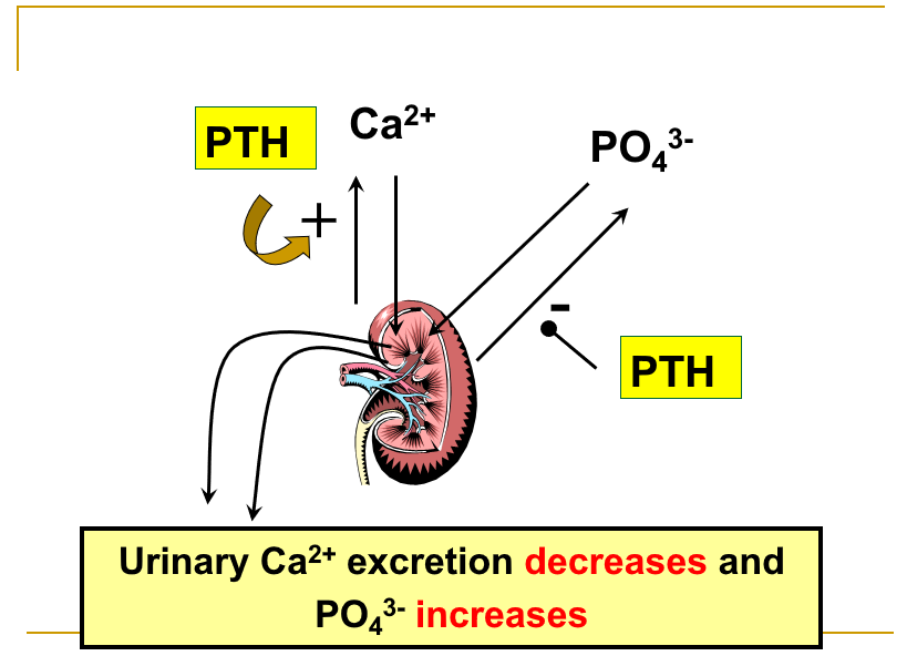
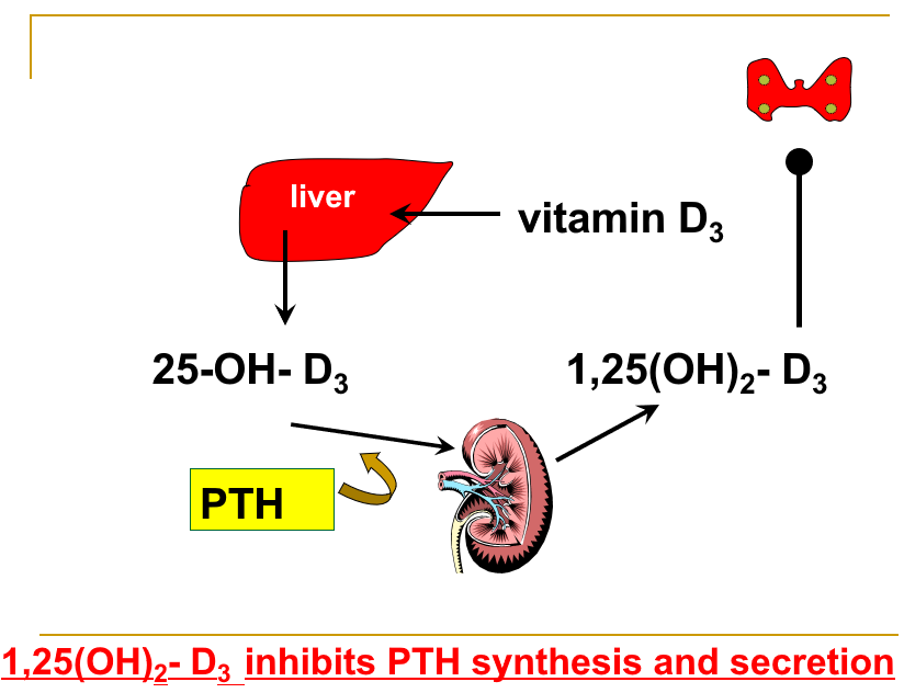
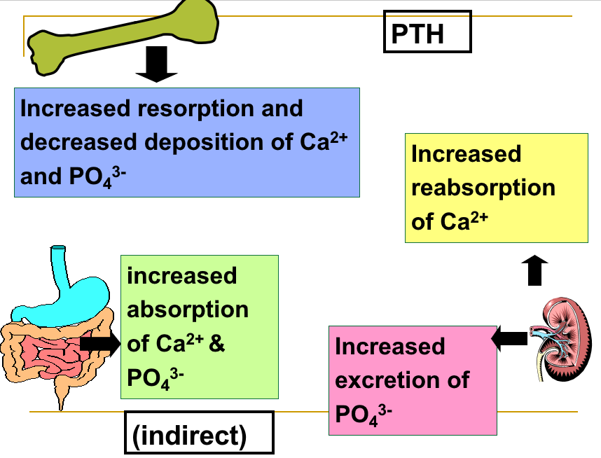
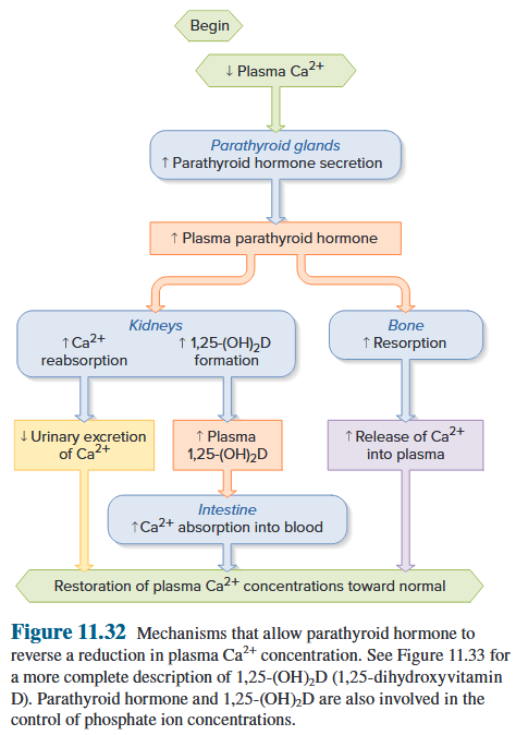
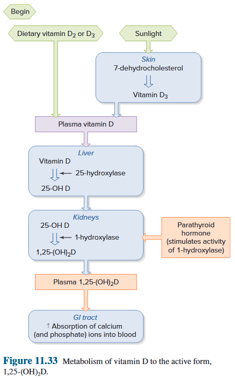
What will be the net effect of PTH on a) blood Ca^2+^ and b) blood PO~4~^3-^ concentrations?
blood calcium concentration ↑ - blood phosphate concentration ↓
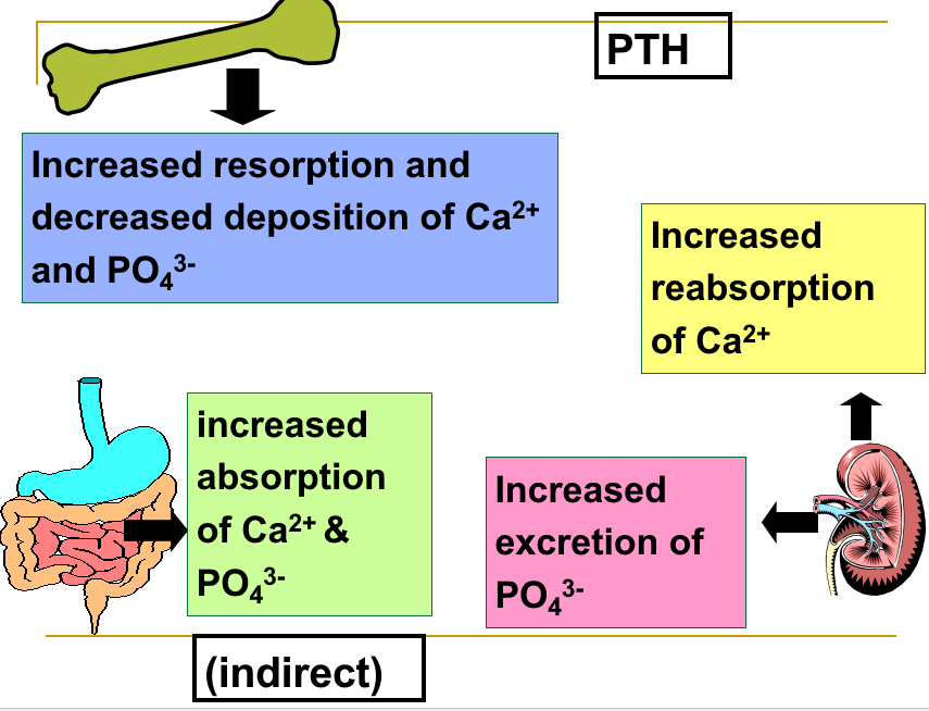
What will be the net effect of calcitonin on blood Ca^2+^ and blood PO43- concentrations?
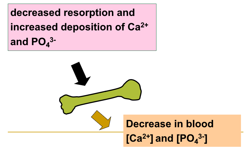
Regulation of PTH secretion
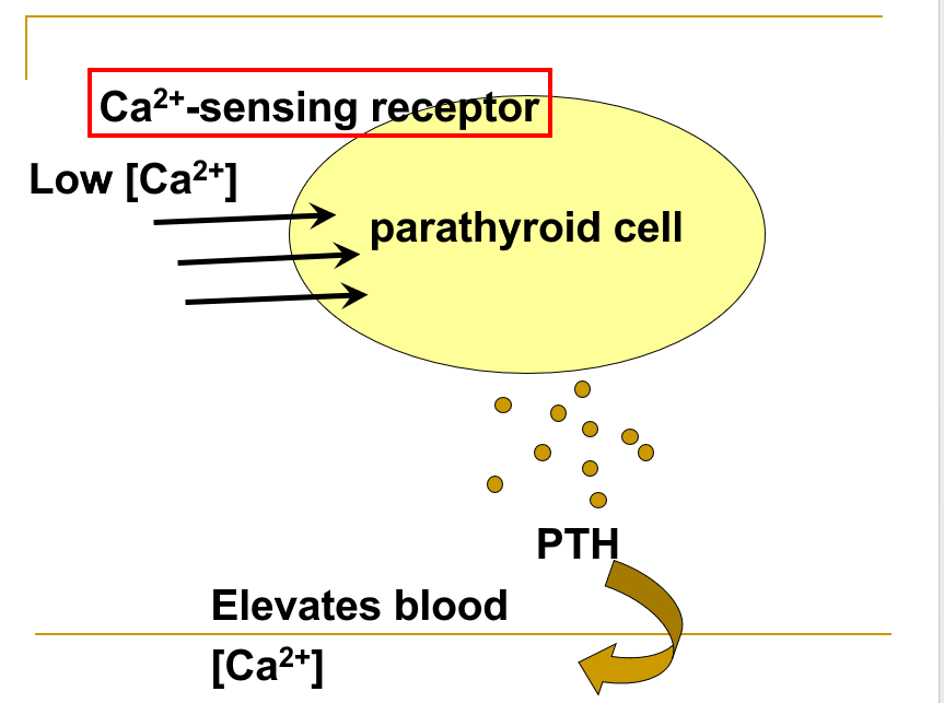
Consequences of PTH abnormalities
Consequences of PTH deficiency: Hyperexcitability of nerve and muscle 神经和肌肉过度兴奋
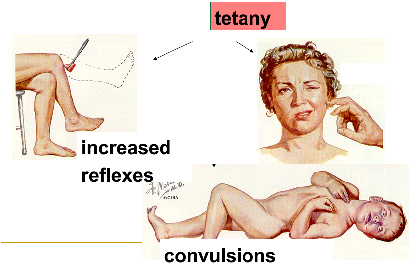
Consequences of oversecretion of PTH
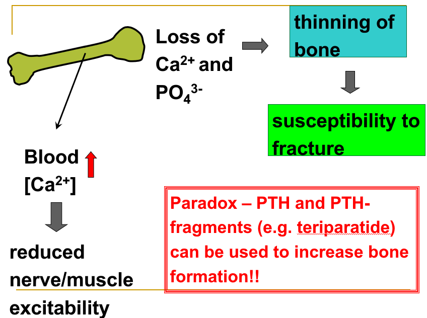
Regulation of calcitonin secretion
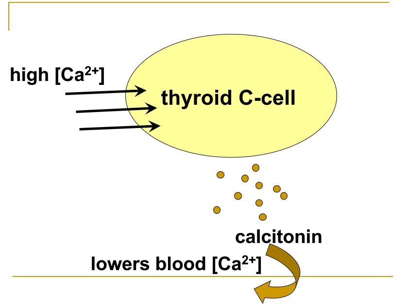
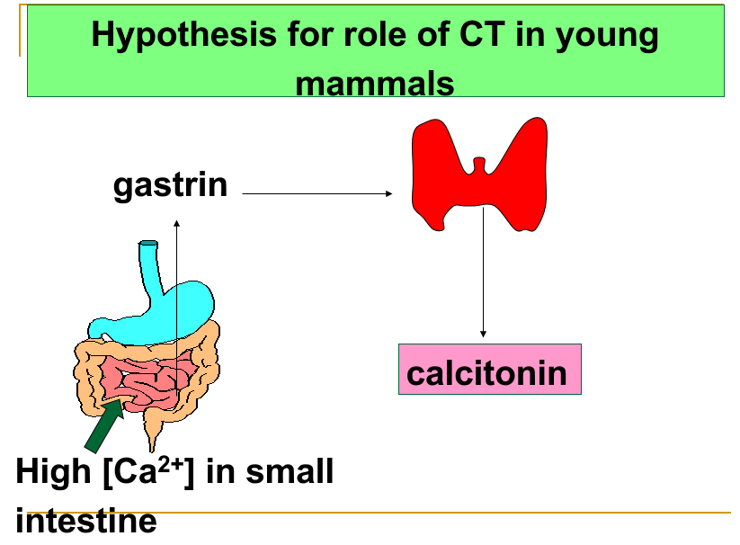
Metabolic Bone Diseases
-
Osteomalacia (adults) and rickets (children): the mineralization of bone is deficient—usually due to inadequate vitamin D intake, absorption, or activation. 骨软化症(成人)和佝偻病(儿童):骨骼矿化不足——通常是由于维生素 D 摄入、吸收或活化不足所致。
-
Osteoporosis: loss of bone density (loss of matrix and minerals).
- Bone resorption exceeds formation.
- It is most common in postmenopausal (estrogen-deficient) women.
- It can be prevented by exercise, adequate Ca and vitamin D intake, and medications (such as bisphosphonates).
骨质疏松症:骨密度损失(基质和矿物质损失)。 1. 骨吸收超过形成。 2. 最常见于绝经后(雌激素缺乏)女性。 3. 可以通过运动、摄入足够的钙和维生素D以及药物(如双膦酸盐)来预防。
Chapter 9 Functions of the Nervous System
9.1 General Principles of Activities of the Nervous System
9.1.1 General Structure and Function of an Individual Neuron
The various structures of the nervous system are intimately interconnected, but for convenience we divide them into two parts: (1) the central nervous system (CNS), composed of the brain and spinal cord; and (2) the peripheral nervous system (PNS), consisting of the nerves that connect the brain and spinal cord with the body’s muscles, glands, sense organs, and other tissues. 神经系统的各种结构是紧密相连的,但为了方便起见,我们将它们分为两部分:(1)中枢神经系统(CNS),由大脑和脊髓组成; (2) 周围神经系统 (PNS),由连接大脑和脊髓与身体肌肉、腺体、感觉器官和其他组织的神经组成。
- The functional unit of the nervous system is the individual cell, or neuron. Neurons operate by generating electrical signals that move from one part of the cell to another part of the same cell or to neighboring cells. In most neurons, the electrical signal causes the release of chemical messengers—neurotransmitters—to communicate with other cells. 神经元通过产生电信号来工作,这些电信号从细胞的一部分移动到同一细胞的另一部分或邻近的细胞。在大多数神经元中,电信号会导致化学信使(神经递质)的释放,以与其他细胞进行交流。
- The other major cell types of the nervous system are nonneuronal cells called glial cells. 神经系统的其他主要细胞类型是称为神经胶质细胞的非神经元细胞。
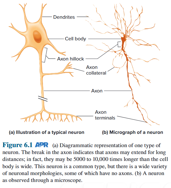
- Cell body - contains nucleus and cellular organelles
- Dendrites - extend from cell body / receive signals from other neurones
- Axon - conducts action potentials → axon terminals
- Nerve terminals - contain, release chemicals (neurotransmitters) → messages to neurones, muscles, glands
Dendrites are a series of highly branched outgrowths of the cell body. 树突是细胞体的一系列高度分支的产物。In the CNS, dendrites and the cell body receive most of the inputs from other neurons. 在中枢神经系统中,树突和细胞体接收来自其他神经元的大部分输入。Branching dendrites increase a cell’s surface area. Knoblike outgrowths called dendritic spines increase the surface area of dendrites still further, and there are often ribosomes present. 分支树突增加了细胞的表面积。称为树突棘的球形突起进一步增加了树突的表面积,并且通常存在核糖体。
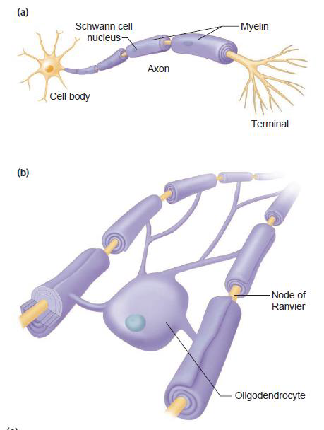
Myelin sheath 髓鞘 Synapse 突触 Oligodendrocyte 寡突细胞 Node of Ranvier 郎飞结
- The axon, sometimes also called a nerve fibre, is a long process that extends from the cell body and carries outgoing signals to its target cells. 轴突有时也称为神经纤维,是一个从细胞体延伸出来的长过程,并将传出信号传递到目标细胞。
Conduction of AP and axoplasmic transport AP 和轴浆运输的传导 - The axons of many neurons are covered by sheaths of myelin which usually consists of 20 to 200 layers of highly modified plasma membrane wrapped around the axon by a nearby supporting cell. 许多神经元的轴突被髓鞘覆盖,髓鞘通常由 20 至 200 层高度修饰的质膜组成,通过附近的支持细胞包裹在轴突周围。
- CNS, these myelin-forming cells are the oligodendrocytes.
PNS, cells called Schwann cells form individual myelin sheaths. - The spaces between adjacent sections of myelin where the axon’s plasma membrane is exposed to extracellular fluid are called the nodes of Ranvier. 轴突质膜暴露于细胞外液的髓磷脂相邻部分之间的空间称为朗飞结。
- Myelin sheath: Wraps round axon → electrical insulation → increase the speed of action potential conduction
Synapse
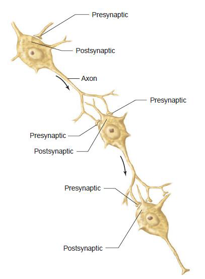
The anatomically specialized junction between two neurons where one neuron alters the electrical and chemical activity of another is called a synapse. 两个神经元之间解剖学上特殊的连接处,其中一个神经元改变另一个神经元的电和化学活动,称为突触。
The signal in synapse is transmitted from one neuron to another by neurotransmitters, a term that also includes the chemicals efferent neurons use to communicate with effector cells. 突触中的信号通过神经递质从一个神经元传递到另一个神经元,该术语还包括传出神经元用于与效应细胞通信的化学物质。
- Presynaptic neuron: A neuron that conducts a signal toward a synapse 突触前神经元:向突触传导信号的神经元
- Postsynaptic neuron: a neuron conducts signals away from a synapse 突触后神经元:神经元从突触传导信号
- the size and shape of the presynaptic and postsynaptic elements can vary greatly 突触前和突触后元件的大小和形状可能有很大差异
9.1.2 Glial Cells and Their General Functions
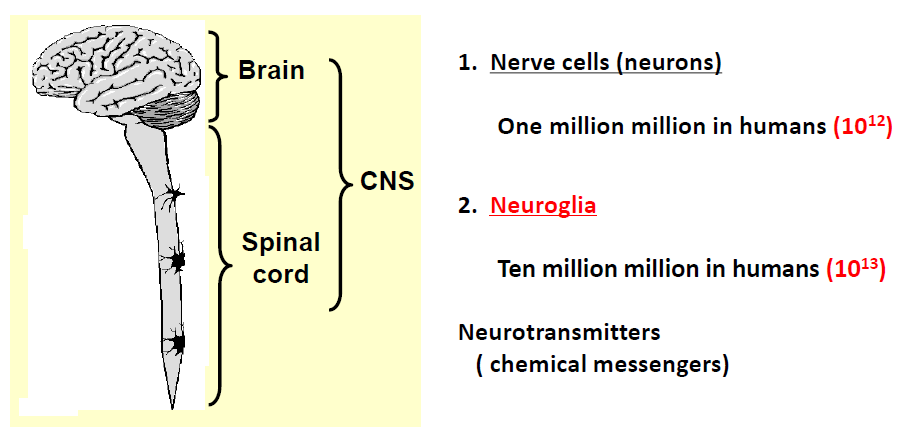
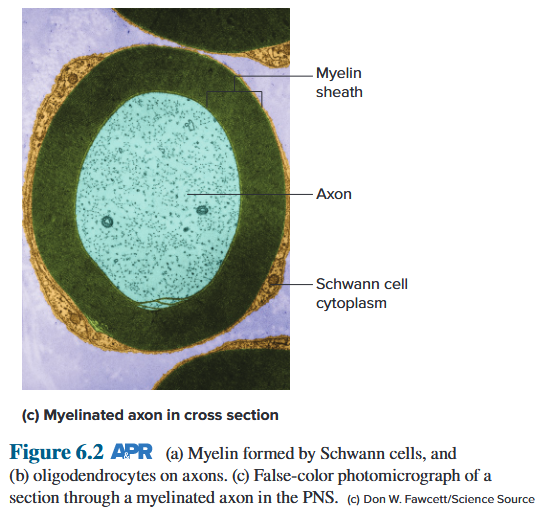
- Microglia: Phagocytes, scavengers
- Ependymal cells: Line ventricles, spinal canal, CSF circulation
- Astrocytes: Line neurones, blood vessels, BBB
- Oligodendrocytes: Myelin sheath in CNS
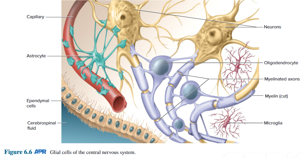
Comparison between neurons and glial cells
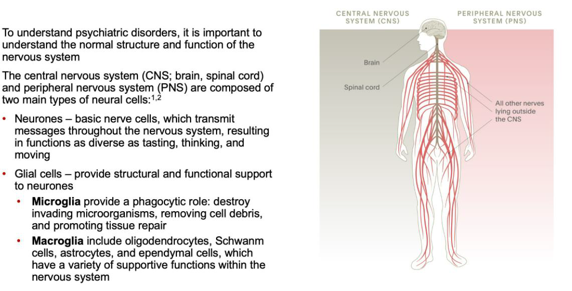
9.1.3 Types of Neuronal Connection
Characteristics of Three Classes of Neurons
- Afferent neurons
- Transmit information into the CNS from receptors at their peripheral endings 将信息从外周末端的受体传输到中枢神经系统
- Single process from the cell body splits into a long peripheral process (axon) that is in the PNS and a short central process (axon) that enters the CNS 来自细胞体的单个突分裂成位于三七总皂苷中的长外周突(轴突)和进入中枢神经系统的短中央突(轴突)
- Efferent neurons
- Transmit information out of the CNS to effector cells, particularly muscles, glands, neurons, and other cells 将信息从中枢神经系统传输到效应细胞,特别是肌肉、腺体、神经元和其他细胞
- Cell body with multiple dendrites and a small segment of the axon are in the CNS; most of the axon is in the PNS 具有多个树突的细胞体和一小段轴突位于中枢神经系统中;大部分轴突位于 PNS 中
- Interneurons
- Function as integrators and signal changers
- Integrate groups of afferent and efferent neurons into reflex circuits
- Lie entirely within the CNS
- Account for > 99% of all neurons
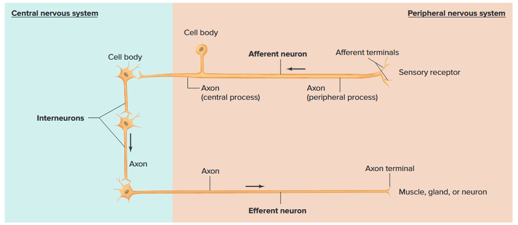
Types of Neuronal Connections in the NS
- Single-line connection is the type of relationship where one presynaptic neuron synapses on only one post synaptic neuron. 单线连接是一种关系类型,其中一个突触前神经元仅与一个突触后神经元发生突触。
- Convergent connection is the type of connection where may presynaptic neurons synapse on one postsynaptic neuron. 会聚连接是突触前神经元可以突触到一个突触后神经元的连接类型。
- Divergent connection is the type of connection where one presynaptic neuron synapses on many postsynaptic neuron. 发散连接是一个突触前神经元突触多个突触后神经元的连接类型。
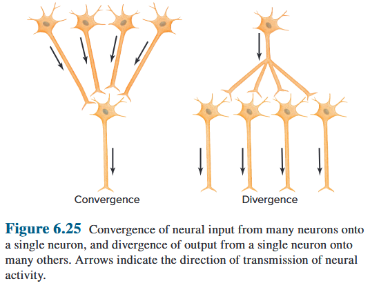
Afferent nerve → Divergence
Efferent nerve → Convergence
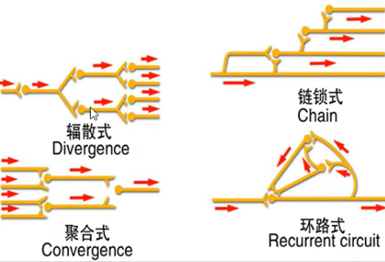
- Divergence and convergence existed at the same time 分歧与趋同同时存在
- All of neurons are excited neuron → Excitation prolong 所有神经元都兴奋神经元→兴奋延长
- Inhibitory neurons exist → reduce/suppress the original activities. 抑制性神经元的存在→减少/抑制原有的活动。
9.1.4 Synaptic Transmission
Activity at synapses can increase or decrease the likelihood that the postsynaptic neuron will fire action potentials by producing a brief, graded potential in the postsynaptic membrane. 突触的活动可以通过在突触后膜中产生短暂的分级电位来增加或减少突触后神经元激发动作电位的可能性。
At a synapse:
- A signal-passing neuron (the presynaptic neuron) 信号传递神经元(突触前神经元)
- The target neuron (the postsynaptic neuron) 目标神经元(突触后神经元)
- The 2 neural membranes create a synaptic cleft that carries out the signaling process (neurotransmission) 2 个神经膜形成突触间隙,执行信号传递过程(神经传递)
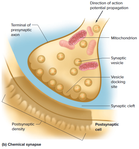
Chemical Synapses
Chemical synapses are specialized junctions through which cells of the nervous system signal to one another and to non-neuronal cells (e.g. muscles or glands). 化学突触是一种特殊的连接点,神经系统的细胞通过它向彼此以及非神经元细胞(例如肌肉或腺体)发出信号。
- Chemical synapse between a motor neuron and a muscle cell is called aneuromuscular junction. 运动神经元和肌肉细胞之间的化学突触称为神经肌肉接头。
- Chemical synapses allow the neurons of the central nervous system to form interconnected neural circuits. 化学突触允许中枢神经系统的神经元形成互连的神经回路。
Mechanisms of Neurotransmitter Release
- Prior to activation, many vesicles are docked on the presynaptic membrane at release regions known as active zones. 在激活之前,许多囊泡停靠在突触前膜上称为活性区的释放区域。
- Process voltage-gated Ca^2+^ channels.
- Depolarization during the action potential opens these Ca^2+^ channels and because the electrochemical gradient favors Ca^2+^ influx, Ca^2+^ flows into the axon terminal. 动作电位过程中的去极化会打开这些 Ca^2+^ 通道,并且由于电化学梯度有利于 Ca^2+^ 流入,Ca^2+^ 流入轴突末端。
- Prior to the arrival of an action potential, vesicles are loosely docked in the active zones by the interaction of a group of proteins, some of which are anchored in the vesicle membrane and others that are found in the membrane of the terminal. These are collectively known as SNARE proteins (soluble N-ethylmaleimide-sensitive factor attachment protein receptors). 在动作电位到达之前,囊泡通过一组蛋白质的相互作用松散地停靠在活性区域中,其中一些蛋白质锚定在囊泡膜上,而其他蛋白质则存在于末端膜中。这些统称为 SNARE 蛋白(可溶性 N-乙基马来酰亚胺敏感因子附着蛋白受体)。
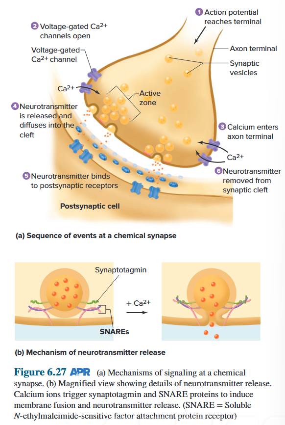
Different classifications
- Electrical Synapse v.s. Chemical Synapse
-
Electrical synapses, the plasma membranes of the presynaptic and postsynaptic cells are joined by gap junctions. 电突触,突触前和突触后细胞的质膜通过间隙连接连接。
-
Chemical Synapse, Gap between 2 neurons where information passes chemically in the form of neurotransmitter molecules. 化学突触,两个神经元之间的间隙,信息以神经递质分子的形式通过化学方式传递。
-
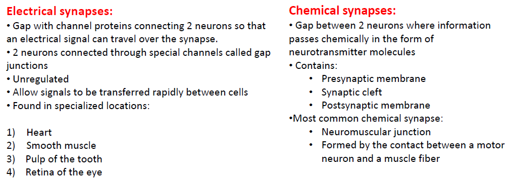
-
Excitatory Synapse v.s. Inhibitory Synapse
The membrane potential of a postsynaptic neuron is brought closer to threshold (depolarized) at an excitatory synapse , and it is either driven farther from threshold (hyperpolarized) or stabilized at its resting potential at an inhibitory synapse. 突触后神经元的膜电位在兴奋性突触处接近阈值(去极化),并且在抑制性突触处被驱动远离阈值(超极化)或稳定在其静息电位。
Activation of the Postsynaptic Cell
Excitatory synapse (EPSP): The membrane potential of a postsynaptic neuron is brought closer to threshold (depolarized) 兴奋性突触 (EPSP):突触后神经元的膜电位接近阈值(去极化)
At an excitatory chemical synapse, the postsynaptic response to the neurotransmitter is a depolarization, bringing the membrane potential closer to threshold. The usual effect of the activated receptor on the postsynaptic membrane at such synapses is to open nonselective channels that are permeable to Na^+^ and K^+^. These ions then are free to move according to the electrical and concentration gradients across the membrane. 在兴奋性化学突触处,对神经递质的突触后反应是去极化,使膜电位更接近阈值。激活的受体对此类突触处的突触后膜的通常作用是打开可渗透 Na^+^ 和 K^+^ 的非选择性通道。然后,这些离子可以根据跨膜的电梯度和浓度梯度自由移动。
Inhibitory synapse (IPSP): The membrane is either driven farther from threshold (hyperpolarized) or stabilized at its resting potential 抑制性突触 (IPSP):膜被驱动远离阈值(超极化)或稳定在其静息电位
At an inhibitory synapse, the activated receptors on the postsynaptic membrane open Cl− or K^+^ channels; Na^+^ permeability is not affected. In those cells that actively regulate intracellular Cl− concentrations via active transport out of the cell, the Cl− equilibrium potential is more negative than the resting potential. Therefore, as Cl− channels open, Cl− enters the cell, producing a hyperpolarization—that is, an IPSP. 在抑制性突触处,突触后膜上激活的受体打开 Cl− 或 K^+^ 通道; Na^+^ 渗透性不受影响。在那些通过主动转运出细胞来主动调节细胞内 Cl− 浓度的细胞中,Cl− 平衡电位比静息电位更负。因此,当 Cl− 通道打开时,Cl− 进入细胞,产生超极化,即 IPSP。
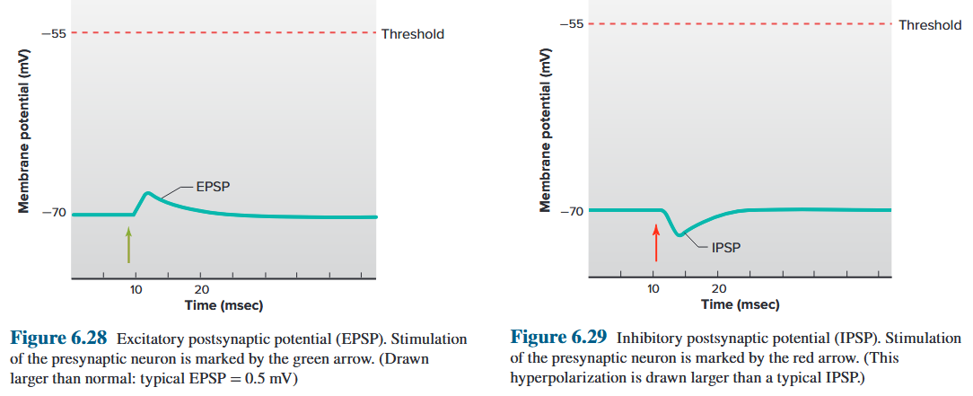
Synaptic Integration
- No interaction occurs
- Temporal summation
- Spatial summation
- Initiation of AP
- Mutual interaction
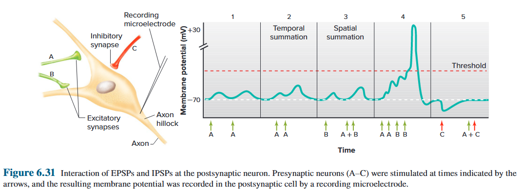
Synaptic Strength
The effectiveness or strength of a given synapse is influenced by both presynaptic and postsynaptic mechanisms.
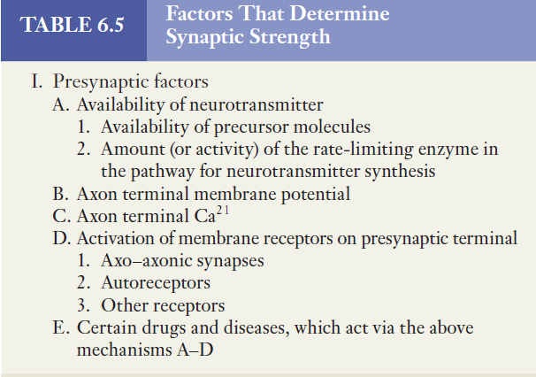
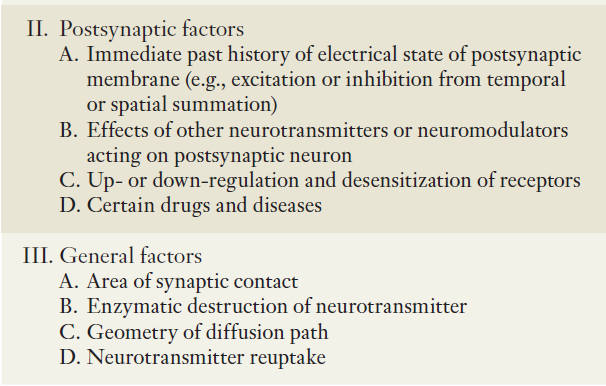
9.1.5 Neurotransmitters and Corresponding Receptors
9.1.5.1 Neurotransmitters
The role of neurotransmitters in eliciting EPSPs and IPSPs. However, certain chemical messengers elicit complex responses that cannot be described as simply EPSPs or IPSPs. The word modulation is used for these complex responses, and the messengers that cause them are called neuromodulators. 神经递质在引发 EPSP 和 IPSP 中的作用。然而,某些化学信使会引起复杂的反应,不能简单地描述为 EPSP 或 IPSP。调制这个词用于描述这些复杂的反应,引起这些反应的信使被称为神经调制器。
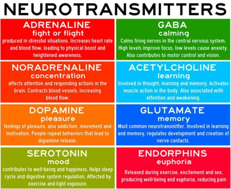
Neural communications
- Excitatory & inhibitory signals
- Chemicals carry the signals (e.g. EP and NE)
- Receptors received the signals
What is a neurotransmitter?
- Chemicals released by a neuron/nerve cells onto a targeted neuron (cells)
- Outside the CNS and PNS, those CM circulated in the blood stream as hormones.
Criteria for Identification of a Substance as a Neurotransmitter
- Synthesised within neurone
- Stored in nerve terminal
- Released by nerve stimulation in Ca^2+^-dependent manner
- Exogenous drug must mimic endogenous neurotransmitter
- Antagonist must inhibit neurotransmitter & exogenous drug
- Specific mechanisms exist for inactivation
Two classes of neurotransmitters by their biochemical structure:
- Non-peptide neurotransmitters
- Neuropeptides
Two classes of neurotransmitters by their function
- Excitatory neurotransmitters
- Cause depolarization of postsynaptic membrane
- Promote action potentials
- Inhibitory neurotransmitters
- Cause hyperpolarization of postsynaptic membranes
- Suppress actions potentials
Classification of neurotransmitters by functions
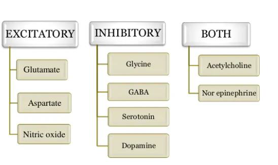
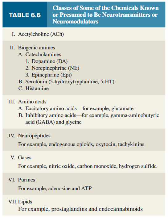
9.1.5.1.1 Acetylcholine and Its Receptors
Acetylcholine (ACh) is a major neurotransmitter in the PNS at the neuromuscular junction (where a motor neuron contacts a skeletal muscle cell; see Chapter 9) and in the brain.
Neurons that release ACh are called cholinergic neurons. The cell bodies of the brain’s cholinergic neurons are concentrated in relatively few areas, but their axons are widely distributed. 释放乙酰胆碱的神经元称为胆碱能神经元。大脑胆碱能神经元的细胞体集中在相对较少的区域,但其轴突分布广泛。
Acetylcholine is synthesized from choline (a common nutrient found in many foods) and acetyl coenzyme A in the cytoplasm of synaptic terminals and stored in synaptic vesicles. After it is released and activates receptors on the postsynaptic membrane, the concentration of ACh at the postsynaptic membrane decreases (thereby stopping receptor activation) due to the action of the enzyme acetylcholinesterase. 乙酰胆碱 (ACh) 是三七总皂苷中神经肌肉接头处(运动神经元与骨骼肌细胞接触的地方;见第 9 章)和大脑中的主要神经递质。释放乙酰胆碱的神经元称为胆碱能神经元。大脑胆碱能神经元的细胞体集中在相对较少的区域,但其轴突分布广泛。乙酰胆碱是由突触末梢细胞质中的胆碱(许多食物中常见的营养素)和乙酰辅酶 A 合成的,并储存在突触小泡中。它被释放并激活突触后膜上的受体后,由于乙酰胆碱酯酶的作用,突触后膜上的ACh浓度降低(从而停止受体激活)。
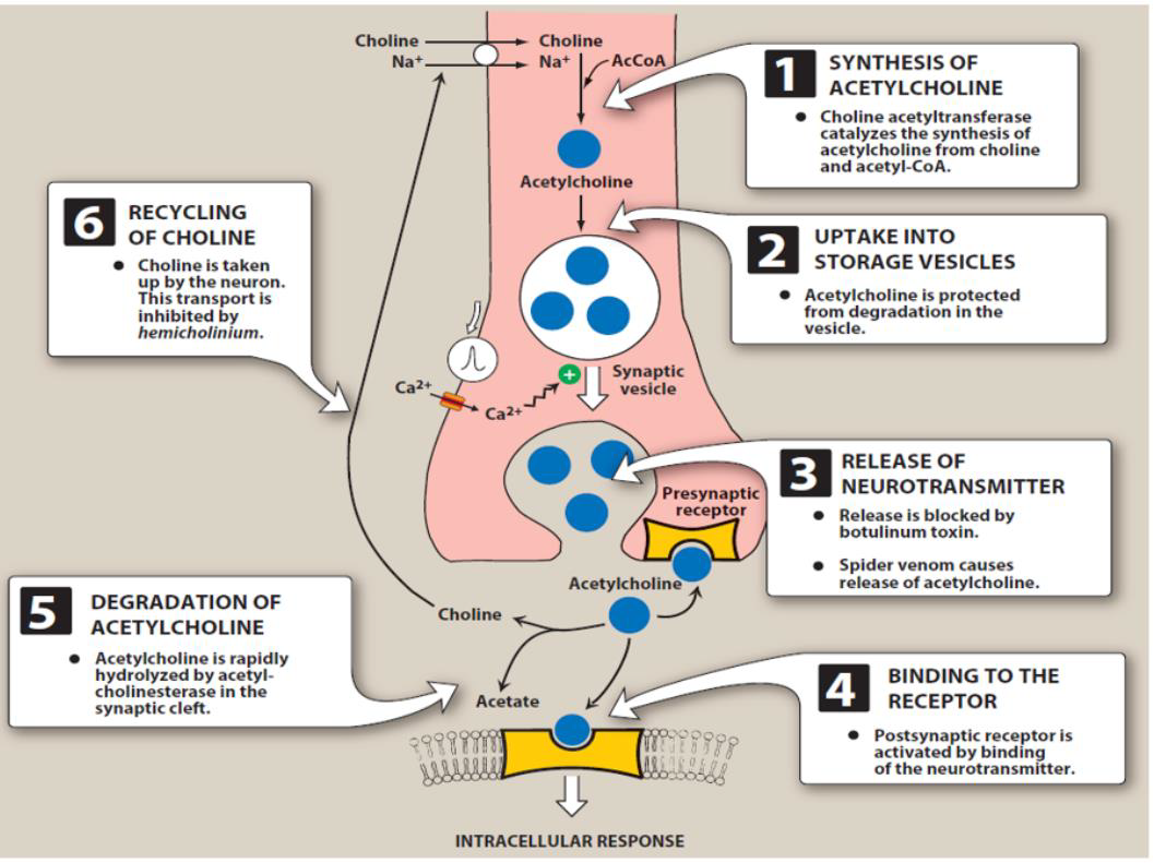
Two types of receptors for Ach:
-
Some ACh receptors respond not only to acetylcholine but to the drug nicotine and have therefore come to be known as nicotinic receptors .
-
Nicotine
Nicotine is an agonist, which means that when it binds to receptors, it brings about a biological response.
Nicotinic-cholinergic receptors are found in many places in the body, including the brain, neuromuscular junctions, the inner part of the adrenal gland, and ganglia (groups of nerve cells).
Nicotine‘s stimulating abilities come from the fact that when it binds to receptors, neurotransmitters like dopamine, acetylcholine, beta-endorphin, norepinephrine, serotonin, and ACTH are released in the body.
-
-
The other general type of cholinergic receptor is stimulated not only by acetylcholine but by the mushroom poison muscarine; therefore, these are called muscarinic receptors .
-
Atropine
Atropine competes with acetylcholine for cholinergic receptor sites on the SA and AV nodes. By blocking acetylcholine, atropine speeds up the heart rate.
Atropine sulfate blocks the action of acetylcholine at parasympathetic sites in smooth muscle, secretory glands, and the central nervous system causing an increase in cardiac output and reduced secretions .
It also reverses the muscarinic effects of cholinergic poisoning by acting as a competitive antagonist of acetylcholine at muscarinic receptors.
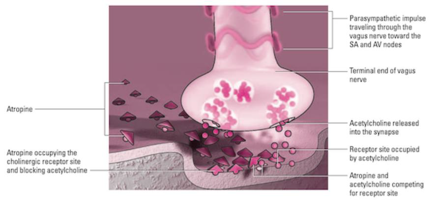
-
Catecholamines
all contain a catechol ring (a six-carbon ring with two adjacent hydroxyl groups) and an amine group, called catecholamines .
Dopamine
Norepinephrine (NE)
Epinephrine
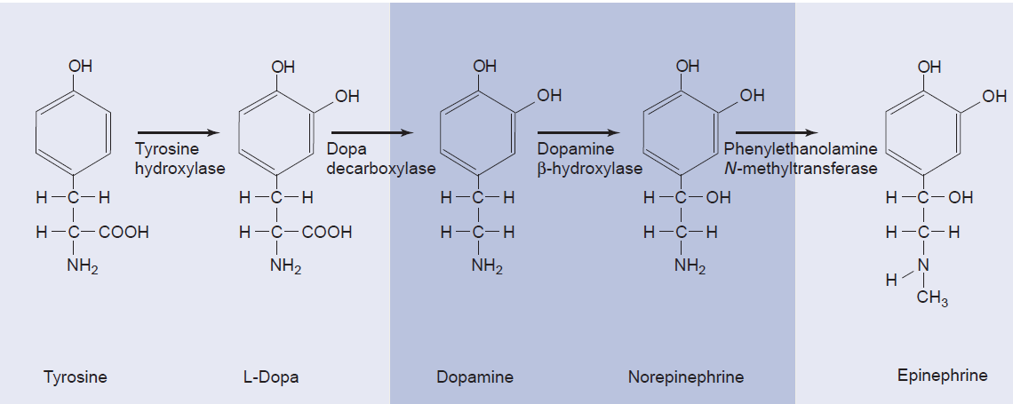
-
Synthesis
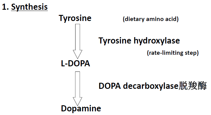
-
Pathway
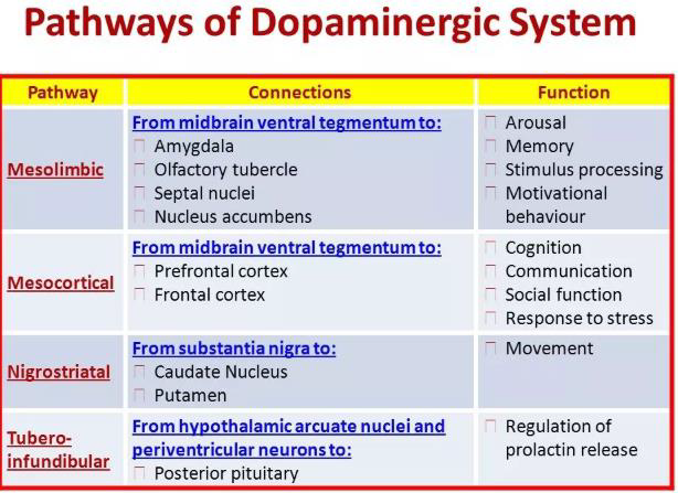
-
Receptors
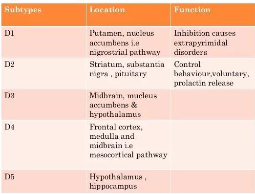
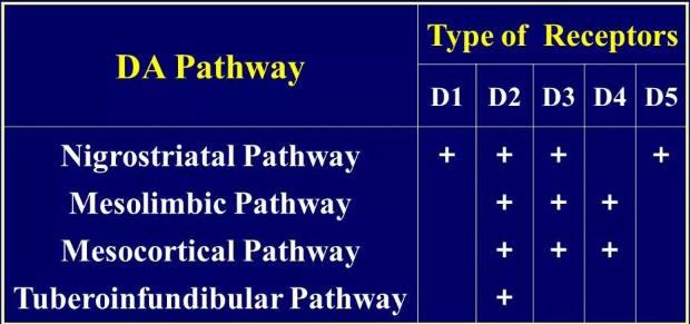
9.1.5.1.2 Glutamate
There are a number of excitatory amino acids, but the most common by far is glutamate, which is estimated to be the primary neurotransmitter at 50% of excitatory synapses in the CNS. 兴奋性氨基酸有很多种,但迄今为止最常见的是谷氨酸,据估计,它是中枢神经系统 50% 兴奋性突触的主要神经递质.
The vast majority are ionotropic, with two important subtypes being found in postsynaptic membranes.
They are designated as AMPA receptors (identified by their binding to α-amino-3–hydroxy-5methyl-4 isoxazolepropionic acid) and NMDA receptors (which bind N-methyl-D-aspartate). 它们被指定为 AMPA 受体(通过与 α-氨基-3-羟基-5甲基-4 异恶唑丙酸的结合来识别)和 NMDA 受体(与 N-甲基-D-天冬氨酸结合)。
NMDA receptors have also been implicated in mediating excitotoxicity.
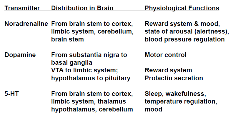
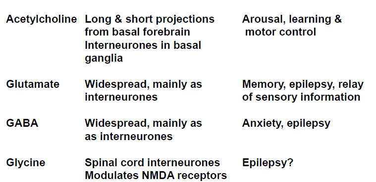
Similarities
- Stored in vesicles, in specific regions
- Ca^2+^-dependent release
- 2nd messengers / $\Delta$ ion channel activity
Differences
-
Synthesized in cell body, part of large precursor protein
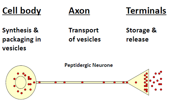
-
Transported to nerve terminal
-
Processed to active peptide
-
Released at lower concentrations
-
Mechanism of inactivation unclear
-
Removed slowly from synaptic cleft
-
Longer action
9.1.5.1.3 Neuropeptides
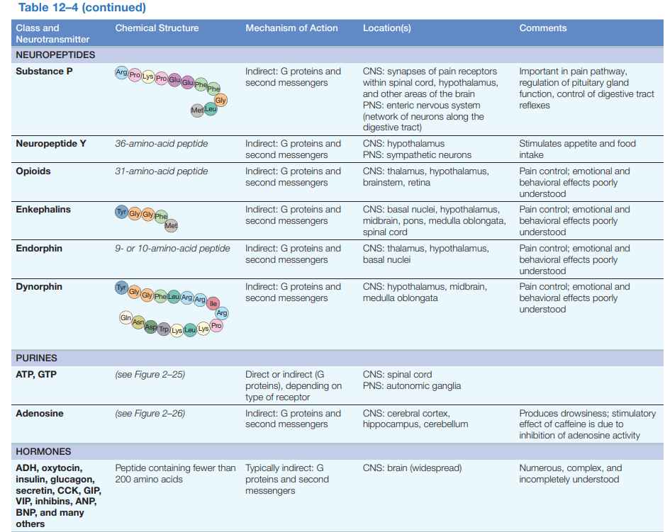
9.1.5.2 Corresponding receptors
Ligand-activated ion channels: These receptors are membrane-spanning ion channel proteins that open directly in response to ligand binding.
Metabotropic receptors: These receptors are not themselves ion channels. Neurotransmitter binding triggers a signaling pathway, which may indirectly open or close channels (or have some other effect entirely).
9.1.5.2.1 Ionotropic receptors
The first class of neurotransmitter receptors areligand-activated ion channels, also known asionotropic receptors.
Ligand-activated ion channels are large protein complexes.
Ionotropic receptors contain a channel which is modulated by the binding of a specific neurotransmitter. Ligand binding causes the opening or closing of the channel, thereby controlling the flow of ions (Na^+^, k+, Ca^2+^, Cl^-^) into the cell.
Types of ionotropic receptors include:
- GABAA receptors
- Glutamate NMDA receptors
- Glutamate Kainate receptors
- Glutamate AMPA receptors
- Glycine receptors
- Nicotinic Acetylcholine receptors (nAChR)
- Serotonin 5-HT3 receptor
Nicotinic receptors
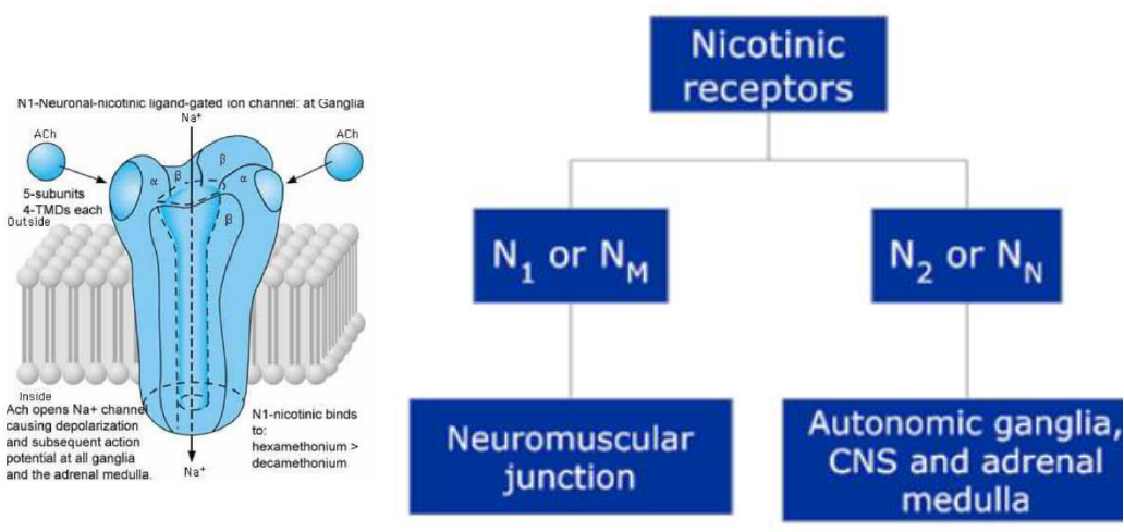
N~1~ or N~M~ receptors: these receptors are located at the neuromuscular junction, acetylcholine receptors of the N~M~ subtype are the only acetylcholine receptors that can be found at the neuromuscular junction.
N~2~ or N~N~ receptors: as mentioned before, nicotinic receptors play a key role in the transmission of cholinergic signals in the autonomic nervous systems.
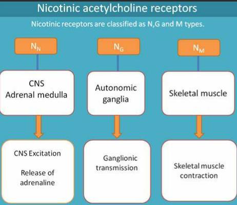
9.1.5.2.2 Metabotropic receptors
Metabotropic receptors do not have a channel that is not opened or closed by ligand binding. When activated they instead modulate pathways controlling the actions of neurotransmitters and ion channels through second messengers.
Types of metabotropic receptors include:
- Adrenergic receptors
- Dopamine receptors
- GABAB receptors
- Glutamate receptors mGluR
- Histamine receptors
- Muscarinic acetylcholine receptors (mAChR)
- Opioid receptors
- Serotonin (5-HT) receptors
Muscarinic acetylcholine receptors (mAChR)
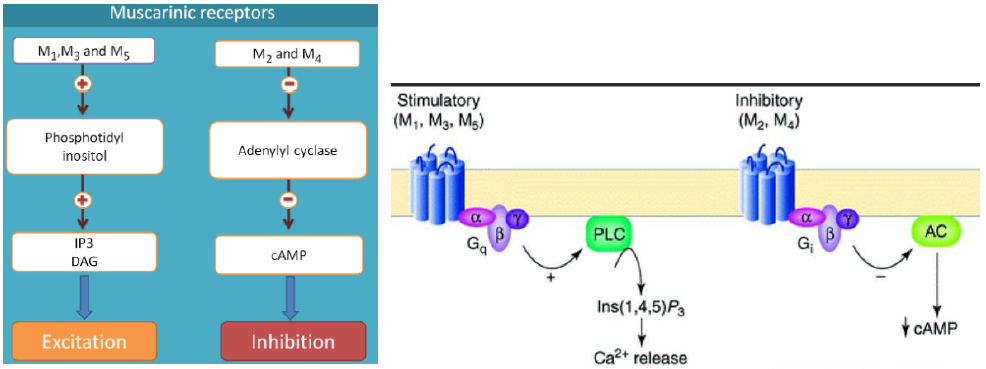
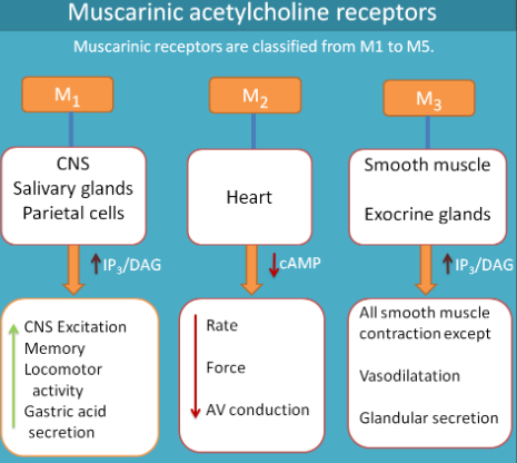
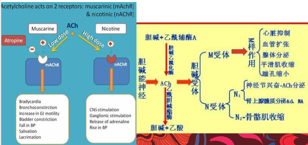
Adrenergic receptors
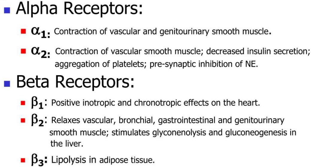
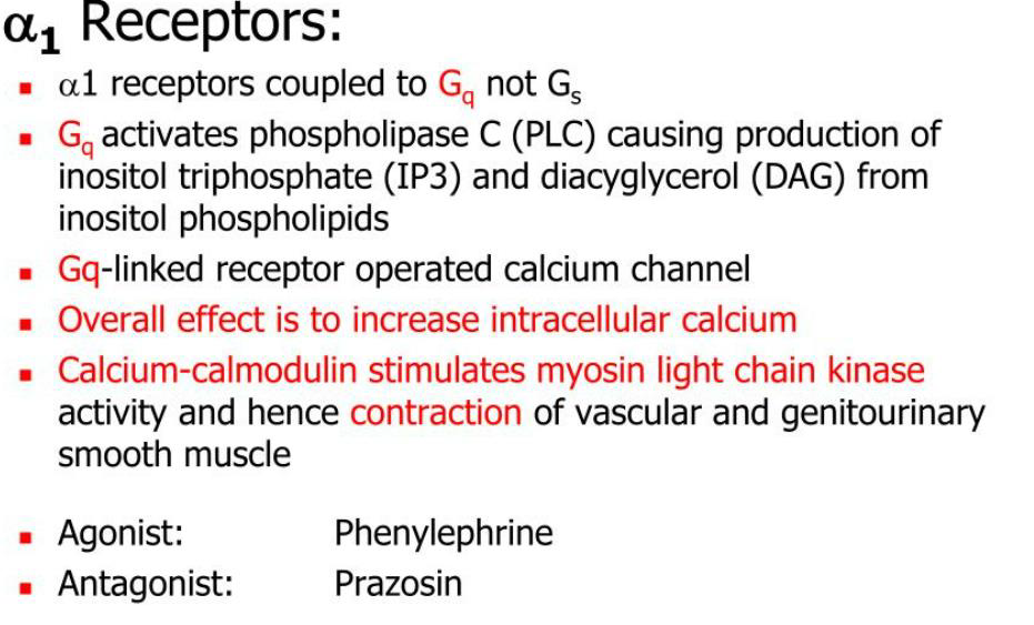
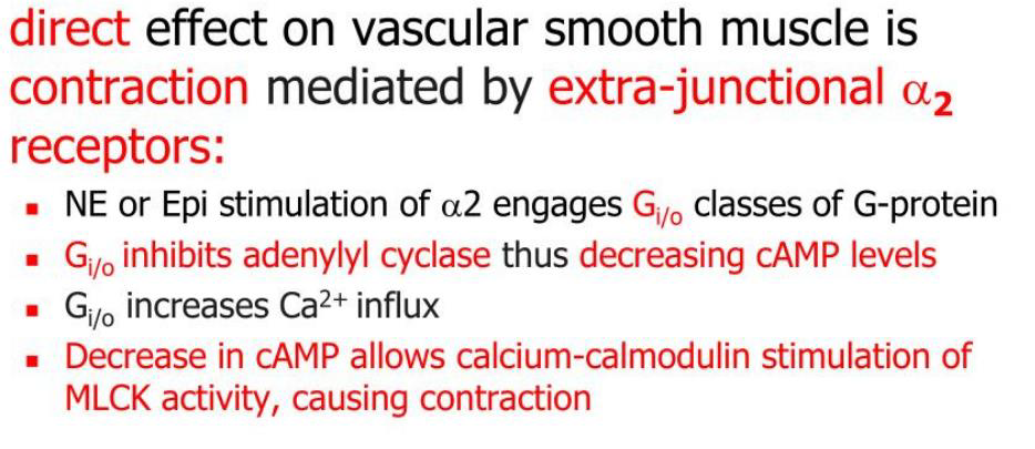
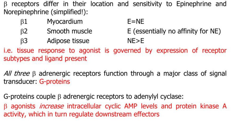
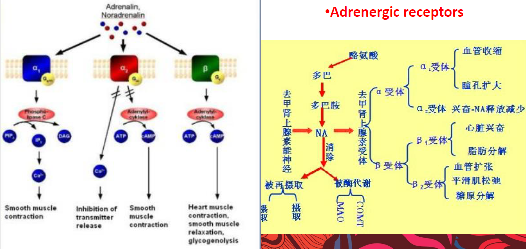
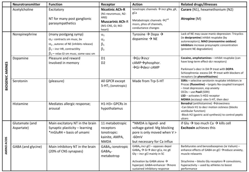
9.2 Sensory Functions of the Nervous System
9.2.1 General Principles
A sensory system is a part of the nervous system that consists of sensory receptors that receive stimuli from the external or internal environment, the neural pathways that conduct information from the receptors to the brain or spinal cord, and those parts of the brain that deal primarily with processing the information. 感觉系统是神经系统的一部分,由从外部或内部环境接收刺激的感觉受体、将信息从受体传导到大脑或脊髓的神经通路以及主要处理信息的大脑部分组成。
Sensory information: no matter if the information reaches consciousness.
Sensation: The information does reach consciousness.
Perception: A person’s awareness of the sensation (and, typically, understanding of its meaning).
Sensations and perceptions occur after the CNS modifies or processes sensory information.
Sensory reception: concept and the MOA
感官信息:无论信息是否到达意识。 感觉:信息确实到达了意识。 感知:一个人对感觉的认识(通常是对其含义的理解)。 感觉和知觉发生在中枢神经系统修改或处理感觉信息之后。 感官接收:概念和 MOA
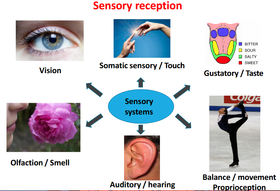
MOA:
- Maintains homeostasis
- Detects & reacts to changes in environment
- Protects body from noxious (damaging) stimuli
维持体内平衡 检测环境变化并做出反应 保护身体免受有害(破坏性)刺激
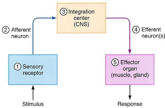
Sensory receptor:
- Structure & classification
- Activation
- Pathways to the brain & central processing
- Pain
9.2.2 Sensory Systems and Receptors
Sensory receptors at the peripheral ends of afferent neurons change this information into graded potentials that can initiate action potentials, which travel into the central nervous system. The receptors are either specialized endings of the primary afferent neurons themselves or separate receptor cells (some of which are actually specialized neurons) that signal the primary afferent neurons by releasing neurotransmitters. 传入神经元外周末端的感觉受体将这些信息转变为分级电位,可以启动动作电位,然后进入中枢神经系统。 这些受体要么是初级传入神经元本身的特殊末端,要么是单独的受体细胞(其中一些实际上是专门的神经元),它们通过释放神经递质向初级传入神经元发出信号。
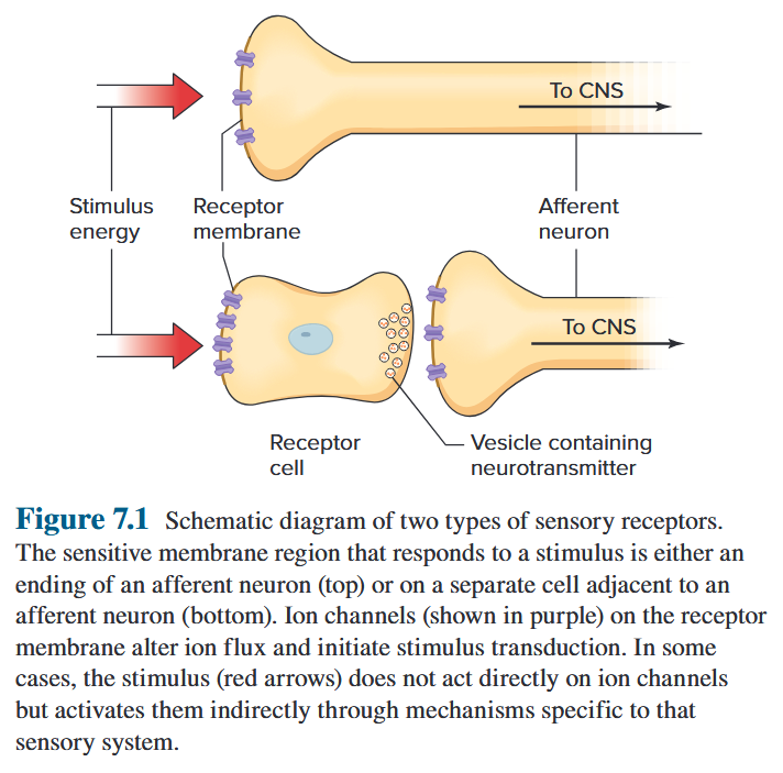
- Sensory Receptor(s) as specialized ending of a peripheral sensory neuron 感觉感受器作为周围感觉神经元的特殊末端

- separate receptor cells that signal the primary afferent neurons by releasing neurotransmitters 通过释放神经递质分离向初级传入神经元发出信号的受体细胞
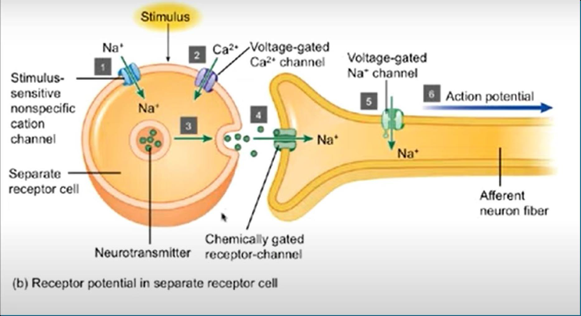
Classification
-
By position:
-
Cutaneous receptors – touch, pressure, temperature, pain
-
Proprioceptors – body position
-
Special senses – sight, hearing, taste, smell
皮肤感受器 – 触觉、压力、温度、疼痛本体感受器 – 身体位置特殊感觉 – 视觉、听觉、味觉、嗅觉
-
-
By the type of stimuli
-
Mechanoreceptors respond to mechanical stimuli
-
Thermoreceptors and photoreceptors
-
Chemoreceptors respond to the binding of particular chemicals to the receptor membrane
-
Nociceptors are a general category of detectors that sense pain due to actual or potential tissue damage
机械感受器对机械刺激做出反应热感受器和光感受器化学感受器对特定化学物质与受体膜的结合做出反应伤害感受器是一类一般的探测器,可以感知由于实际或潜在的组织损伤而产生的疼痛
-
Special Sense Organs
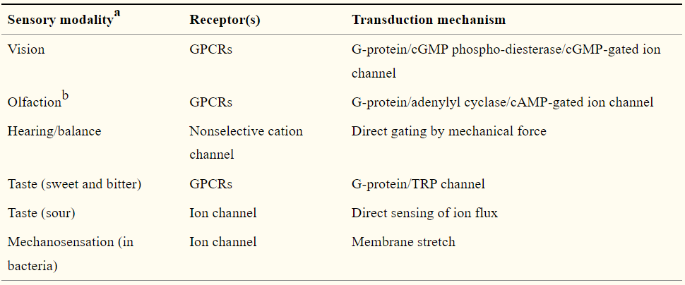
Stimulus
The energy or chemical that impinges upon and activates a sensory receptor is known as a stimulus. 撞击并激活感觉受体的能量或化学物质称为刺激。
The type of stimulus to which a particular receptor responds in normal functioning is known as its adequate stimulus. 特定受体在正常功能中响应的刺激类型被称为其充分刺激。
Sensory threshold is the weakest stimulus that an organism can sense. 感觉阈值是生物体能够感觉到的最弱的刺激。
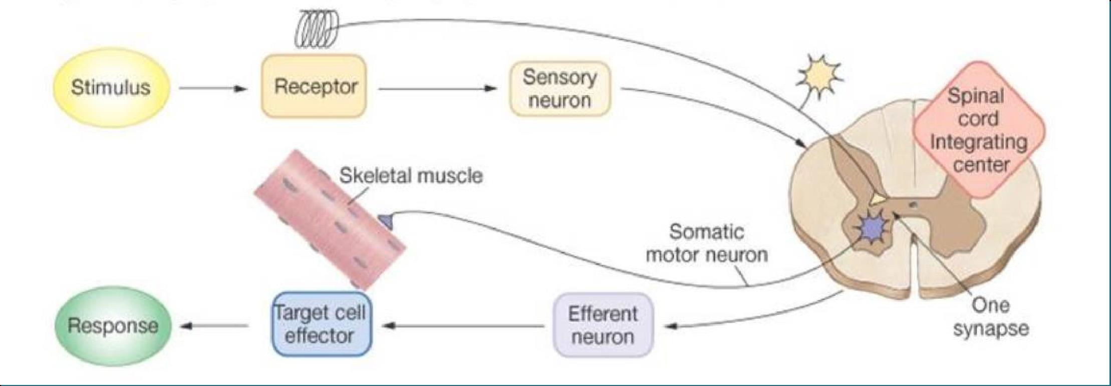
Detectors: they detect the changes in the external of internal environment
Transducers: they transform and form of energy into a receptor potential
9.2.3 Sensory Transduction and Receptor Potential
The process where a chemical stimulus is transformed into an electrical response is known as sensory transduction. 化学刺激转化为电反应的过程称为感觉转导。
The gating of these ion channels allows a change in ion flux across the receptor membrane, which in turn produces a change in the membrane potential. This change is a graded potential called a receptor potential. 这些离子通道的门控允许穿过受体膜的离子通量发生变化,进而产生膜电位的变化。这种变化是一种分级电位,称为受体电位。
Receptor potentials are depolarizing graded potentials that activate sensory neurons. - No refractory period, summation to rapidly successive stimuli possible - Converted in AP at sensory neuron level - 1) if receptor is a part of afferent neuron; voltage gated Na^+^ channels change local current flow. - 2) if receptor is next to afferent neuron; neurotransmitter release depolarized afferent neuron. 受体电位是激活感觉神经元的去极化分级电位。 - 无不应期,可能对快速连续刺激求和 - 在感觉神经元水平上转换为 AP - 1) 如果受体是传入神经元的一部分;电压门控Na^+^通道改变局部电流。 - 2)如果受体靠近传入神经元;神经递质释放去极化传入神经元。
The magnitude of the receptor potential determines the frequency of the action potentials, it does not determine the amplitude of those action potentials. 受体电位的大小决定了动作电位的频率,但并不决定这些动作电位的幅度。
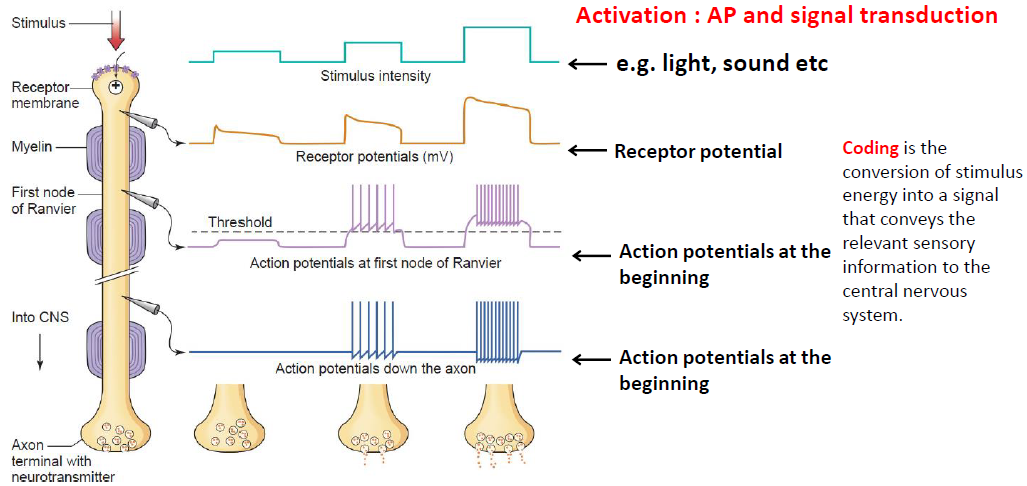
Response to a stimulus can be altered in 3 ways:
- Receptor adaptation: refer toa decrease in receptor sensitivity, which results in a decrease in action potential frequency in an afferent neuron despite the continuous presence of a stimulus. 受体适应:指受体敏感性降低,导致传入神经元动作电位频率降低,尽管刺激持续存在。
- Neuron Habituation: Neurons will reduce their response to repeated stimuli by depressing synaptic activity. 神经元习惯:神经元会通过抑制突触活动来减少对重复刺激的反应。
- Neuron Sensitivity: Neurons will increase their responsiveness to stimuli following strong or noxious stimulus. 神经元敏感性:神经元在强烈或有害的刺激后会增加对刺激的反应性。
Rapidly adapting receptors generate a receptor potential and action potentials at the onset of a stimulus but very quickly cease responding. (AKA: Rate receptor or phasic receptor) Sample: Cutaneous receptors (touch receptor in skin) 快速适应的受体在刺激开始时产生受体电位和动作电位,但很快就会停止响应。又名:速率受体或相位受体,样本:皮肤受体(皮肤中的触摸受体)
Slowly adapting receptors maintain a persistent or slowly decaying receptor potential during a constant stimulus, initiating action potentials in afferent neurons for the duration of the stimulus. (AKA: Tonic receptor) 缓慢适应的受体在持续刺激期间保持持续或缓慢衰减的受体电位,在刺激期间启动传入神经元的动作电位。又名:强直受体。
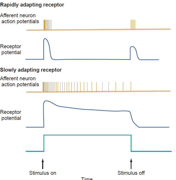
9.2.4 Primary Sensory Coding
A single afferent neuron with all its receptor endings makes up a sensory unit. 单个传入神经元及其所有受体末端构成一个感觉单元。
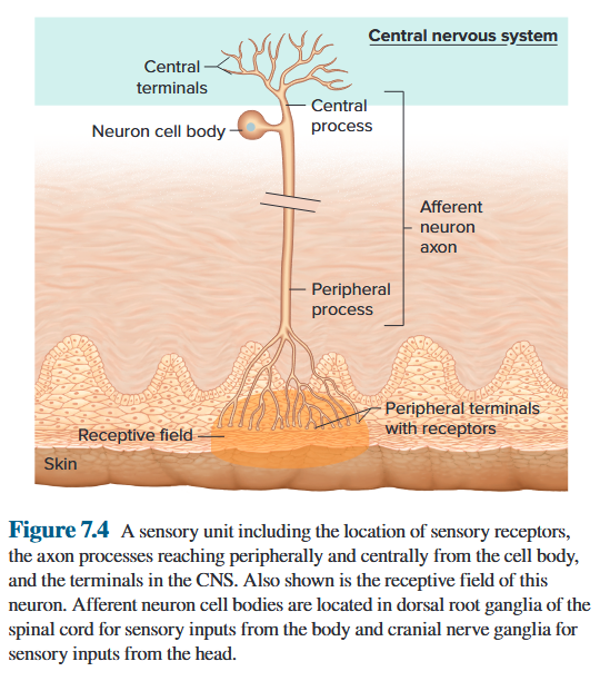
Stimulus Type
Another term for stimulus type (heat, cold, sound, or pressure, for example) is stimulus modality. The type of sensory receptor a stimulus activates plays the primary role in coding the stimulus modality. 刺激类型(例如热、冷、声音或压力)的另一个术语是刺激形态。刺激激活的感觉受体类型在编码刺激方式中起主要作用。
Stimulus Intensity
As the strength of a local stimulus increases, receptors on adjacent branches of an afferent neuron are activated, resulting in a summation of their local currents. Figure 7.5 shows an experiment in which increased stimulus intensity to the receptors of a sensory unit is reflected in increased action potential frequency in its afferent neuron. 随着局部刺激强度的增加,传入神经元相邻分支上的受体被激活,从而产生其局部电流的总和。图 7.5 显示了一个实验,其中感觉单元受体刺激强度的增加反映在其传入神经元动作电位频率的增加。
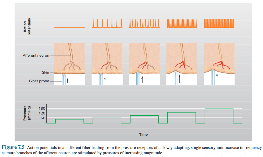
Stimulus Location
Stimulus location is coded by the site of a stimulated receptor, as well as by the fact that action potentials from each receptor travel along unique pathways to a specific region of the CNS associated only with that particular modality and body location. 刺激位置由受刺激受体的位点以及来自每个受体的动作电位沿着独特的路径行进到仅与特定模式和身体位置相关的中枢神经系统特定区域的事实进行编码。
The precision, or acuity, with which we can locate and discern one stimulus from an adjacent one depends upon the amount of convergence of neuronal input in the specific ascending pathways. The greater the convergence, the less the acuity. 我们定位和辨别一种刺激与相邻刺激的精确度或敏锐度取决于特定上行通路中神经元输入的收敛量。收敛性越大,锐度越低。
Locating sensations from internal organs is less precise than from the skin because there are fewer afferent neurons in the internal organs and each has a larger receptive field. 定位内脏器官的感觉不如皮肤精确,因为内脏器官中的传入神经元较少,并且每个神经元都有较大的感受野。
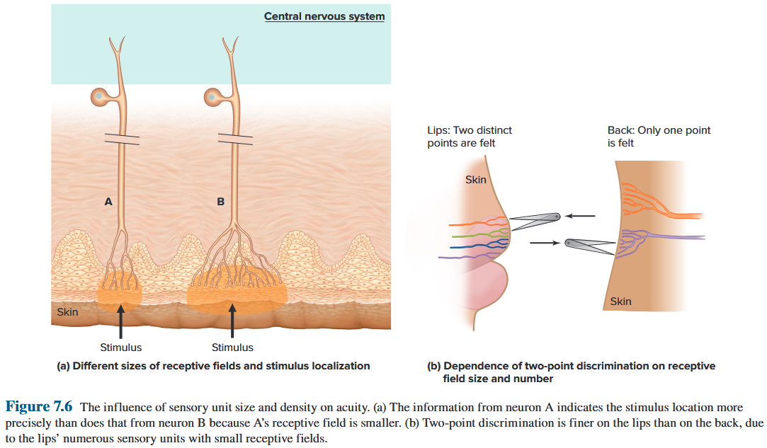
Importance of Receptive Field Overlap
A stimulus activates more receptors and generates more action potentials in its associated afferent neuron if it occurs at the center of the receptive field. A high frequency of impulses in the single afferent nerve fibre of Figure 7.7 could mean either that a moderately intense stimulus was applied to the center at point A or that a stronger stimulus was applied near the periphery at point B. 如果刺激发生在感受野的中心,则会激活更多的受体并在其相关的传入神经元中产生更多的动作电位。 图 7.7 的单个传入神经纤维中的高频脉冲可能意味着在 A 点的中心施加了中等强度的刺激,或者在 B 点的外围附近施加了更强的刺激。
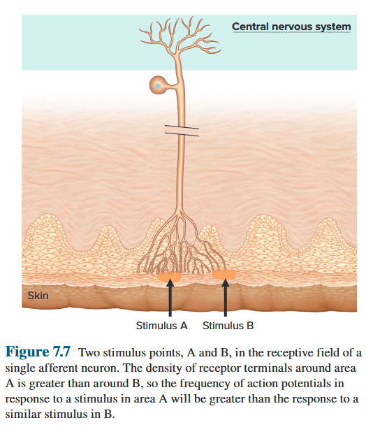
Neurons A and C, stimulated near the edges of their receptive fields where the receptor density is low, fire action potentials less frequently than does neuron B, stimulated at the center of its receptive field. A high action potential frequency in neuron B occurring simultaneously with lower frequencies in A and C provides the brain with a more accurate localization of the stimulus near the center of neuron B’s receptive field. 神经元 A 和 C 在其感受野边缘附近受到刺激,那里的受体密度较低,其激发动作电位的频率低于在感受野中心受到刺激的神经元 B。神经元 B 中的高动作电位频率与 A 和 C 中的较低频率同时发生,为大脑提供了神经元 B 感受野中心附近刺激的更准确定位。
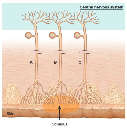
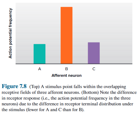
Receptors in skin
Touch-Pressure receptors:
- Merkel’s disk
- Ruffini’s ending
- Messner’s corpuscle
- Pacinian corpuscle (Sensitive to pressure, vibration)
Pain receptor:
- Free nerve ending (Sensitive to pain, temperature)
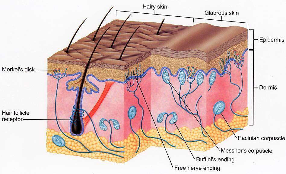
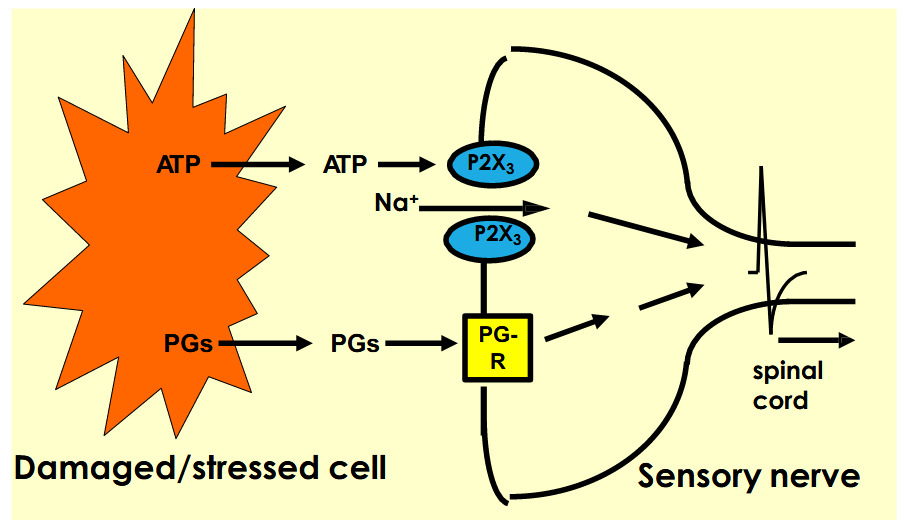
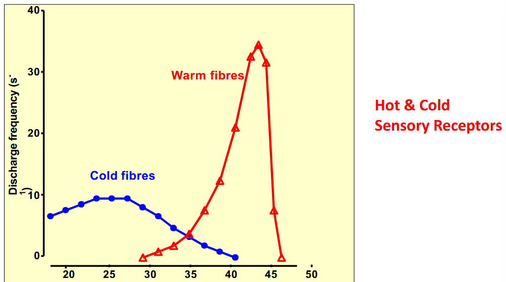
9.2.5 Ascending Neural Pathways in Sensory Systems
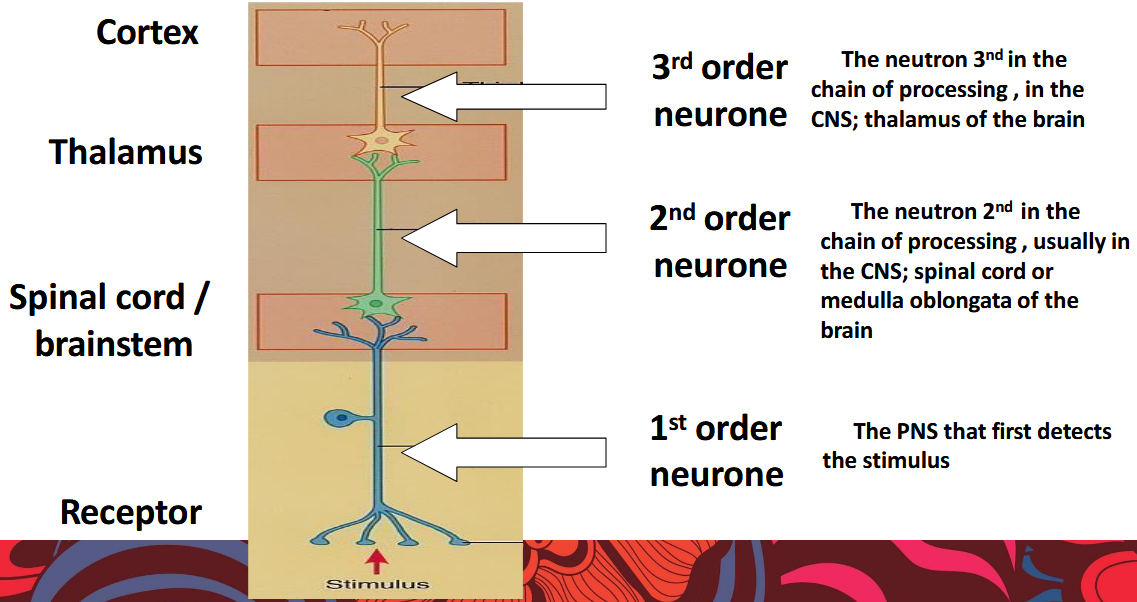
Afferent sensory pathways are generally formed by chains of three or more neurons connected by synapses. 传入感觉通路通常由通过突触连接的三个或更多神经元链形成。
Some pathways terminate in parts of the cerebral cortex responsible for conscious recognition of the incoming information; others carry information not consciously perceived. Sensory pathways are also called ascending pathways because they project “up” to the brain. 有些通路终止于负责有意识识别传入信息的大脑皮层部分;其他人则携带着无意识感知的信息。感觉通路也称为上升通路,因为它们“向上”投射到大脑。
The ascending pathways in the spinal cord and brain that carry information about single types of stimuli are known as the specific ascending pathways. 脊髓和大脑中携带有关单一类型刺激的信息的上行通路被称为特定上行通路。
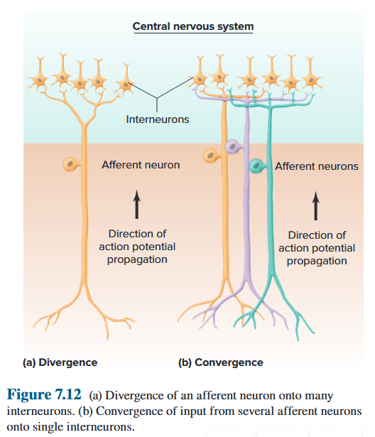
Specific ascending pathways from the taste buds pass to the gustatory cortex adjacent to the region of the somatosensory cortex where information from the face is processed. 从味蕾出发的特定上升路径传递到与处理来自面部信息的体感皮层区域相邻的味觉皮层。
The pathways serving olfaction project to portions of the limbic system and the olfactory cortex , which is located on the undersurface of the frontal and temporal lobe. 嗅觉通路投射到边缘系统和嗅觉皮层的部分,嗅觉皮层位于额叶和颞叶的下表面。
In contrast to the specific ascending pathways, neurons in the nonspecific ascending pathways are activated by sensory units of several different types and therefore signal general information. 与特定的上升通路相反,非特定的上升通路中的神经元被几种不同类型的感觉单元激活,因此发出一般信息的信号。
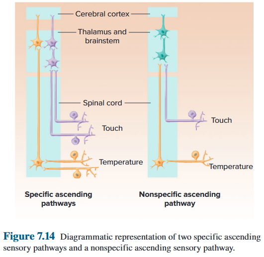
Law of specific nerve energies 特定神经能量定律
Each sensory receptor organ and its nerve fibre receive and transmit just one modality of sensation, regardless of how it is stimulated; moreover, each type of sensation depends not on any special character of the different sensory nerves, but instead on the part of the brain in which their impulses terminate. 每个感觉受体器官及其神经纤维仅接收和传输一种感觉方式,无论它是如何受到刺激的;此外,每种类型的感觉并不取决于不同感觉神经的任何特殊特征,而是取决于它们的冲动终止的大脑部分。
Lateral inhibition 侧向抑制
The phenomenon of lateral inhibition is an important mechanism enabling the localization of a stimulus site for some sensory systems. 侧抑制现象是实现某些感觉系统刺激部位定位的重要机制。
In lateral inhibition, information from afferent neurons whose receptors are at the edge of a stimulus is strongly inhibited compared to information from the stimulus’s center. 在侧抑制中,与来自刺激中心的信息相比,来自其受体位于刺激边缘的传入神经元的信息受到强烈抑制。
Lateral inhibition is utilized to the greatest degree in the pathways providing the most accurate localization. 侧向抑制在提供最准确定位的通路中得到最大程度的利用。
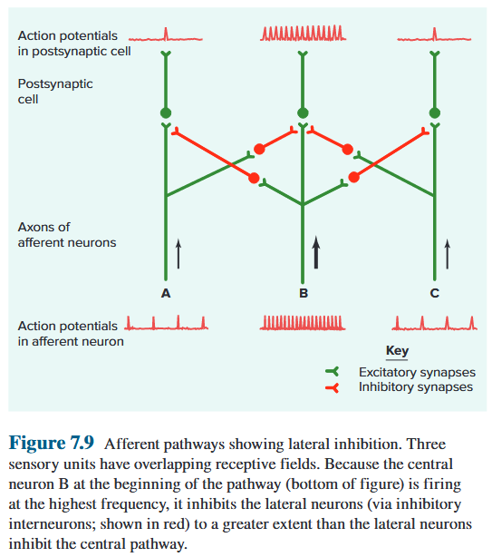
Central Control of Afferent Information 传入信息的中央控制
The inhibitory controls may be exerted directly by synapses on the axon terminals of the primary afferent neurons (an example of presynaptic inhibition) or indirectly via interneurons that affect other neurons in the sensory pathways. 抑制控制可以通过初级传入神经元轴突末端的突触直接施加(突触前抑制的一个例子),也可以通过影响感觉通路中其他神经元的中间神经元间接施加。
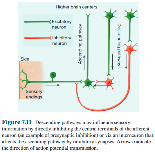
The specific ascending pathways that transmit information from somatic receptors project to the somatosensory cortex. 将信息从体细胞受体传递到体感皮层的特定上升路径。
- Somatic receptors are those carrying information from the skin, skeletal muscle, bones, tendons, and joints. 体细胞受体是那些携带来自皮肤、骨骼肌、骨骼、肌腱和关节信息的受体。
- The somatosensory cortex is a strip of cortex that lies in the parietal lobe of the brain just posterior to the central sulcus, which separates the parietal and frontal lobes. 体感皮层是位于大脑顶叶中央沟后方的一条皮层,中央沟将顶叶和额叶分开。
- The specific ascending pathways from the eyes connect to a different primary cortical receiving area, the visual cortex, which is in the occipital lobe. 从眼睛开始的特定上升路径连接到不同的主要皮质接收区域,即位于枕叶的视觉皮层。
- The specific ascending pathways from the ears go to the auditory cortex, which is in the temporal lobe. 从耳朵到位于颞叶的听觉皮层的特定上升路径。
- The cortical association areas are not considered part of the sensory pathways, but they play a role in the progressively more complex analysis of incoming information. 皮质关联区域不被认为是感觉通路的一部分,但它们在对传入信息进行逐渐复杂的分析中发挥着作用。
- The region of association cortex closest to the primary sensory cortical area processes the information in fairly simple ways and serves basic sensory-related functions. 最接近初级感觉皮层区域的联合皮层区域以相当简单的方式处理信息并提供基本的感觉相关功能。
- Axons from neurons of the parietal and temporal lobes go to association areas in the frontal lobes and other parts of the limbic system. 顶叶和颞叶神经元的轴突进入额叶和边缘系统其他部分的关联区域。
- Further perceptual processing involves not only arousal, attention, learning, memory, language, and emotions but also comparison of the information presented via one type of sensation with that presented through another. 进一步的感知处理不仅涉及唤醒、注意力、学习、记忆、语言和情感,还涉及通过一种感觉呈现的信息与通过另一种感觉呈现的信息的比较。
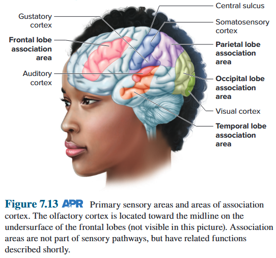
9.2.6 Association Cortex and Perceptual Processing 关联皮层和感知处理
Neural pathways of the somatic and visceral sensory system 躯体和内脏感觉系统的神经通路
In the somatosensory cortex, the endings of the axons of the specific somatic pathways are grouped according to the peripheral location of the receptors that give input to the pathways. 在体感皮层中,特定体细胞通路的轴突末端根据向通路提供输入的受体的外周位置进行分组。
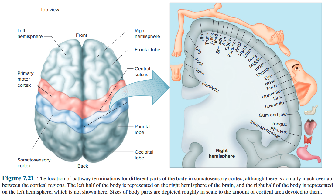
Factors That Affect Perception 影响感知的因素
- Sensory receptor mechanisms (e.g., adaptation) and processing of the information along afferent pathways can influence afferent information. 感觉受体机制(例如适应)和沿着传入途径的信息处理可以影响传入信息。
- Factors such as emotions, personality, experience, and social background can influence perceptions. 情绪、个性、经历和社会背景等因素都会影响感知。
- Not all information entering the central nervous system gives rise to conscious sensation. 并非所有进入中枢神经系统的信息都会引起有意识的感觉。
- We lack suitable receptors for many types of potential stimuli. 我们缺乏针对多种潜在刺激的合适受体。
- Damaged neural networks may give faulty perceptions as in the phenomenon known as phantom limb. 受损的神经网络可能会产生错误的感知,如幻肢现象。
- Some drugs alter perceptions. 有些药物会改变认知。
- Various types of mental illness can alter perceptions of the world, like the hallucinations that can occur in the disease schizophrenia. 各种类型的精神疾病可以改变对世界的看法,例如精神分裂症中可能出现的幻觉。
9.2.7 Specific Sensory Systems
9.2.7.1 Somatic and Visceral Sensations 躯体和内脏感觉
Sensation from the skin, skeletal muscles, bones, tendons, and joints— somatic sensation — is initiated by a variety of sensory receptors collectively called somatic receptors. 皮肤、骨骼肌、骨骼、肌腱和关节的感觉(躯体感觉)是由各种感觉受体(统称为躯体受体)发起的。
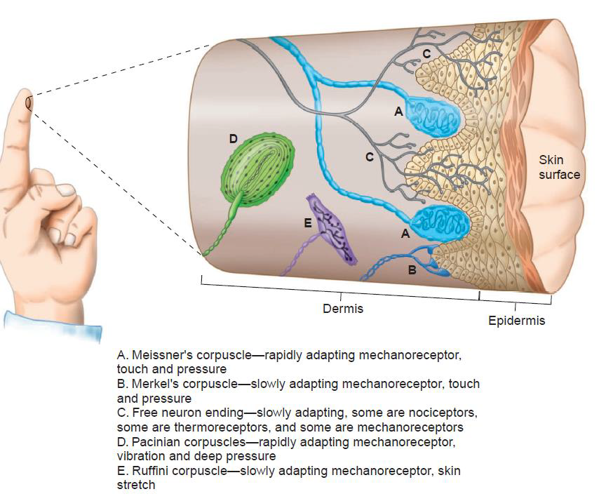
Activation of somatic receptors gives rise to the sensations of touch, pressure, awareness of the position of the body parts and their movement, temperature, and pain. 体细胞感受器的激活会产生触觉、压力、身体部位的位置及其运动、温度和疼痛的感觉。
The receptors for visceral sensations, which arise in certain organs of the thoracic and abdominal cavities, are the same types as the receptors that give rise to somatic sensations. 胸腔和腹腔某些器官中产生的内脏感觉受体与引起躯体感觉的受体类型相同。
Touch and Pressure
Stimulation of different types of mechanoreceptors in the skin leads to a wide range of touch and pressure experiences—hair bending, deep pressure, vibrations, and superficial touch. 刺激皮肤中不同类型的机械感受器会导致各种触摸和压力体验——头发弯曲、深层压力、振动和浅表触摸。
Skin mechanoreceptors adapt at different rates. 皮肤机械感受器以不同的速率适应。
Activation of rapidly adapting receptors gives rise to the sensations of touch, movement, and vibration. 快速适应受体的激活引起触摸、运动和振动的感觉。
Slowly adapting receptors give rise to the sensation of pressure. 慢慢适应的感受器会产生压力感。
Proprioception
Proprioception: the senses of posture and movement are complex. (Temperature; Pain) 本体感觉:姿势和运动的感觉很复杂。 (温度;疼痛)
The major receptors responsible for these senses are the muscle-spindle stretch receptors and Golgi tendon organs. These mechanoreceptors occur in skeletal muscles and the fibrous tendons that connect them to bone. 负责这些感觉的主要受体是肌梭牵张受体和高尔基腱器官。这些机械感受器存在于骨骼肌和将骨骼肌连接到骨骼的纤维肌腱中。
Temperature
The actual temperature sensors are ion channels in the plasma membranes of the axon terminals that belong to a family of proteins called transient receptor potential (TRP) proteins. 实际的温度传感器是轴突末端质膜中的离子通道,属于瞬时受体电位 (TRP) 蛋白家族。
Different channels have overlapping temperature ranges, which is somewhat analogous to the overlapping receptive fields of tactile receptors. 不同的通道具有重叠的温度范围,这有点类似于触觉感受器的重叠感受野。
Pain and Itch
Most stimuli that cause, or could potentially cause, tissue damage elicit a sensation of pain. Receptors for such stimuli are known as nociceptors. Nociceptors, like thermoreceptors, are free axon terminals of small-diameter afferent neurons with little or no myelination. 大多数引起或可能引起组织损伤的刺激都会引起疼痛感。 这种刺激的感受器被称为伤害感受器。伤害感受器与温度感受器一样,是小直径传入神经元的自由轴突末端,很少或没有髓鞘形成。
Noxious stimulus: activate nociceptors → transmit to brain → perception of pain
Incoming nociceptive afferents activate interneurons, it may lead to the phenomenon of referred pain, in which the sensation of pain is experienced at a site other than the injured or diseased tissue. 传入的伤害性传入神经元会激活中间神经元,可能会导致牵涉痛现象,即在受伤或患病组织以外的部位感受到疼痛。
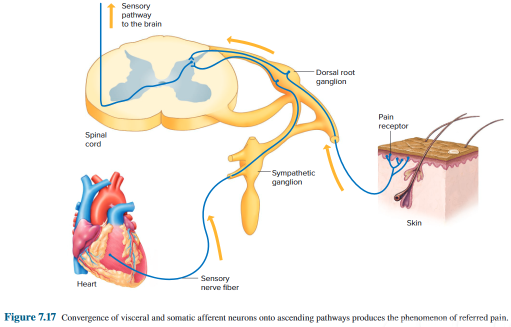
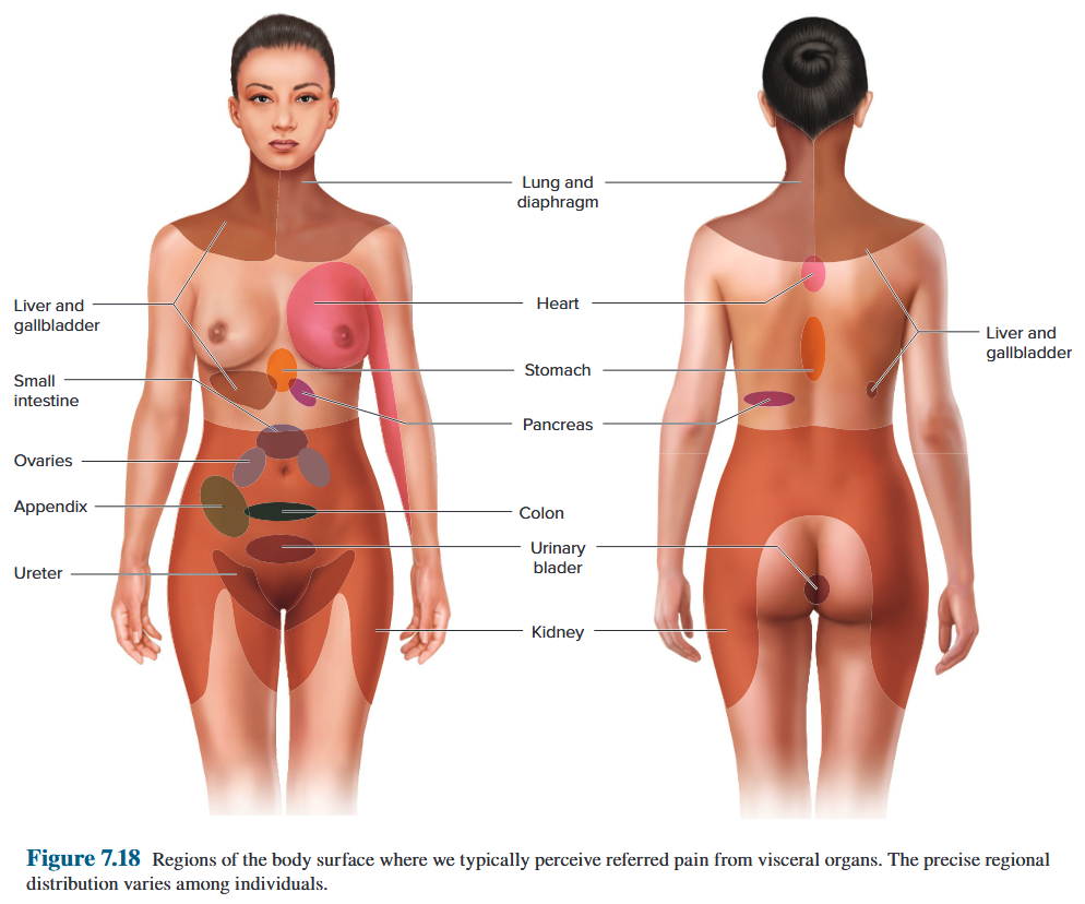
The primary afferents having nociceptor endings synapse on ascending neurons after entering the central nervous system. Glutamate and the neuropeptide substance P are among the neurotransmitters released at these synapses. 进入中枢神经系统后,具有伤害感受器末端突触的初级传入神经元上行。谷氨酸和神经肽 P 物质是这些突触释放的神经递质之一。
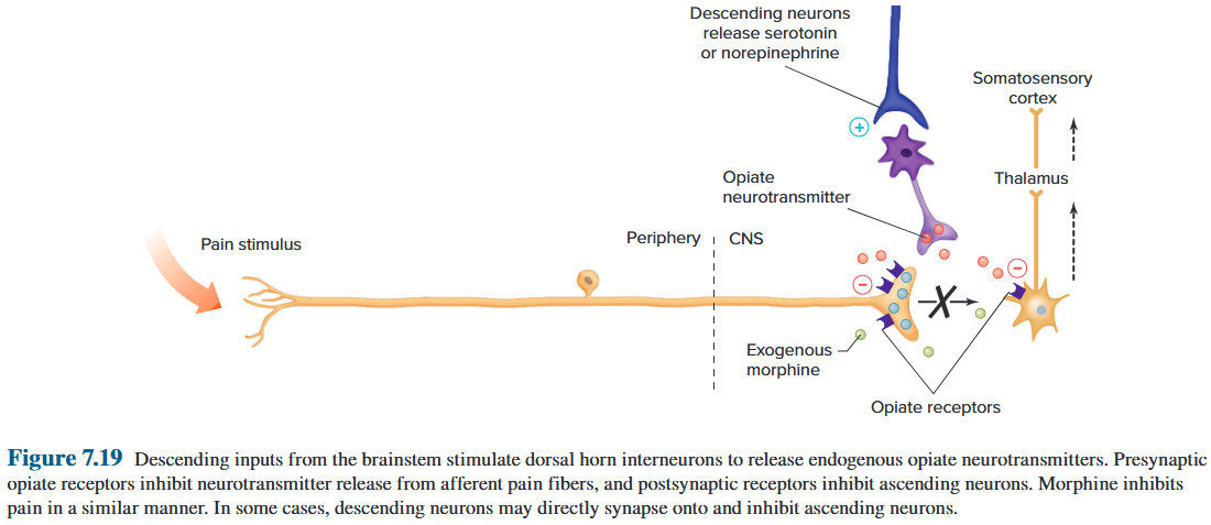
9.2.7.2 Neural Pathways of the Somatic and Visceral Sensory System
Levels of integration: first order
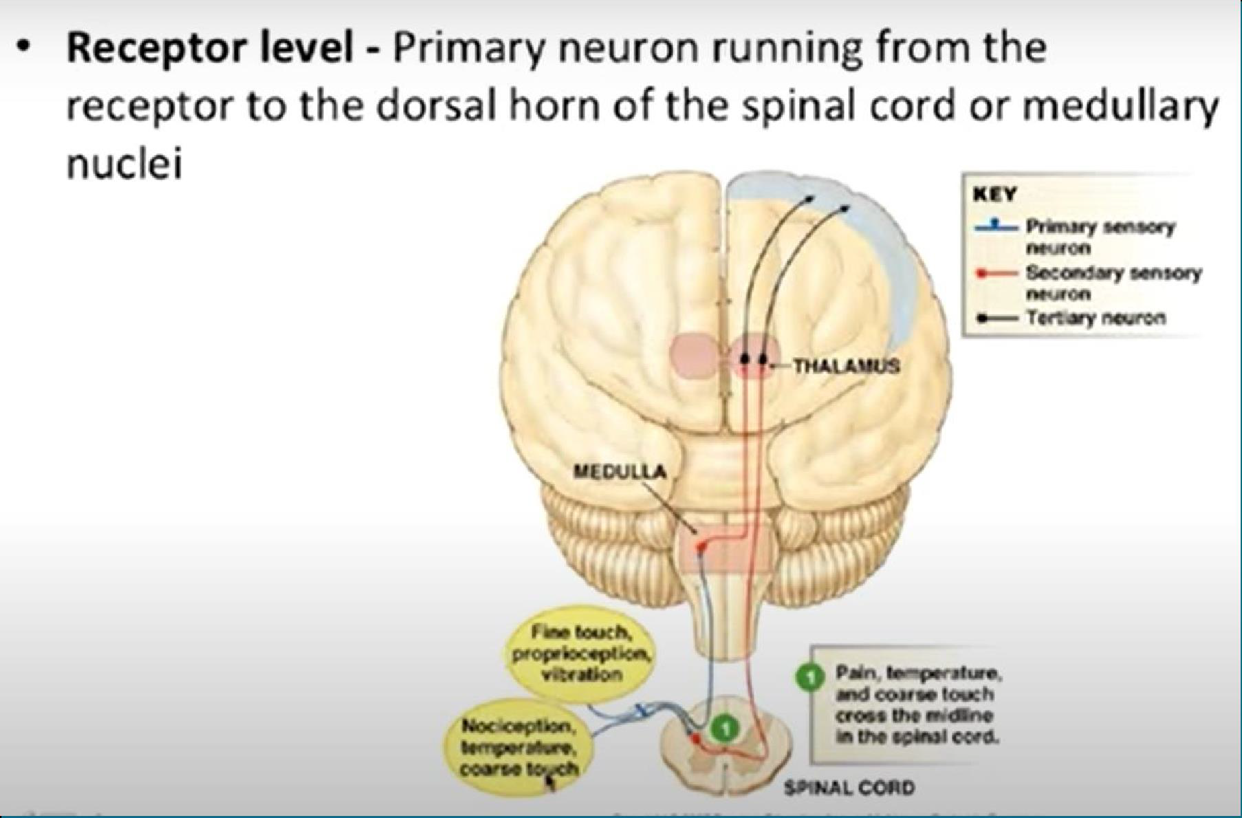
Levels of integration: second order
Levels of integration: third order
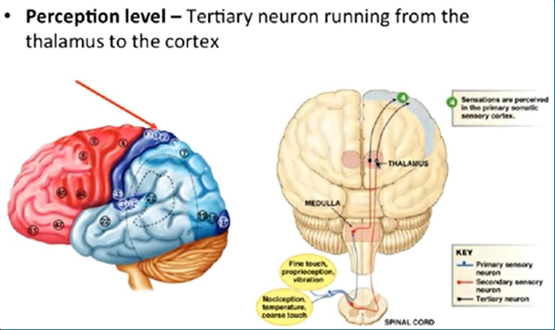
Sensory Pathways to the Brain
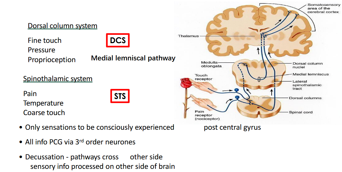
Pain pathway
The 4 Steps of the Pain Pathway:
- Transduction: How a Mechanical Stimulus in Tissues Becomes an Electrical Signal in Nerves 传导:组织中的机械刺激如何变成神经中的电信号
- Transmission: Propagation of the Electrical Signal From Nerves to the Brain 传输:电信号从神经到大脑的传播
- Modulation: Altering or Blocking the Pain Signal as It Travels Through Spinal Cord, Medulla, Pons, and Midbrain to the Cerebral Cortex 调制:改变或阻止疼痛信号通过脊髓、延髓、脑桥和中脑到达大脑皮层
- Perception: How the Brain Interprets the Signal and Produces “Pain” 感知:大脑如何解释信号并产生“疼痛”
Transmission of Pain
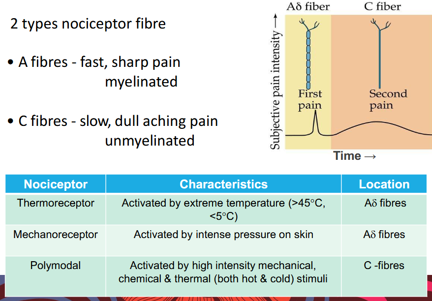
Spinothalamic Pathway (lateral spinothalamic tract) 脊髓丘脑通路(脊髓丘脑外侧束)
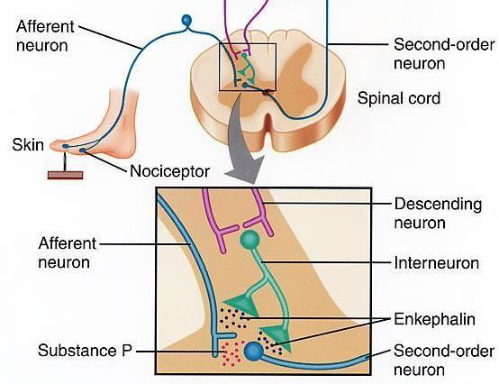
Spinal Cord - Modulation of Pain Carried by Aδ fibres 脊髓 - Aδ 纤维带来的疼痛调节
- Aδ fibres synapse in different area of spinal cord
- No interneurons that release enkephalins → 5-HT ineffective 没有释放脑啡肽的中间神经元 → 5-HT 无效
- No opiate receptors → morphine ineffective 没有阿片受体 → 吗啡无效
- Sharp pain harder to treat 剧烈疼痛更难治疗
Spinal Cord - Modulation of Pain Carried by C fibres 脊髓 - C 纤维带来的疼痛调节
- Substance P
- Enkephalin

CNS - Perception of Pain 中枢神经系统 - 疼痛感知
Modulated by previous experiences, genetics, (sensitisation?), mood, etc.
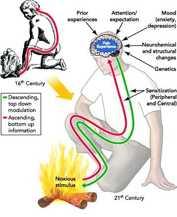
9.3 Control of Body Movement
9.3.1 Motor Control Hierarchy
9.3.1.1 Voluntary and Involuntary Movement
Voluntary movement (随意运动):
- the movement is accompanied by a conscious awareness of what we are doing and why we are doing it 该运动伴随着对我们正在做什么以及为什么这样做的有意识的认识
- our attention is directed toward the action or its purpose 我们的注意力集中在该动作或其目的上,
Involuntary (非随意运动):
- describes actions that do not have these characteristics 描述不具有这些特征的操作
- Unconscious, automatic, and reflex often serve as synonyms for involuntary 无意识、自动和反射常常被视为非自愿的同义词
the motor system, the term reflex has a more precise meaning. 对于运动系统,术语反射具有更精确的含义。
9.3.1.2 Classification of movement
- Reflexes: fixed, automatic movements triggered in response to a specific sensory stimulus 响应特定的感官刺激而触发的固定、自动运动
- usually fairly rapid
- little voluntary control, but can be modulated
- e.g., patellar tendon, eye blink, etc.
- Postural Movements: Combination of reflex and volition used to maintain an upright position with respect to gravity 反射和意志的结合,用于相对于重力保持直立位置
- e.g.,vestibulospinal reflexes 前庭脊髓反射
- Rhythmic Movements (节律运动): also a combination of reflex and volition 也是反射和意志的结合
- initiation and termination is voluntary, but the actual movement is more stereotyped
- e.g., walking, running, chewing, etc.
- Voluntary Movements: purposeful, goal-directed movements 有目的、有目标的运动
- initiated entirely from within the CNS
- performance improves with practice
- reflex and postural movements are often initiated that compensate for the effects of the intended action on other parts of the body 通常会发起反射和姿势运动,以补偿预期动作对身体其他部位的影响
9.3.1.3 Sample: Voluntary movement
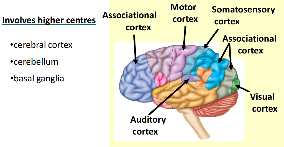
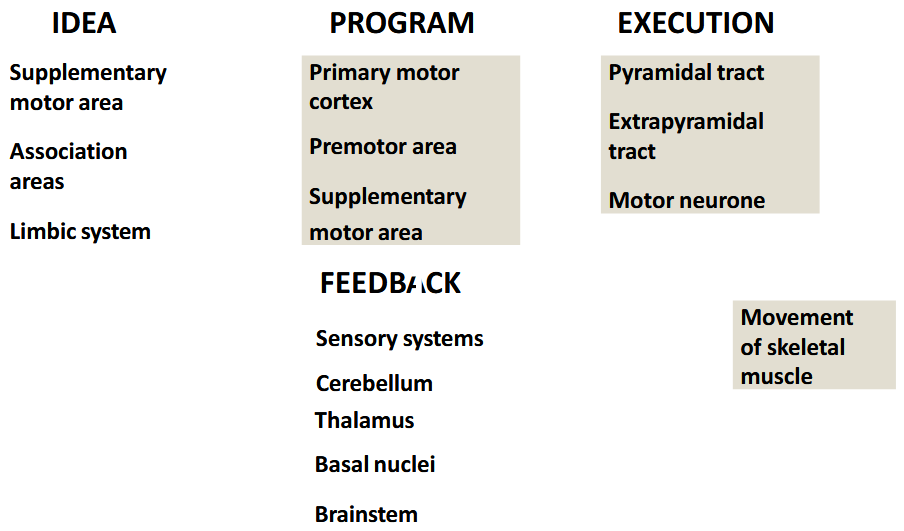
9.3.1.4 Hierarchy 等级制度
Highest level
To begin a consciously planned movement, a general intention such as “pick up sweater” or “write signature” or “answer telephone” is generated at the highest level of the motor control hierarchy. These higher centers include many regions of the brain, including sensorimotor areas and others involved in memory, emotions, and motivation. 为了开始有意识地计划的运动,在运动控制层次结构的最高层产生诸如“拿起毛衣”或“写签名”或“接听电话”之类的一般意图。这些高级中心包括大脑的许多区域,包括感觉运动区域和其他涉及记忆、情绪和动机的区域。
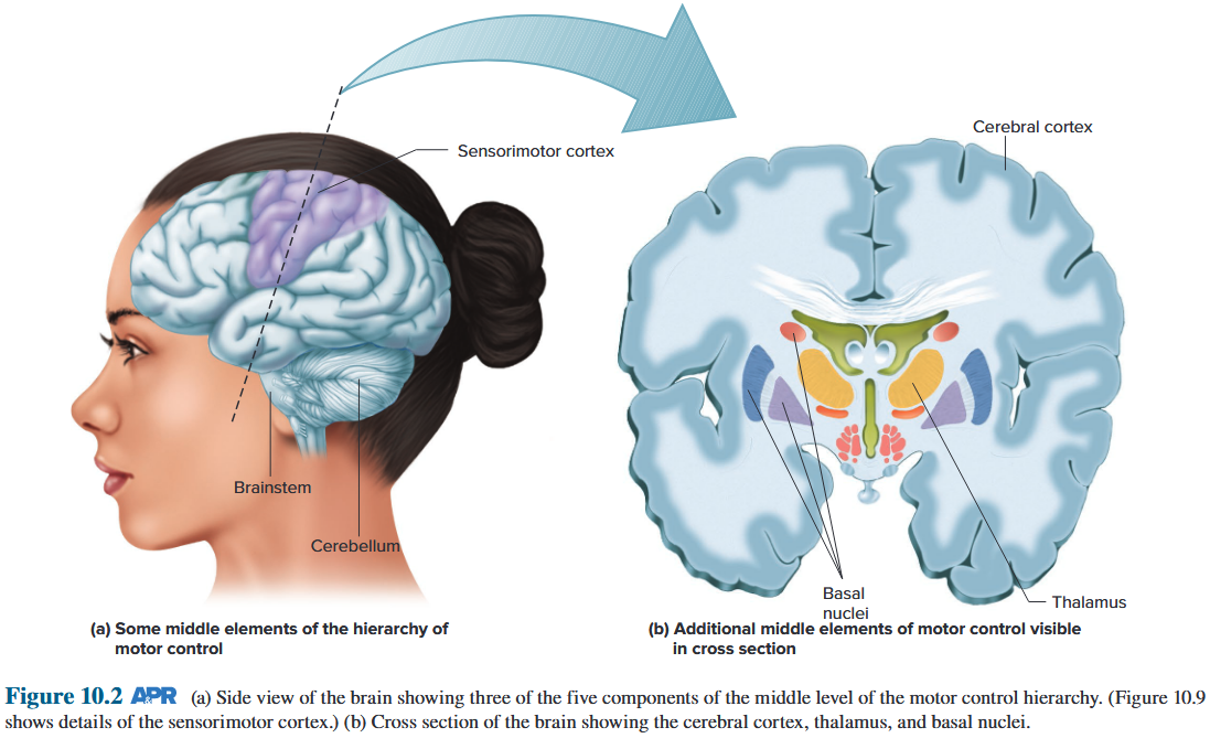
Middle level
Information is relayed from these higher-center “command” neurons to parts of the brain that make up the middle level of the motor control hierarchy 信息从这些较高中心的“命令”神经元传递到构成运动控制层次结构中层的大脑部分
The middle-level structures specify the individual postures and movements needed to carry out the intended action. The middle-level hierarchical structures are located in parts of the cerebral cortex as well as in the cerebellum, subcortical nuclei, and brainstem. Neurons of the middle level of the hierarchy integrate all of this afferent information with the signals from the command neurons to create a motor program - defined as the pattern of neural activity required to properly perform the desired movement. The information determined by the motor program is transmitted via descending pathways to the local level of the motor control hierarchy.
中层结构指定了执行预期动作所需的个人姿势和动作。 中层层次结构位于大脑皮层的部分区域以及小脑、皮层下核团和脑干。 层次结构中层的神经元将所有这些传入信息与来自命令神经元的信号整合起来,以创建一个运动程序——定义为正确执行所需运动所需的神经活动模式。 由运动程序确定的信息通过下降路径传输到运动控制层次结构的本地级别。
Local level
The descending pathways to the local level arise only in the sensorimotor cortex and brainstem. Other brain areas, notably the basal nuclei (also referred to as the basal ganglia), thalamus, and cerebellum, exert their effects on the local level only indirectly via the descending pathways from the cerebral cortex and brainstem. 通往局部水平的下行通路仅出现在感觉运动皮层和脑干中。其他大脑区域,特别是基底核(也称为基底神经节)、丘脑和小脑,仅通过来自大脑皮层和脑干的下行通路间接在局部水平发挥作用。
Afferent information about the position of the body and its parts in space is called proprioception. 有关身体及其部位在空间中的位置的传入信息称为本体感觉。
Reflex circuits acting entirely at the local level are also important in refining ongoing movements. 完全在局部发挥作用的反射回路对于完善正在进行的运动也很重要。
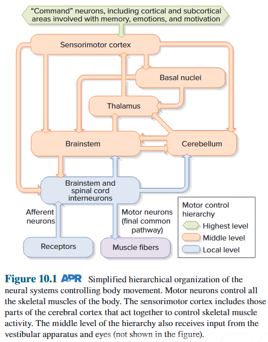
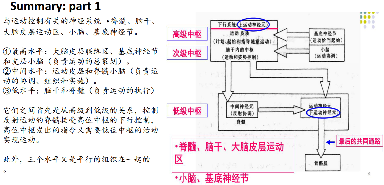
9.3.2 Local Control of Motor Neuron
9.3.2.1 Spinal Shock and Spinal Animal
Spinal shock Defined as a state of transient physiologic (rather than anatomic) reflex depression of cord function below the level of injury, with associated loss of sensorimotor functions is a reversible reduction or loss in sensory and motor function after an acute SCI. 脊髓休克定义为损伤水平以下脊髓功能的短暂生理(而非解剖)反射抑制状态,并伴有感觉运动功能丧失,是急性 SCI 后感觉和运动功能的可逆性降低或丧失。
An immediate loss of reflex function, called areflexia, below the level of injury
- Slow heart rate
- Low blood pressure
- Flaccid paralysis of skeletal muscles
- Loss of somatic sensations
- Urinary bladder dysfunction
心率缓慢 低血压 骨骼肌弛缓性麻痹 躯体感觉丧失 膀胱功能障碍
Spinal shock may begin within an hour after injury and last from several minutes to several months, after which reflex activity gradually returns. 脊髓休克可能在受伤后一小时内开始,持续几分钟到几个月,之后反射活动逐渐恢复。
9.3.2.2 Motor Neurons in Spinal Cord
Motor neurons are located in the central nervous system (CNS), specifically in the motor cortex, brainstem and spinal cord. Motor neurons are also known as efferent neurons, meaning they carry information from the CNS to muscles, and other peripheral systems such as organs and glands. 运动神经元位于中枢神经系统 (CNS),特别是运动皮层、脑干和脊髓。运动神经元也称为传出神经元,这意味着它们将信息从中枢神经系统传递到肌肉以及其他外周系统(例如器官和腺体)。
Motor neurons can be divided into two types of neurons, depending on their function: upper motor neurons and lower motor neurons. These neurons form a variety of controlled and complex circuits throughout the body, controlling both voluntary and involuntary movements. 运动神经元根据其功能可分为两类:上运动神经元和下运动神经元。这些神经元在全身形成各种受控且复杂的电路,控制自主和非自主运动。
The “Lower neurons” in the spinal cord are not only affected by the local circuits in the spinal cord, they are also controlled and coordinated by many “Upper neurons” in the motor areas of the cerebral cortex and the brainstem. 脊髓中的“下层神经元”不仅受脊髓内局部回路的影响,还受到大脑皮层和脑干运动区的许多“上层神经元”的控制和协调。
The final common path: a motoneuron that forms the terminal step of one or more reflex circuits transmitting their stimuli to an effector end organ. 最后的共同路径:运动神经元,形成一个或多个反射回路的最终步骤,将其刺激传递到效应器末端器官。
Lower motor neurons, located in the spinal cord, communicate signals directly to the skeletal muscles, organs, and glands. They receive information from the upper motor neurons, either directly or via interneurons, and stimulate their activity, extending their fibers all the way to their appropriate destinations. 它们直接或通过中间神经元从上运动神经元接收信息,并刺激它们的活动,将它们的纤维一直延伸到适当的目的地。
There are a few subtypes of lower motor neurons:
- somatic neurons
- special visceral efferent neurons
- general visceral motor neurons
Interneurons
Interneurons comprise 90% of spinal cord neurons, and they are of several types. Some are near the motor neuron they synapse upon and thus are called local interneurons. 中间神经元占脊髓神经元的 90%,它们有多种类型。有些靠近它们突触的运动神经元,因此被称为局部中间神经元。 中间神经元很重要
The interneurons are important elements of the local level of the motor control hierarchy, integrating inputs not only from higher centers and peripheral receptors but from other interneurons as well. 中间神经元是运动控制层次局部水平的重要元素,不仅整合来自高级中枢和外周受体的输入,还整合来自其他中间神经元的输入。
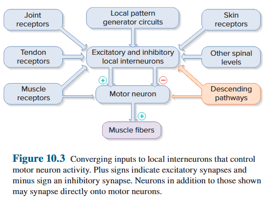
Somatic motor neurons originate in the central nervous system, specifically the brainstem and project their axons to skeletal muscles. Somatic motor neurons can be further divided into alpha, beta, and gamma motor neurons. 体细胞运动神经元起源于中枢神经系统,特别是脑干,并将其轴突投射到骨骼肌。 体细胞运动神经元可进一步分为α、β和γ运动神经元。
Alpha motor neurons are relatively large motor neurons: α运动神经元是相对较大的运动神经元
- nervate skeletal muscle 支配骨骼肌
- cause the muscle contractions that generates movement 引起肌肉收缩产生运动
- Specifically, they innervate extrafusal muscle fibres, which are standard muscle fibres 具体来说,它们支配梭外肌纤维,这是标准的肌纤维
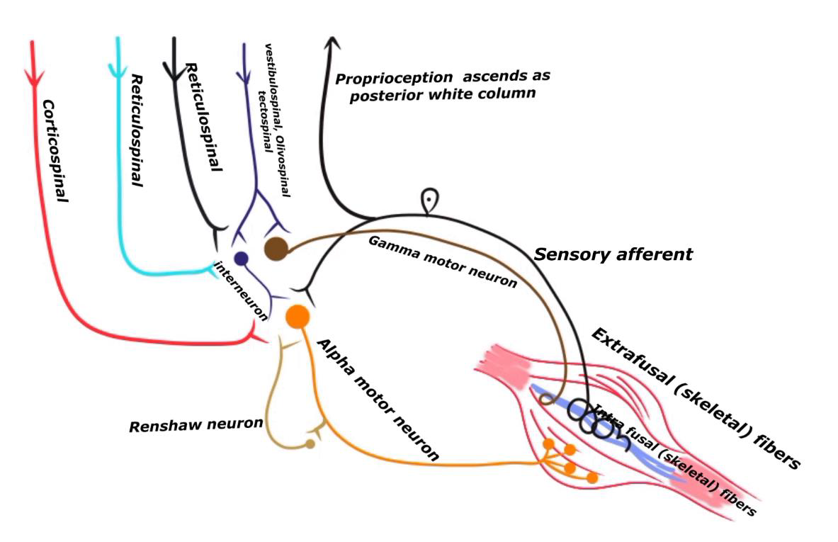
Gamma motor neurons respond to stretch receptors of the skeletal muscle, also known as muscle spindles. Although known as a motor neuron, gamma motor neurons do not cause any motor function directly. Instead, they are thought to be activated alongside the alphas to fine-tune the muscle contraction. 伽玛运动神经元对骨骼肌的拉伸感受器(也称为肌梭)做出反应。 虽然被称为运动神经元,但伽马运动神经元并不直接引起任何运动功能。相反,它们被认为与阿尔法一起被激活以微调肌肉收缩。
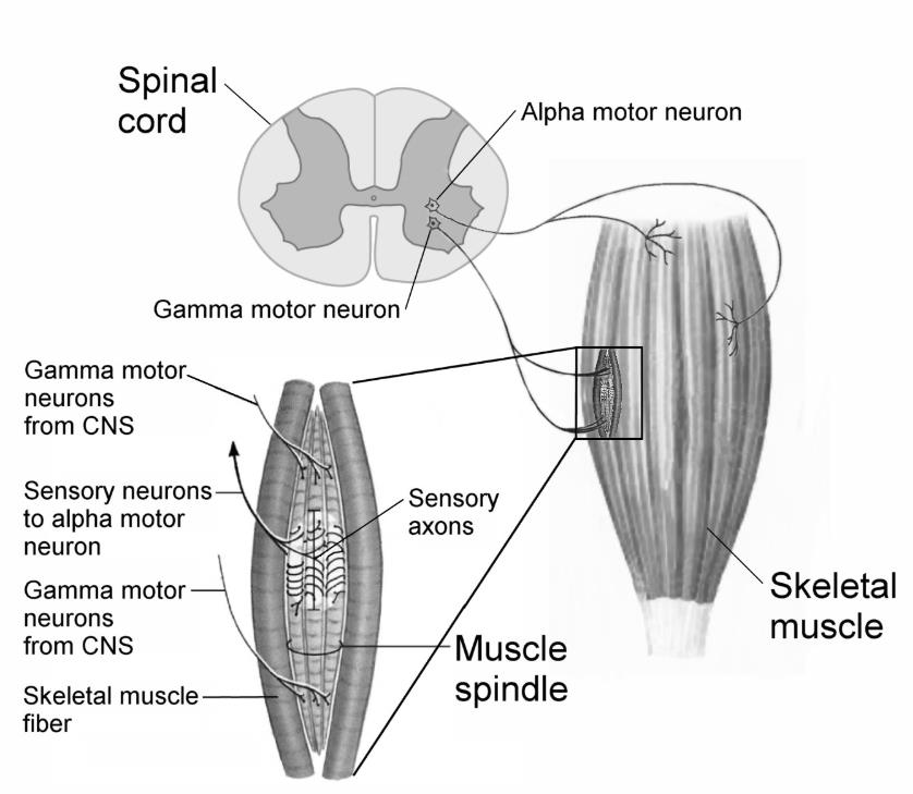
Beta motor neurons are not as well categorized as alpha motor neurons, but are understood to also innervate extrafusal muscle fibres, as well as intrafusal fibres, which serve as specialized sensory organs and are innervated by both motor and sensory fibres. β 运动神经元不像 α 运动神经元那样分类,但被认为也支配梭外肌纤维和梭内纤维,后者作为专门的感觉器官,并受运动纤维和感觉纤维的支配。
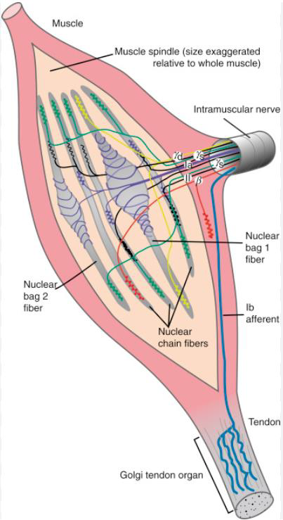
Alpha motor neurons (αMNs) at the neuromuscular junction innervate extrafusal muscle fibres that generate force-producing contractions, whereas gamma motor neurons (γMNs) innervate intrafusal muscle fibres to regulate muscle spindle sensitivity and beta motor neurons (βMNs) innervate both intrafusal and extrafusal muscle fibres. 神经肌肉接头处的α运动神经元(αMN)支配梭外肌纤维,产生产生力的收缩,而γ运动神经元(γMN)支配梭内肌纤维以调节肌梭敏感性,β运动神经元(βMN)支配梭内和梭外肌肉纤维。
9.3.2.3 Local Afferent Input
The afferent fibres carry information from sensory receptors located in three places: 传入纤维携带来自位于三个位置的感觉受体的信息:(1)由运动神经元控制的骨骼肌; (2)附近的其他肌肉,例如具有对抗作用的肌肉;(3)受肌肉活动影响的身体部位的肌腱、关节和皮肤
- in the skeletal muscles controlled by the motor neurons; 在由运动神经元控制的骨骼肌中;
- in other nearby muscles, such as those with antagonistic actions; 其他附近的肌肉,例如具有对抗性动作的肌肉;
- in the tendons, joints, and skin of body parts affected by the action of the muscle. 受肌肉活动影响的身体部位的肌腱、关节和皮肤。
9.3.2.4 Length-Monitoring Systems
These receptors consist of peripheral endings of afferent nerve fibres wrapped around modified muscle fibres, several of which are enclosed in a connective-tissue capsule. The entire apparatus is collectively called a muscle spindle (肌梭).
The modified muscle fibers within the spindle are known as intrafusal fibers (梭内肌纤维). The skeletal muscle fibers that form the bulk of the muscle and generate its force and movement are the extrafusal fibres (梭外肌纤维).
这些受体由包裹着改良肌纤维的传入神经纤维的外周末梢组成,其中一些肌纤维被封闭在结缔组织囊中。整个装置统称为肌梭。 梭内肌纤维被称为梭内肌纤维。构成肌肉主体并产生力量和运动的骨骼肌纤维是梭外肌纤维。
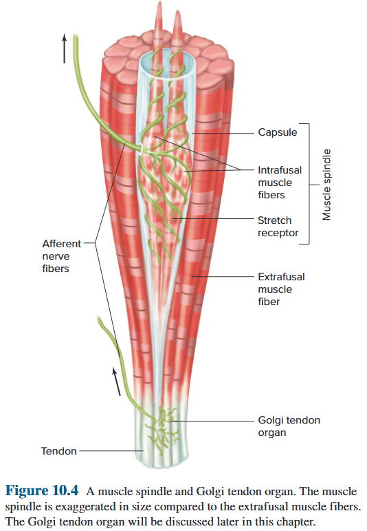
An external force stretching the muscle also pulls on the intrafusal fibers, stretching them and activating their receptor endings. 拉伸肌肉的外力也会拉动梭内纤维,拉伸它们并激活它们的受体末端。
The more or the faster the muscle is stretched, the greater the rate of receptor firing. In contrast, if action potentials along motor neurons cause contraction of the extrafusal fibers, the resultant shortening of the muscle removes tension on the spindle and slows the rate of firing in the stretch receptor. 肌肉拉伸得越多或越快,受体发射的速率就越大。相反,如果沿着运动神经元的动作电位引起梭外纤维收缩,由此产生的肌肉缩短会消除纺锤体上的张力并减慢牵张感受器的放电速率。
A mechanism called alpha–gamma coactivation prevents this loss of information. Extrafusal fibres of a muscle are activated by large-diameter alpha motor neurons, and the two ends of intrafusal muscle fibres are activated by smaller-diameter neurons called gamma motor neurons. 一种称为α-γ共激活的机制可以防止这种信息丢失。肌肉的梭外纤维由大直径的α运动神经元激活,梭内肌纤维的两端由称为γ运动神经元的较小直径的神经元激活。
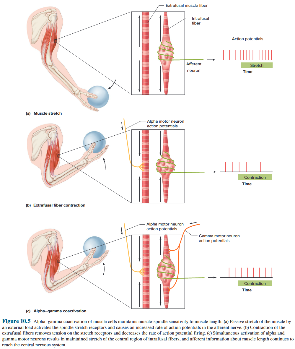
9.2.3.5 Reflex
Path A makes excitatory synapses directly onto motor neurons that return to the muscle that was stretched, thereby completing a reflex arc known as the stretch reflex (牵张反射).
This reflex is probably most familiar in the form of the knee-jerk reflex (膝跳反射), part of a routine medical examination.
Tapping the patellar tendon stretches the extensor muscle, causing (paths A and C) compensatory contraction of this and other extensor muscles, (path B) relaxation of flexor muscles, and (path D) information about muscle length to go to the brain.
The afferent nerve fibres in the stretched muscle synapse directly on the motor neurons to that muscle without any interneurons, this portion of the stretch reflex is called monosynaptic. Stretch reflexes have the only known monosynaptic reflex arcs. All other reflex arcs are polysynaptic.
路径 A 使兴奋性突触直接作用到运动神经元上,运动神经元返回到被拉伸的肌肉,从而完成称为牵张反射的反射弧。 这种反射可能以膝跳反射(膝跳反射)的形式最为人们所熟悉,它是常规医学检查的一部分。 敲击髌腱会拉伸伸肌,导致(路径 A 和 C)该伸肌和其他伸肌的代偿性收缩,(路径 B)屈肌的放松,以及(路径 D)有关肌肉长度的信息进入大脑。 被拉伸的肌肉中的传入神经纤维突触直接作用于运动神经元到该肌肉,没有任何中间神经元,这部分牵张反射称为单突触。牵张反射具有唯一已知的单突触反射弧。所有其他反射弧都是多突触的。
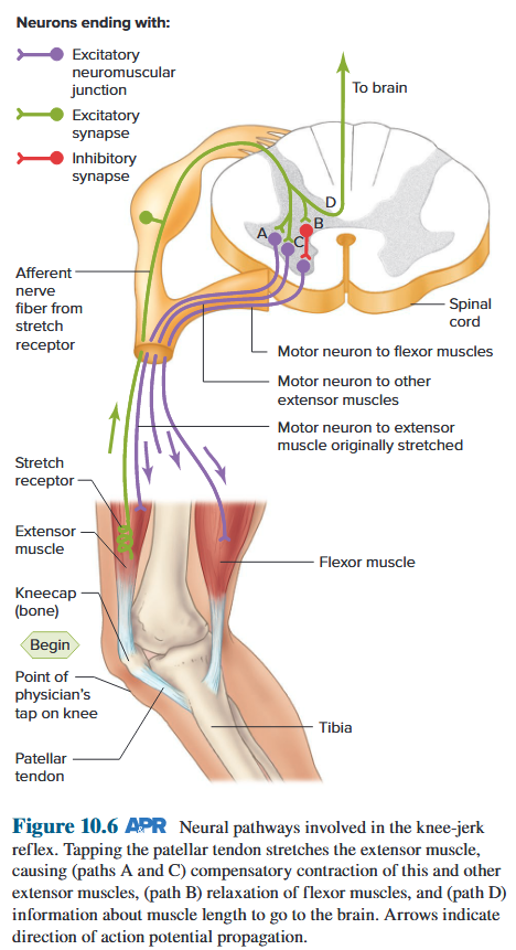
9.2.3.6 Tension-Monitoring Systems
The tension depends on muscle length, the load on the muscles, and the degree of muscle fatigue. Feedback is necessary to inform the motor control systems of the tension actually achieved. 张力取决于肌肉长度、肌肉负荷以及肌肉疲劳程度。反馈对于告知运动控制系统实际达到的张力是必要的。
The receptors employed in this tension-monitoring system are the Golgi tendon organs, which are endings of afferent nerve fibres that wrap around collagen bundles in the tendons near their junction with the muscle. 这种张力监测系统中使用的受体是高尔基肌腱器官,它是传入神经纤维的末端,包裹在肌腱与肌肉连接处附近的胶原蛋白束周围。
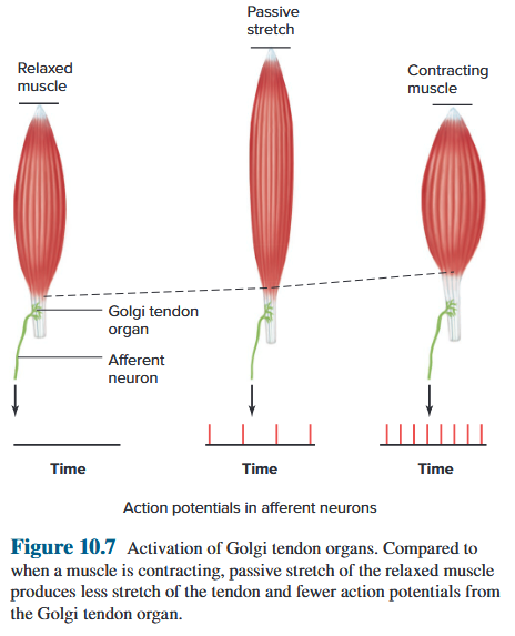
9.2.3.6 Tension-Monitoring Systems
This reciprocal innervation is the opposite of that produced by the muscle-spindle afferents. This difference reflects the different functional roles of the two systems: 这种相互神经支配与肌梭传入神经产生的神经支配相反。 这种差异反映了两个系统不同的功能作用: → 肌梭提供肌肉长度的局部稳态控制 → 高尔基腱器官提供肌肉张力的局部稳态控制。
- The muscle spindle provides local homeostatic control of muscle length
- the Golgi tendon organ provides local homeostatic control of muscle tension.
9.2.3.7 The Withdrawal Reflex
The action moves the affected limb away from the harmful stimulus and is thus known as a withdrawal reflex (屈肌反射).
The same stimulus causes just the opposite response in the contralateral leg (on the opposite side of the body from the stimulus); motor neurons to the extensors are activated while the flexor muscle motor neurons are inhibited.
This crossed-extensor reflex (对侧促肌反射) enables the contralateral leg to support the body’s weight as the injured foot is lifted by flexion.
这一动作使受影响的肢体远离有害刺激,因此被称为屈肌反射。 相同的刺激只会在对侧腿(与刺激相反的身体一侧)引起相反的反应;伸肌运动神经元被激活,而屈肌运动神经元被抑制。 当受伤的脚通过屈曲抬起时,这种交叉伸肌反射(对侧促肌反射)使对侧腿能够支撑身体的重量
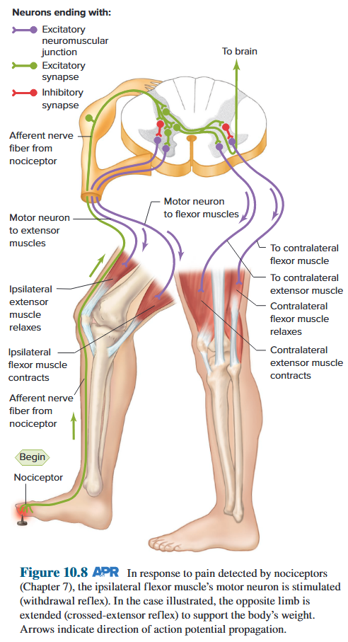
9.3.3 Muscle Tone / Maintenance /walking
9.3.3.1 Muscle Tone
Even when a skeletal muscle is relaxed, there is a slight and uniform resistance when it is stretched by an external force. This resistance is known as muscle tone. Muscle tone is due both to the passive elastic properties of the muscles and joints and to the degree of ongoing alpha motor neuron activity. 即使骨骼肌处于放松状态,当受到外力拉伸时,也会产生轻微且均匀的阻力。这种阻力称为肌张力。 肌肉张力既取决于肌肉和关节的被动弹性特性,也取决于正在进行的α运动神经元活动的程度。
Maintenance of Upright Posture and Balance
people can operate under conditions of unstable equilibrium because complex interacting postural reflexes maintain their balance. 人们可以在不稳定平衡的条件下进行操作,因为复杂的相互作用的姿势反射维持着他们的平衡。
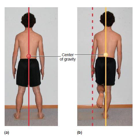
9.3.3.2 Regulation on Muscle Tone by brainstem
Reticular formation: a set of interconnected nuclei that are located throughout the brainstem. 网状结构:位于整个脑干的一组相互连接的核团。
- Somatic motor
- Cardiovascular control
- Pain modulation
- Sleep and consciousness
- Habituation
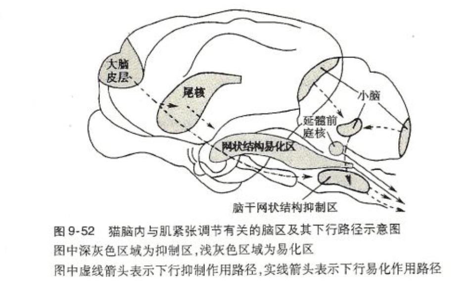
- Feedforward adjustment: Adjust posture in advance to prevent random movements from interfering with body balance. 前馈调整:提前调整姿势,防止随机动作干扰身体平衡。
- Feedback adjustment: When the balance is disturbed, the form is fixed Rapid response to correct accidental disturbances of cerebral cortex and reticular formation involved in feedforward regulation of postural balance The vestibular system plays a decisive role in feedback regulation of postural balance. 反馈调节:平衡受到干扰时,形态固定 快速反应,纠正参与姿势平衡前馈调节的大脑皮层和网状结构的意外干扰 前庭系统在姿势平衡反馈调节中起决定性作用
Abnormal Muscle Tone
Spasticity is a form of hypertonia in which the muscles do not develop increased tone until they are stretched a bit, and after a brief increase in tone, the contraction subsides for a short time.
Rigidity is a form of hypertonia in which the increased muscle contraction is continual and the resistance to passive stretch is constant.
痉挛是张力亢进的一种形式,其中肌肉在稍微拉伸之前不会出现张力增加,在张力短暂增加后,收缩会短暂消退。 强直是肌张力亢进的一种形式,其中肌肉收缩持续增加,被动拉伸的阻力恒定。
9.3.4 The brain motor center and the descending pathways they control
9.3.4.1 Functional anatomy
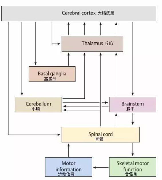
9.3.4.2 Sensorimotor cortex
Sensorimotor cortex is used to include all those parts of the cerebral cortex that act together to control muscle movement.
A large number of neurons that give rise to descending pathways for motor control come from two areas of sensorimotor cortex on the posterior part of the frontal lobe: the primary motor cortex and the premotor area. the supplementary motor cortex, which lies mostly on the surface on the frontal lobe where the cortex folds down between the two hemispheres, the somatosensory cortex, and parts of the parietal-lobe association cortex.
感觉运动皮层包括大脑皮层中共同作用以控制肌肉运动的所有部分。 产生运动控制下行通路的大量神经元来自额叶后部感觉运动皮层的两个区域:初级运动皮层和前运动区域。辅助运动皮层,主要位于额叶表面,皮层在两个半球、体感皮层和顶叶联合皮层之间向下折叠。
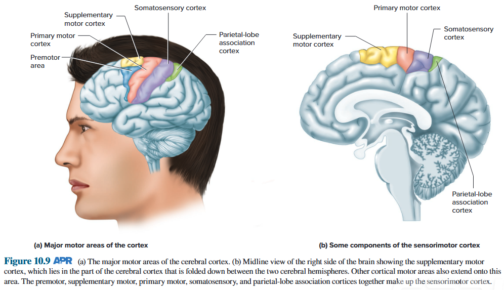
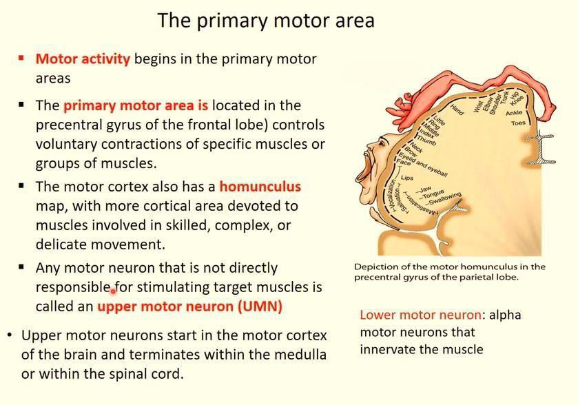
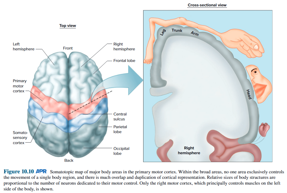
9.3.4.3 Subcortical and Brainstem Nuclei
They form a link in some of the looping parallel circuits through which activity in the motor system is transmitted from a specific region of sensorimotor cortex to the basal nuclei, from there to the thalamus, and then back to the cortical area where the circuit started. 它们在一些循环并行电路中形成一个链接,通过该链接,运动系统中的活动从感觉运动皮层的特定区域传输到基底核,从那里传输到丘脑,然后返回到电路开始的皮质区域。
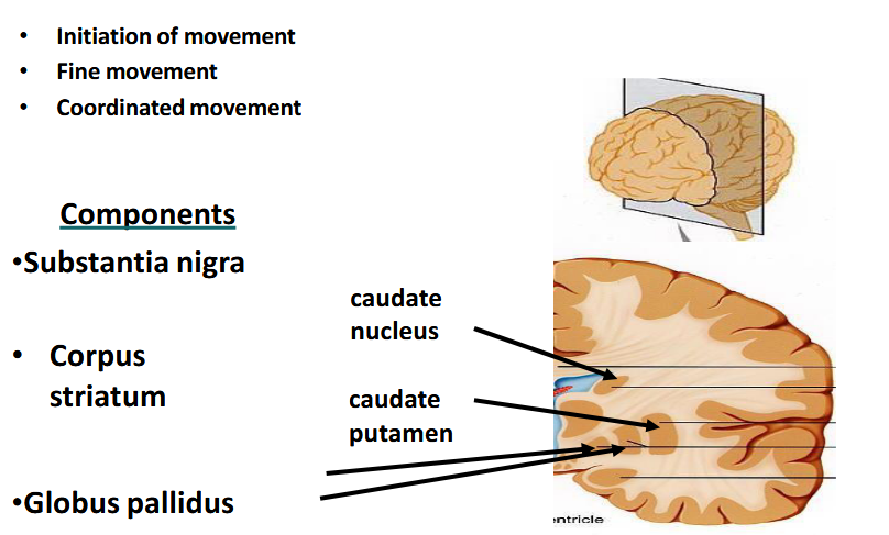
9.3.4.4 Modulation of Motor Activity by Basal Ganglia
Substantia nigra
Project: Inhibitory dopaminergic neurons to corpus striatum
Receive: Inhibitory GABAergic neurons from corpus striatum
Corpus striatum
Project: Inhibitory GABAergic neurones to substantia nigra
Receive: Inhibitory dopaminergic neurons from substantia nigra
Internal: Inhibitory cholinergic interneurones
Project: Inhibitory GABAergic neurones to globus pallidus
黑质项目:纹状体的抑制性多巴胺能神经元 接收:纹状体的抑制性 GABA 能神经元 纹状体项目:黑质的抑制性 GABA 能神经元 接收:黑质的抑制性多巴胺能神经元 内部:抑制性胆碱能中间神经元 项目:球状体的抑制性 GABA 能神经元
Globus pallidus
Receive: Inhibitory GABAergic neurones from corpus striatum
Project: Inhibitory GABAergic neurones to thalamus
Thalamus
Receive: Inhibitory GABAergic neurones from globus pallidus
Project: Excitatory glutamatergic neurones to cortex
Cortex
Receive: Excitatory glutamatergic neurones from thalamus
Project: Excitatory glutamatergic neurones to corpus
Striatum
Project: Excitatory glutamatergic neurones to spinal cord
苍白球接收:来自纹状体的抑制性 GABA 神经元 项目:丘脑的抑制性 GABA 神经元 丘脑接收:来自苍白球的抑制性 GABA 神经元 项目:皮质的兴奋性谷氨酸神经元 皮质接收:来自丘脑的兴奋性谷氨酸神经元 项目:纹状体的兴奋性谷氨酸神经元 项目:来自丘脑的兴奋性谷氨酸能神经元:对脊髓的兴奋性谷氨酸能神经元绳索
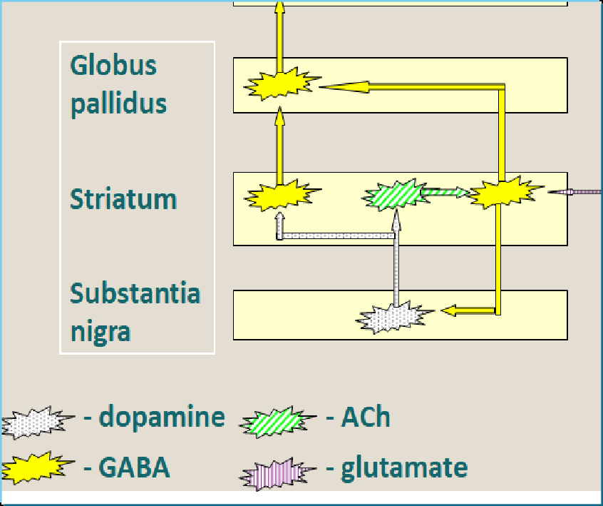
9.3.4.5 Cerebellum
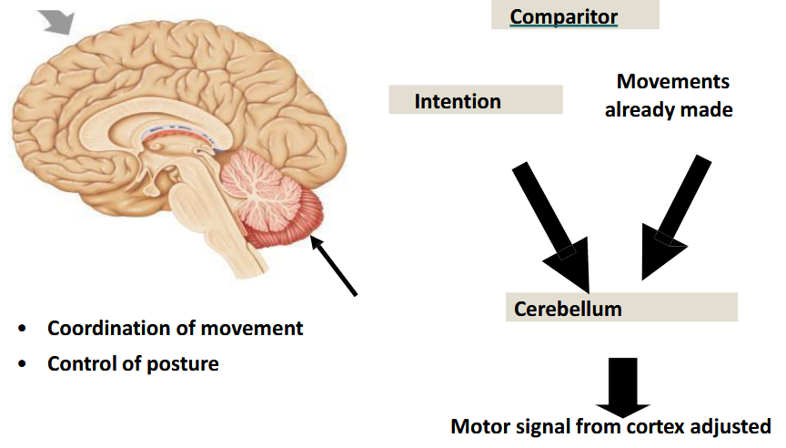
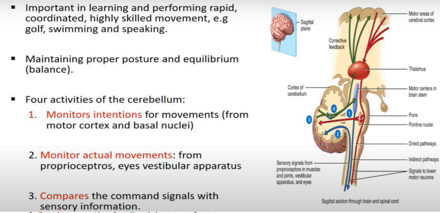
- Sends corrective feedback in case of any discrepancy through thalamus to upper motor neuron.
Cerebellum Dysfunction 小脑功能障碍
Ataxia: • Poor balance • Unsteady walking • Tendency to fall over 共济失调 • 平衡能力差 • 行走不稳 • 容易摔倒
Intention tremor: • Poor adjustment of motor signal • Oscillating movements 意向性震颤 • 运动信号调节不良 • 振荡运动
9.3.4.5 Upper motor neuron
The descending pathways and neurons of the motor cortex are often referred to as the upper motor neurons. Abnormalities due to their dysfunction are classified, therefore, as upper motor neuron disorders.
The upper motor neurons are located in either the motor cortex of the brain or the brainstem and are the main neurons which initiate voluntary movement throughout the body by connecting the cerebral cortex to the brain stem and spinal cord. There are a few tracts, or pathways that upper motor neurons can travel, which serve different functions: pyramid, extrapyramidal( rubrospinal, tectospinal, and reticulospinal tracts) tracts.
运动皮层的下行通路和神经元通常被称为上运动神经元。因此,由于其功能障碍而导致的异常被归类为上运动神经元疾病。
脑运动中枢及其控制的下行通路 上运动神经元位于大脑运动皮层或脑干,是通过连接大脑皮层到脑干和脊髓。 上运动神经元可以通过一些束或通路,它们具有不同的功能:锥体束、锥体外系(红核脊髓束、顶盖脊髓束和网状脊髓束)。
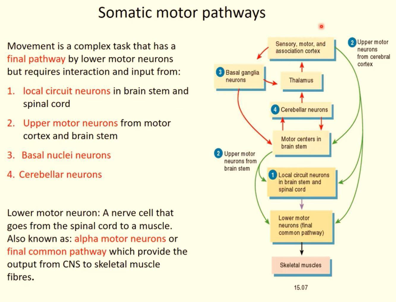
9.3.4.6 Descending pathways
The pathways are of two types:
- corticospinal pathways, originated in the cerebral cortex.
- brainstem pathways, originated in the brainstem.
End at synapses on alpha and gamma motor neurons or on interneurons. The ultimate effect of the descending pathways on the alpha motor neurons may be excitatory or inhibitory.
这些通路有两种类型: → 皮质脊髓通路,起源于大脑皮层。 → 脑干通路,起源于脑干。 → 终止于α和γ运动神经元或中间神经元的突触 → 下行通路对α运动神经元的最终影响可能是兴奋性的或抑制性的。
Corticospinal Pathway 皮质脊髓通路
The corticospinal pathways are also called the pyramidal tracts or pyramidal system because of their triangular shape as they pass along the ventral surface of the medulla oblongata. 皮质脊髓通路也称为锥体束或锥体系统,因为它们沿着延髓的腹侧表面通过时呈三角形。
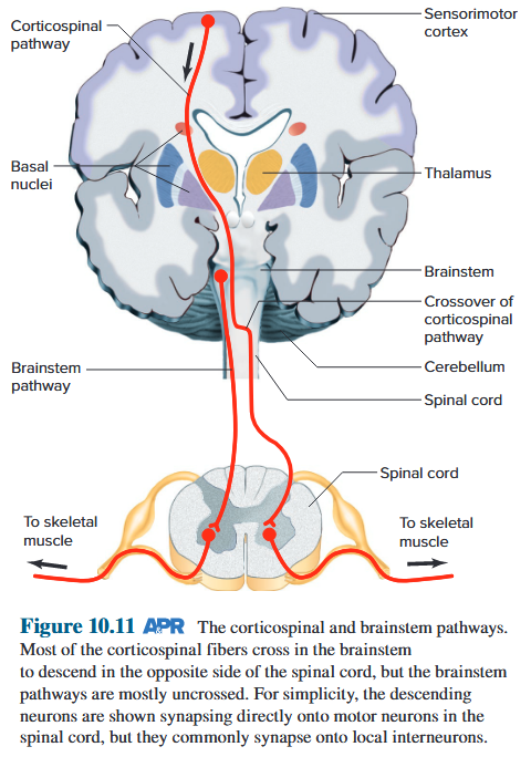
Voluntary Motor Impulses 随意运动的神经传导机制
- Arise in motor area of cerebral cortex
- Synapse w/ motor neurons in spinal cord
- Axons form pyramidal tracts
- Cross over in medulla oblongata (decussation)
- Density of muscle innervation proportional to complexity of movement
• 出现在大脑皮层的运动区 • 脊髓中带有运动神经元的突触 • 轴突形成锥体束 • 在延髓中交叉(交叉) • 肌肉神经支配的密度与运动的复杂性成正比
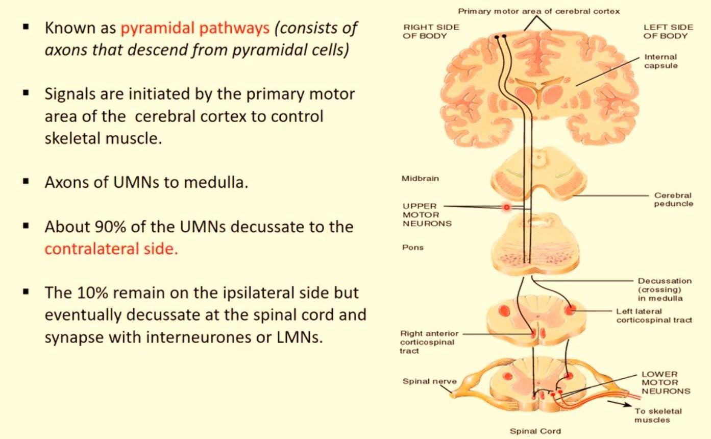
Brainstem Pathway
Axons from neurons in the brainstem also form pathways that descend into the spinal cord to influence motor neurons. These pathways are sometimes referred indirect pathways, to distinguish them from the corticospinal (pyramidal) pathways. 脑干神经元的轴突也形成下降到脊髓的通路,影响运动神经元。 这些通路有时被称为间接通路,以区别于皮质脊髓(锥体)通路。
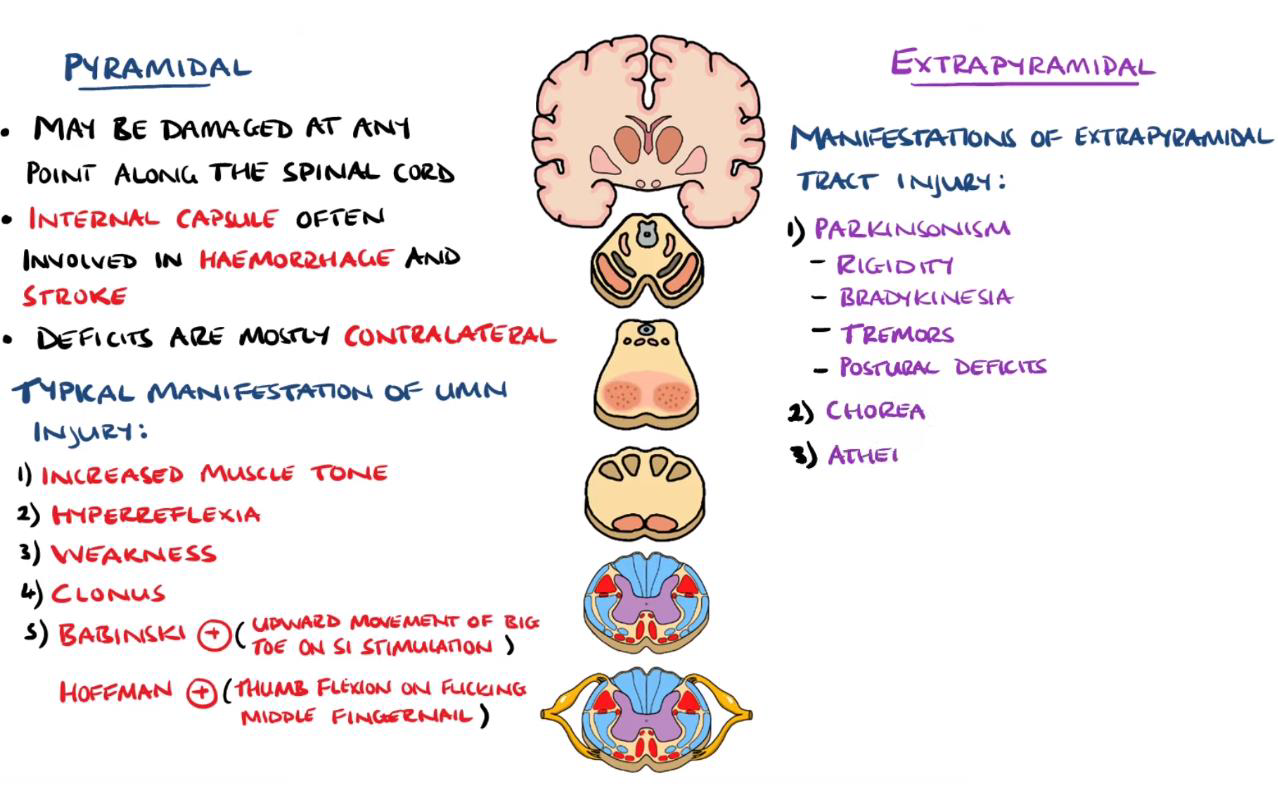
Some of the descending fibres affect afferent systems. They do this via 1) presynaptic synapses on the terminals of afferent neurons as these fibres enter the central nervous system; 2) synapses on interneurons in the ascending pathways. 一些下行纤维影响传入系统。 他们通过以下方式做到这一点:1)传入神经元末端的突触前突触,当这些纤维进入中枢神经系统时; 2)上行通路中中间神经元上的突触。
Parkinson disease
Parkinson disease (formerly called Parkinson’s disease ), the input to the basal nuclei is diminished, the interplay of the facilitory and inhibitory circuits is unbalanced, and activation of the motor cortex (via the basal nuclei–thalamus limb of the circuit just mentioned) is reduced. 帕金森病(以前称为帕金森病),基底核的输入减少,促进和抑制回路的相互作用不平衡,运动皮层的激活(通过刚刚提到的回路的基底核-丘脑肢)减少。
80% dopaminergic neurones in nigrostriatal pathway (substantia nigra to striatum) ➔ inhibition of striatal GABAergic output ➔ inhibitory output from globus pallidus to thalamus ➔ excitatory output from thalamus onto cortex ➔ excitatory output from cortex to spinal cord
黑质纹状体通路中的 80% 多巴胺能神经元(黑质到纹状体) ➔ 抑制纹状体 GABA 能输出 ➔ 从苍白球到丘脑的抑制性输出 ➔ 从丘脑到皮质的兴奋性输出 ➔ 从皮质到脊髓的兴奋性输出
9.4 Autonomic Nervous System
9.4.1 Structures of the sympathetic and parasympathetic nerves
A special case occurs in the gastrointestinal tract, where autonomic neurons innervate a nerve network in the wall of the intestinal tract. This network is called the enteric nervous system. The synapse between the two neurons is outside the CNS in a cell cluster called an autonomic ganglion. The neurons passing between the CNS and the ganglia are called preganglionic neurons; those passing between the ganglia and the effector cells are postganglionic neurons.
胃肠道中存在一种特殊情况,自主神经元支配肠道壁的神经网络。这个网络称为肠神经系统。两个神经元之间的突触位于中枢神经系统之外的一个称为自主神经节的细胞簇中。 在中枢神经系统和神经节之间传递的神经元称为节前神经元; 在神经节和效应细胞之间传递的神经元是节后神经元。
Anatomical and physiological differences within the autonomic nervous system are the basis for its further subdivision into sympathetic and parasympathetic divisions. 自主神经系统内的解剖学和生理学差异是其进一步细分为交感神经和副交感神经的基础。The neurons of the sympathetic and parasympathetic divisions leave the CNS at different levels— the sympathetic fibers from the thoracic and lumbar regions of the spinal cord, and the parasympathetic fibers from the brainstem and the sacral portion of the spinal cord. 交感神经和副交感神经的神经元在不同的水平离开中枢神经系统——交感纤维来自脊髓的胸椎和腰部区域,副交感纤维来自脑干和脊髓的骶部。The sympathetic division is also called the thoracolumbar division, and the parasympathetic division is called the craniosacral division. 交感神经部又称胸腰部,副交感神经部称为颅骶部。
Differ in the location of ganglia. Most of the sympathetic ganglia lie close to the spinal cord and form the two chains of ganglia—one on each side of the cord—known as the sympathetic trunks. 神经节位置不同。大多数交感神经节靠近脊髓,形成两条神经节链(脊髓两侧各一条),称为交感干。
Increased sympathetic activity to occur body-wide when circumstances warrant activation. The parasympathetic system exhibits less divergence. 当环境需要激活时,全身交感神经活动就会增加。副交感神经系统表现出较小的分歧。
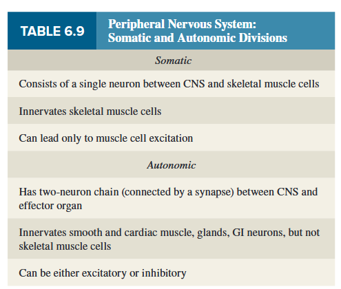
9.4.2 Neurotransmitters and the corresponding receptors of the autonomic nervous system
In both the sympathetic and parasympathetic divisions, acetylcholine is the neurotransmitter released between pre and postganglionic neurons in autonomic ganglia, and the postganglionic cells have predominantly nicotinic acetylcholine Receptors. At many autonomic synapses, one or more co-transmitters are stored and released with the major neurotransmitter. These include ATP, dopamine, and several of the neuropeptides, all of which seem to play a relatively small role. 在交感神经和副交感神经中,乙酰胆碱是自主神经节中节前和节后神经元之间释放的神经递质,节后细胞主要具有烟碱乙酰胆碱受体。 在许多自主神经突触中,一种或多种辅助递质与主要神经递质一起储存和释放。 其中包括 ATP、多巴胺和几种神经肽,所有这些似乎都发挥着相对较小的作用。
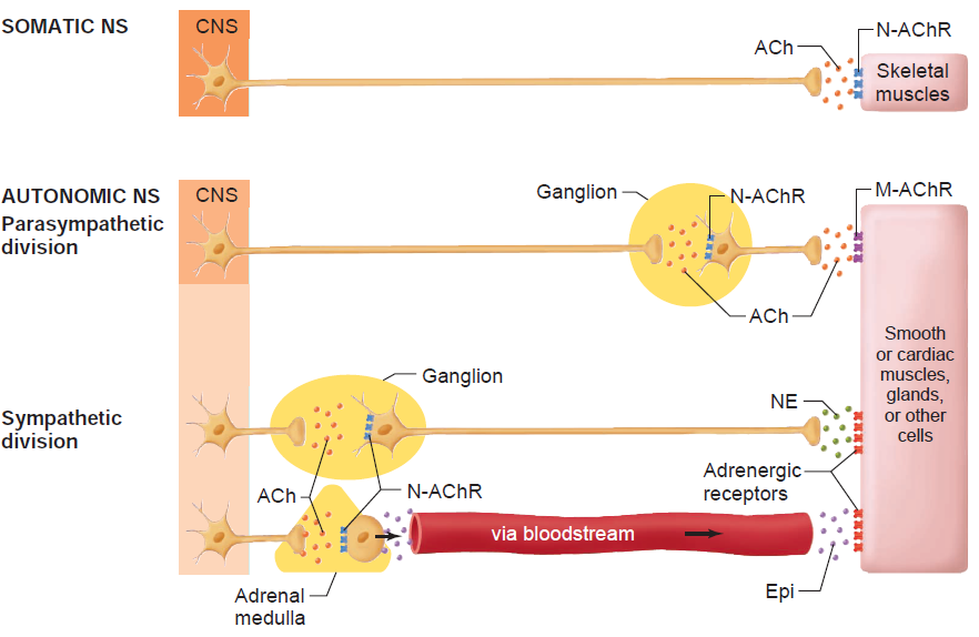
9.4.2.1 ACh and its receptor
Acetylcholine is the chief neurotransmitter in the parasympathetic nervous system and functions in both the central nervous system, CNS, and the peripheral nervous system, PNS.
Major function: commonly associated with memory, cognition, and sleep. → myasthenia gravis, and Parkinson’s disease, Alzheimer’s disease (acetylcholine low)
乙酰胆碱是副交感神经系统中的主要神经递质,在中枢神经系统 (CNS) 和周围神经系统 (PNS) 中发挥作用。 主要功能: 通常与记忆、认知和睡眠有关。 → 重症肌无力、帕金森病、阿尔茨海默病 ( 乙酰胆碱低)
ACh synthesis involves two chemical groups: choline and acetyl coenzyme A (AcCoA) from glucose. 乙酰胆碱的合成涉及两种化学基团:胆碱和来自葡萄糖的乙酰辅酶 A (AcCoA)。
Acetylcholine is synthesized from acetyl coenzyme A and choline by the enzyme choline acetyltransferase. In the nervous system, this enzyme is thought to exist primarily in the nerve termina l cytoplasm. Coenzyme A is synthesized in mitochondria and accesses choline acetyltransferase following transport across the mitochondrial membrane into the cytoplasm. 乙酰胆碱是由乙酰辅酶A和胆碱通过胆碱乙酰转移酶合成的。在神经系统中,这种酶被认为主要存在于神经末梢细胞质中。辅酶 A 在线粒体中合成,并通过线粒体膜转运至细胞质后接触胆碱乙酰转移酶。
Two types of cholinergic receptors 两种类型的胆碱能受体
- Nicotinic acetylcholine receptors : Nicotinic acetylcholine receptor signaling in the brain helps to activate skeletal muscles. 烟碱乙酰胆碱受体:大脑中的烟碱乙酰胆碱受体信号传导有助于激活骨骼肌。
- Muscarinic acetylcholine receptors: Cholinergic muscarinic receptors are located all around the body, from the heart and gut to glands and the brain. 毒蕈碱乙酰胆碱受体:胆碱能毒蕈碱受体遍布身体各处,从心脏和肠道到腺体和大脑。
Muscarinic receptors
There are five types of muscarinic receptors:
M1: The M1 receptor is in the peripheral ganglia.
M2: The M2 receptor is a "specific subtype of muscarinic receptor found in the lower brain, the heart and in smooth muscle-containing organs.
M3: The M3 receptor is found in smooth muscle-containing organs.
M4: The M4 receptor is a specific subtype of muscarinic receptor found in the corpus striatum and the lung.
M5: a specific subtype of muscarinic receptor found in a variety of locations including the salivary glands and the substantia nigra and ventral tegmental area of the brain
Nicotinic receptors
N1 receptors are in the neuromuscular junction
N2 receptors in ganglia.
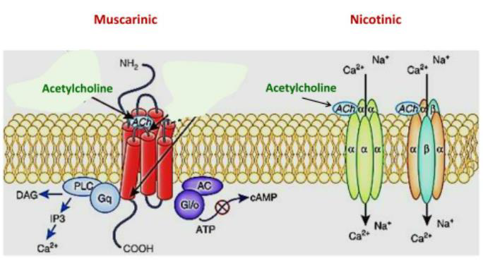
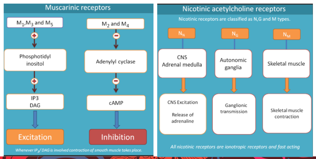
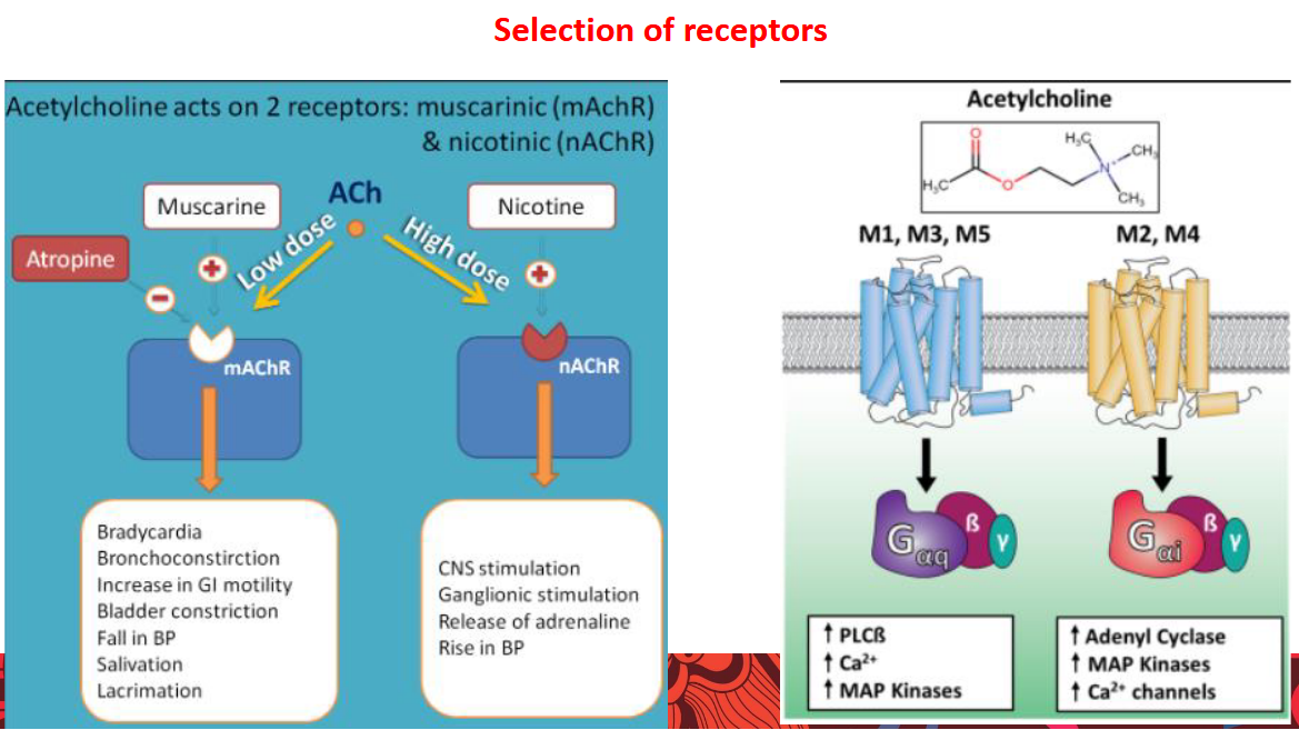
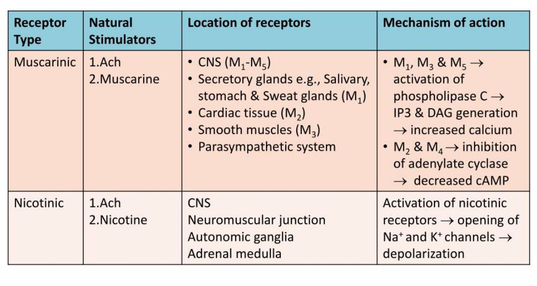
9.4.2.2 NE and its receptor
Norepinephrine (noradrenaline)
Substance that is released predominantly from the ends of sympathetic nerve fibres and that acts to increase the force of skeletal muscle contraction and the rate and force of contraction of the heart. 去甲肾上腺素(去甲肾上腺素)主要从交感神经纤维末端释放的物质,其作用是增加骨骼肌收缩的力量以及心脏收缩的速度和力量。
Major Function: The actions of norepinephrine are vital to the fight-or-flight response, whereby the body prepares to react to or retreat from an acute threat. 主要功能:去甲肾上腺素的作用对于战斗或逃跑反应至关重要,通过这种反应,身体准备对严重威胁做出反应或撤退。
Fight or flight response: (acute stress response): The physiological reaction that occurs when in the presence of something mentally or physically terrifying. This response is triggered by the release of hormones that prepare your body to either stay and deal with a threat or to run away to safety. 战斗或逃跑反应:(急性应激反应):当存在精神或身体上令人恐惧的事物时发生的生理反应。这种反应是由荷尔蒙的释放引发的,这些荷尔蒙会让您的身体做好准备,要么留下来应对威胁,要么逃往安全的地方。
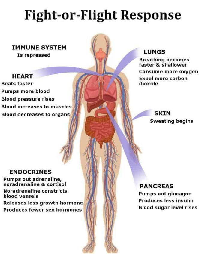
In response to acute stress, the body’s sympathetic nervous system is activated due to the sudden release of hormones. The sympathetic nervous systems stimulate the adrenal glands triggering the release of catecholamines, which include adrenaline and noradrenaline. This results in an increase in heart rate, blood pressure, and breathing rate. After the threat is gone, it takes between 20 to 60 minutes for the body to return to its pre-arousal levels. 为了应对急性压力,身体的交感神经系统会因激素的突然释放而被激活。交感神经系统刺激肾上腺,引发儿茶酚胺的释放,其中包括肾上腺素和去甲肾上腺素。这会导致心率、血压和呼吸频率加快。威胁消失后,身体需要 20 到 60 分钟才能恢复到觉醒前的水平。
Adrenergic receptors are divided into alpha (α1 and α2) and beta (β1, β2, and β3) types and are localized in various autonomic effector tissues.
- The αs are classified into α1 (a receptor coupled to Gq protein) and α2 (a receptor that couples to a Gi protein)
- α1 has 3 subtypes: α1A, α1B and α1D
- α2 has 3 subtypes: α2A, α2B and α2C
- The βs are divided into β1, β2 and β3. All three bind to Gs proteins, but the β2 and β3 receptors also bind to Gi proteins.
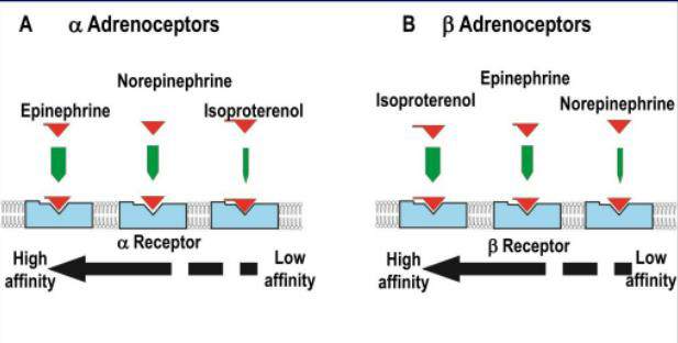
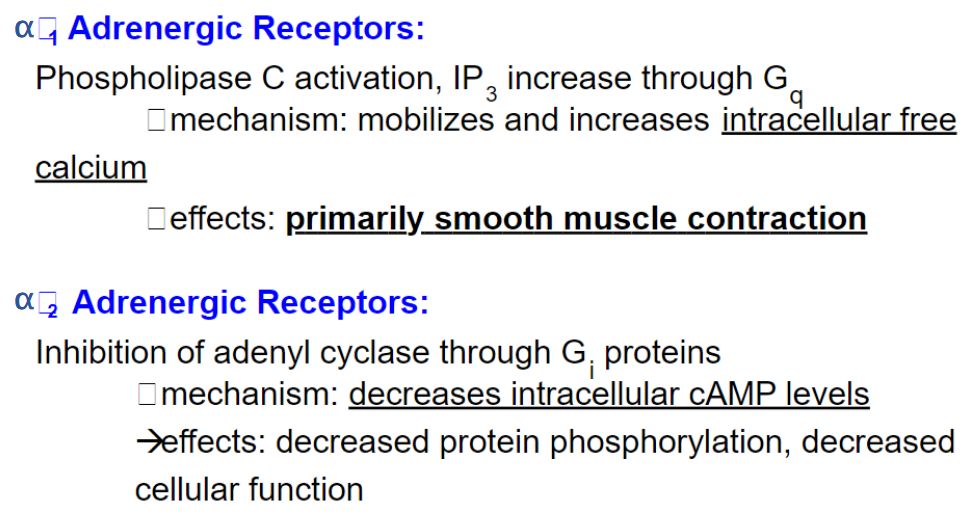
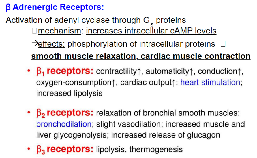
9.4.2.3 Neural transmitters and their distribution
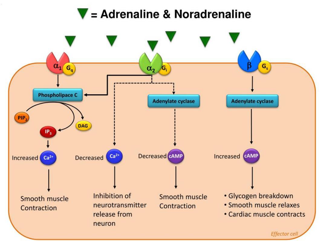
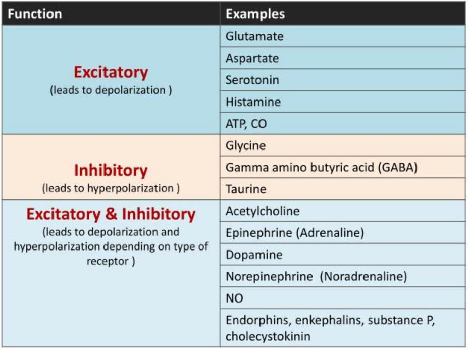
The heart and many glands and smooth muscles are innervated by both sympathetic and parasympathetic fibers; that is, they receive dual innervation. These two divisions are usually activated reciprocally; that is, as the activity of one division increases, the activity of the other decreases.
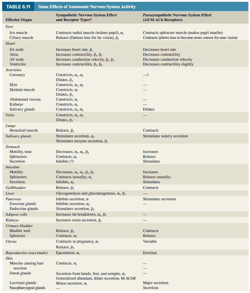
9.4.2.4 Summary (AchR and AR–mediated effect)
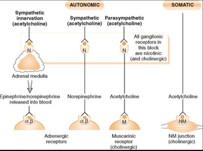
9.4.3 Function of autonomic nervous system
The autonomic nervous system (ANS) maintains blood pressure, regulates the rate of breathing, influences digestion, urination, and modulates sexual arousal. The sympathetic portion of the ANS controls reactions like the stress response and the fight-or-flight reaction. The parasympathetic portion of the ANS controls responses related to eating, growth, and reproduction. 自主神经系统 (ANS) 维持血压、调节呼吸频率、影响消化、排尿并调节性唤起。 ANS 的交感神经部分控制应激反应和战斗机逃跑反应等反应。 ANS 的副交感神经部分控制与进食、生长和繁殖相关的反应。
The autonomic nervous system can be contrasted to the somatic nervous system, which is controlled voluntarily. Where the autonomic nervous system controls things like heart rate and digestion, the somatic nervous system controls things like muscle movements. Breathing is a function which can switch between the autonomic and somatic nervous systems; this is why you are able to hold your breath, but also why you never have to think about breathing. 自主神经系统可以与自主控制的躯体神经系统形成对比。自主神经系统控制心率和消化等,而躯体神经系统控制肌肉运动等。呼吸是一种可以在自主神经系统和躯体神经系统之间切换的功能;这就是为什么你能够屏住呼吸,也是为什么你永远不必考虑呼吸。
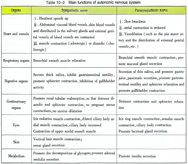
9.4.3.1 Differences on function between both sympathetic and parasympathetic nerves
Except blood vessels inside skin and muscle, slave glands and medulla in kidney, organs accept the regulation from both sympathicus and parasympathicus nerves. They are only be regulated by sympathicus nerves. 除皮肤和肌肉内的血管、肾内的奴隶腺和髓质外。它们仅受交感神经调节。
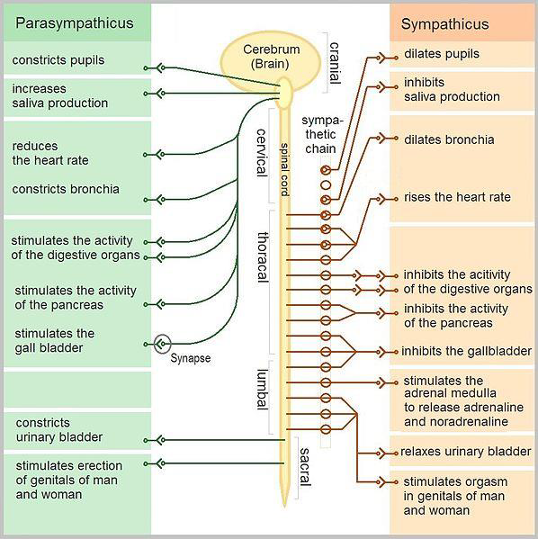
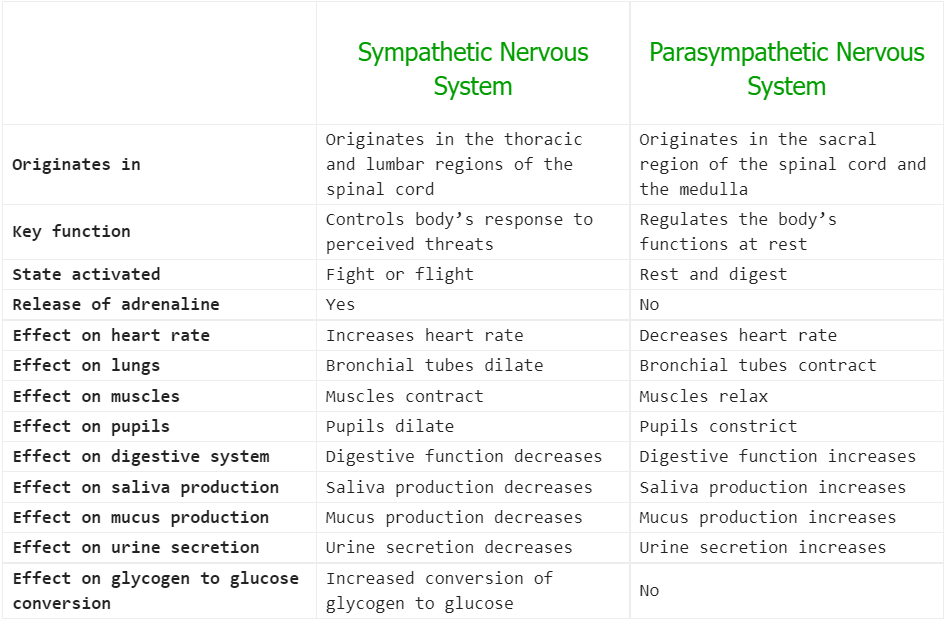
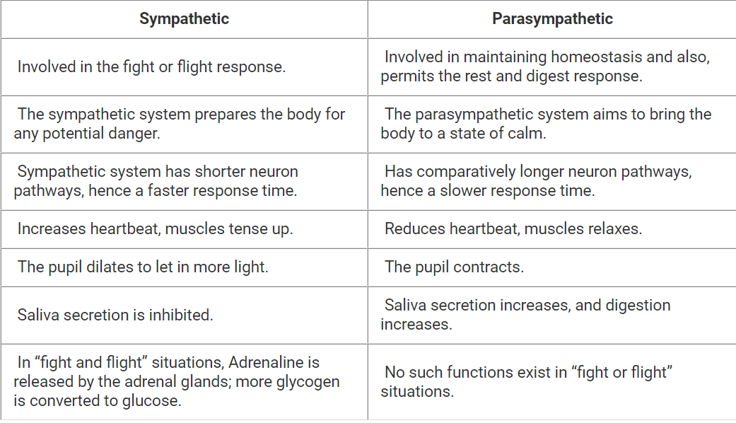
9.4.3.2 Fight-Flight and Feed-Breed
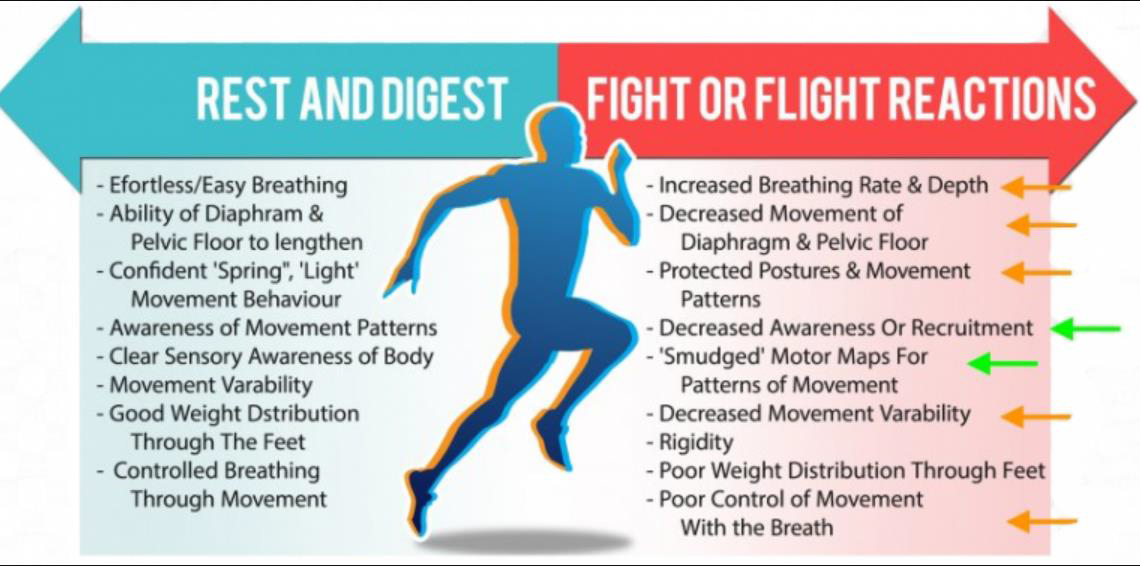
Sympathetic Autonomic Nervous System: It is the part of the autonomic nervous system located near the thoracic and lumbar regions in the spinal cord. Its primary function is to stimulate the body’s fight-or-flight response. It does this by regulating the heart rate, rate of respiration, pupillary response and more. 交感自主神经系统:它是位于脊髓中胸椎和腰椎区域附近的自主神经系统的一部分。它的主要功能是刺激身体的战斗或逃跑反应。它通过调节心率、呼吸频率、瞳孔反应等来实现这一点。
Parasympathetic Autonomic Nervous System: It is located in between the spinal cord and the medulla. It primarily stimulates the body’s “rest and digest” and “feed and breed” responses. 副交感自主神经系统:位于脊髓和延髓之间。它主要刺激身体的“休息和消化”和“进食和繁殖”反应。
The autonomic nervous system comprises two parts- the sympathetic and parasympathetic nervous systems. The sympathetic nervous system activates the fight or flight response during a threat or perceived danger, and the parasympathetic nervous system restores the body to a state of calm. 自主神经系统包括两部分——交感神经系统和副交感神经系统。交感神经系统在威胁或感知到危险时激活战斗或逃跑反应,副交感神经系统使身体恢复到平静状态。
9.4.4 Central control of autonomic nervous system activity
9.4.4.1 Central Autonomic Nervous System
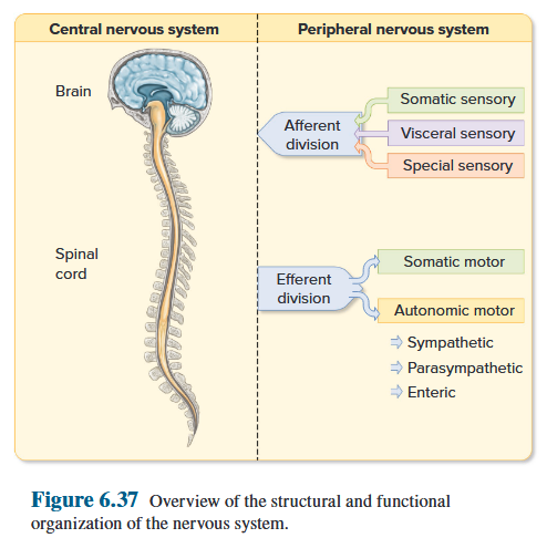
Extensive interconnection occurs between sites receiving visceral inputs and that control autonomic efferent outputs, between sites for the control of sympathetic versus parasympathetic nervous system output, and between sites for autonomic control and somatic, endocrine and limbic circuitry. Collectively, this set of interconnections is termed the central autonomic network. 广泛的互连发生在接收内脏输入和控制自主传出输出的站点之间、控制交感神经系统与副交感神经系统输出的站点之间、以及自主控制站点和躯体、内分泌和边缘电路之间。总的来说,这组互连被称为中央自治网络。
Spinal Cord
Spinal cord can achieve primary regulation of visceral activities. 脊髓可实现内脏活动的初级调节。
Spinal shock: the response that complete loss of motor, sensory, reflex, and autonomic functions below the level of injury. 损伤水平以下运动、感觉、反射和植物神经功能完全丧失的反应。
The regions of gray matter projecting toward the back of the body are called the dorsal horns , whereas those oriented toward the front are the ventral horns. Groups of afferent fibers that enter the spinal cord from the peripheral nerves enter on the dorsal side of the cord via the dorsal roots. The axons of efferent neurons leave the spinal cord on the ventral side via the ventral roots. 向身体后部突出的灰质区域称为背角,而朝向身体前方的灰质区域称为腹角。从周围神经进入脊髓的传入纤维组通过背根进入脊髓的背侧。传出神经元的轴突通过腹侧根离开腹侧的脊髓。
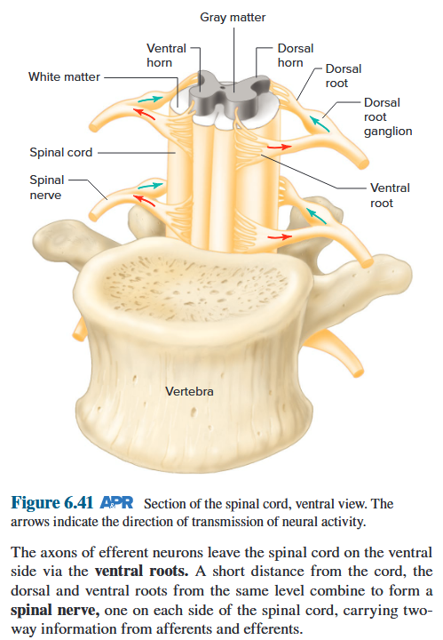
Brainstem
Running through the core of the brainstem and consisting of loosely arranged neuron cell bodies intermingled with bundles of axons is the reticular formation , the one part of the brain absolutely essential for life. 贯穿脑干核心、由松散排列的神经元细胞体和轴突束混合而成的网状结构,是大脑对生命绝对必需的部分。
The reticular formation is involved in motor functions, cardiovascular and respiratory control, and the mechanisms that regulate sleep and wakefulness and that focus attention. 网状结构涉及运动功能、心血管和呼吸控制,以及调节睡眠和觉醒以及集中注意力的机制。
The brainstem contains nuclei involved in processing information for 10 of the 12 pairs of cranial nerves . These are the peripheral nerves that connect directly with the brain and innervate the muscles, glands, and sensory receptors of the head, as well as many organs in the thoracic and abdominal cavities. 脑干包含参与处理 12 对脑神经中的 10 对信息的神经核。这些是直接与大脑连接的周围神经,支配头部的肌肉、腺体和感觉感受器,以及胸腔和腹腔中的许多器官。
Hypothalamus
The hypothalamus can be roughly divided into four areas:
The anterior area 前区
The medial area 内侧区
The lateral area 外侧区
The posterior regions 后区
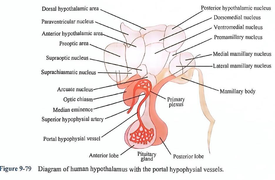
Hypothalamus stimulation produces autonomic responses, instead, the autonomic responses triggered in the hypothalamus are part of more complex functional activities such as eating and drinking, and emotions such as fear and rage.
The hypothalamus is not a mere sympathetic and parasympathetic center, but rather a higher level of regulation of visceral activity linked to other physiological activities that regulate important physiological processes such as body temperature, nutrient intake, water balance, endocrine, emotional response, biological rhythms.
下丘脑刺激会产生自主反应,相反,下丘脑触发的自主反应是更复杂的功能活动的一部分,例如饮食,以及恐惧和愤怒等情绪。 下丘脑不仅仅是交感和副交感中枢,而是与其他生理活动相关的内脏活动的更高水平的调节,调节重要的生理过程,如体温、营养摄入、水平衡、内分泌、情绪反应、生物节律。
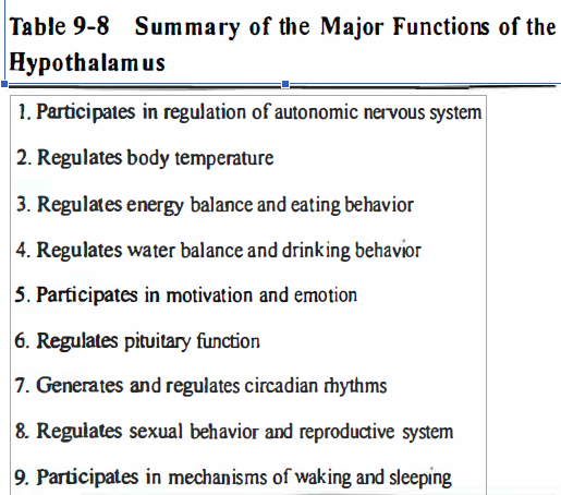
9.4.4.2 Hypothalamus: Regulating Body Temperature
The center of the regulating body temperature is in the hypothalamus.
Thermoregulatory center can sense the temperature when the blood temperature is above or below a certain level (As level is called the set point, normal about 37 °C), then to regulate the heat and cooling activities to maintain body temperature relatively stable. 调节体温的中枢位于下丘脑。 → 当血液温度高于或低于某一水平(该水平称为设定点,正常约为37°C)时,体温调节中枢可以感知温度,然后调节散热和散热活动,保持体温相对稳定。
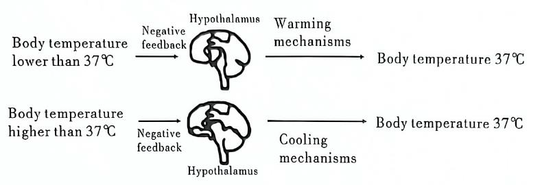
Hypothalamus: Regulating Energy Balance and Eating Behavior
Two areas regulates appetite for food
-
“feeding center” located in the lateral hypothalamic area
-
"satiety center’’ in the ventromedial nucleus
-
Stimulation of the feeding center evokes eating behavior in conscious animals
-
Stimulation of the satiety center causes cessation of eating.
-
Feeding center is chronically active and that its activity is transiently inhibited by activity in the satiety center after the ingestion of food.
9.4.4.3 Hypothalamus: Regulating Water Balance and Drinking Behavior
The most important hormone, vasopressin, synthesized in the hypothalamus and secreted from the neurohypophysis, exerts the regulatory effect via water reabsorption in the renal tubules and collecting ducts so as to control the water excretion. 最重要的激素加压素由下丘脑合成,由神经垂体分泌,通过肾小管和集合管的水分重吸收发挥调节作用,从而控制水分的排泄。
Osmolality acts via osmoreceptors located in the anterior hypothalamus, causing an increase in vasopressin synthesis and secretion, in tum, the increased vasopressin acts on the CNS producing thirst, ultimately results in an increase in water intake. 渗透压通过位于下丘脑前部的渗透压感受器起作用,导致加压素合成和分泌增加,反过来,增加的加压素作用于中枢神经系统,产生口渴,最终导致水摄入量增加。
The effect of extracellular fluid volume depletion on thirst is mediated in part via the renin-angiotensin system. Secretion of renin is increased by hypovolemia, thus the circulating angiotensin II is increased. Angiotensin II then acts on some circumventricular organs, such as the subfornical organ (SFO), organum vasculosurn of the lamina terminalis (OVLT), the specialized receptor areas in the diencephalon, to stimulate the neural areas concerned with thirst. 细胞外液容量减少对口渴的影响部分是通过肾素-血管紧张素系统介导的。血容量不足时肾素的分泌增加,因此循环血管紧张素II增加。然后,血管紧张素 II 作用于一些脑室周围器官,如穹窿下器官 (SFO)、终板血管器官 (OVLT)、间脑中的特殊受体区域,以刺激与口渴有关的神经区域。
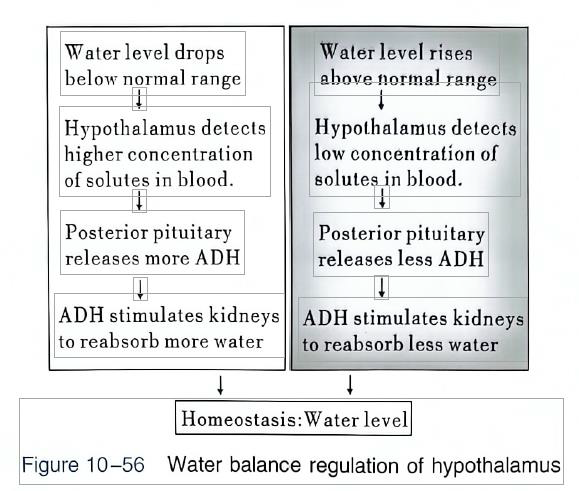
9.4.4.4 Hypothalamus: Participating in Motivation and Emotion
Motivation can lead to hormonal, autonomic, and behavioral responses.
Primary motivated behavior: In homeostatic goal-directed behavior, specific body "needs“ are satisfied. 主要动机行为:在稳态目标导向行为中,特定的身体“需求”得到满足
Secondary motivated behavior: Factors that can be lumped together under the term “incentives”. 次要动机行为:可以归为“激励”一词的因素
The concepts of reward and punishment are inseparable from motivation. Rewards are things that organisms work for or things that make the behavior that leads to them occur more often-in other words, positive reinforcement. Punishments are the opposite. 奖励和惩罚的概念与动机密不可分。奖励是有机体为之努力的东西,或者是使导致它们的行为更频繁地发生的东西,换句话说,是积极的强化。惩罚则相反。
Emotion can be considered in terms of a relation between an individual and the environment based on the individual’s evaluation of the environment ,disposition toward the environment, and the actual physical response to it. 情感可以被认为是个人与环境之间的关系,基于个人对环境的评价、对环境的倾向以及对环境的实际身体反应。
The reticular activating system, which rises in the brainstem and comprises several components. 网状激活系统位于脑干中,由几个组成部分组成。
The component involved in motivation is known as the mesolimbic dopamine pathway: meso- because it arises in the midbrain (mesencephalon) area of the brainstem; limbic because it sends its fibers to areas of the limbic system, such as the prefrontal cortex, the nucleus accumbens, and the undersurface of the frontal lobe. 参与动机的成分被称为中脑边缘多巴胺通路:中观-因为它出现在脑干的中脑(中脑)区域;中观-因为它出现在脑干的中脑(中脑)区域;边缘系统,因为它将其纤维发送到边缘系统的区域,例如前额皮质、伏隔核和额叶的下表面。
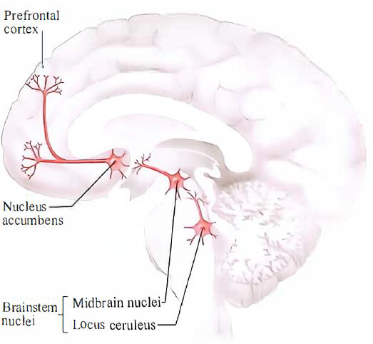
9.4.4.5 Hypothalamus: Regulating Body Temperature
Distinguish → the anatomical sites where the emotional value of a stimulus is determined → the hormonal, autonomic, and outward expressions and displays of response to the stimulus (so-called emotional behavior) → the conscious experience, or inner emotions, such as feelings of fear, love, anger, joy, anxiety, hope, and so on. 区分 → 确定刺激的情感价值的解剖部位 → 对刺激的反应的荷尔蒙、自主和外在表达和表现(所谓的情感行为) → 意识体验或内在情感,例如恐惧、爱、愤怒、喜悦、焦虑、希望等等。
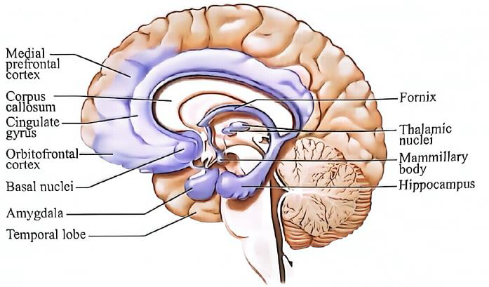
9.4.4.6 Hypothalamus: Regulating Pituitary Function
These substances are: thyrotropin-releasing hormone (TSH), gonadotropin-releasing hormone (GuRH), growth hormone releasing-inhibiting hormone (GHIH) growth hormone-releasing hormone(GRH), corticotropin releasing hormone (CRH), melanocyte-stimulating hormone (MSH), MSH release-inhibiting factor (MIF), prolactin release factor (PRF), prolactin release-inhibiting factor (PIF) and so on. 这些物质是:促甲状腺激素释放激素(TSH)、促性腺激素释放激素(GuRH)、生长激素释放抑制激素(GHIH)、生长激素释放激素(GRH)、促肾上腺皮质激素释放激素(CRH)、黑素细胞刺激激素(MSH)、MSH释放抑制因子(MIF)、催乳素释放因子(PRF)、催乳素释放抑制因子(PIF)等等。
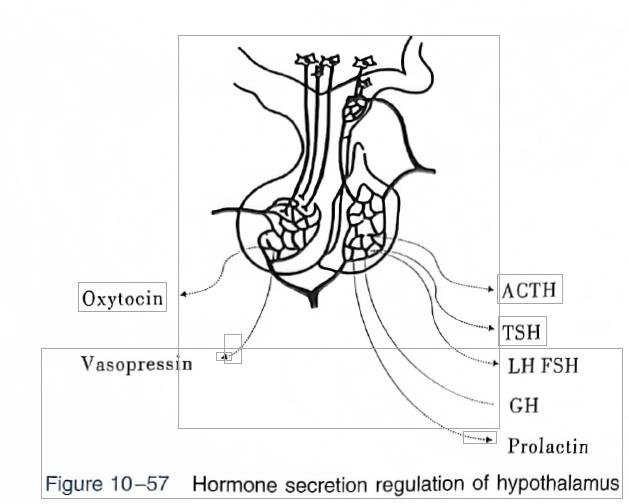
9.4.4.7 Hypothalamus: Generating and Regulating Circadian Rhythm
Supracluasmatic nucleus of the hypothalamus may be the center of all the rhythms. In embryonic period in mice, when the suprachiasmatic nucleus has not yet been established with the surrounding tissue, the circadian rhythm of its metabolism and discharge activity already exists. Destruction of the mouse suprachiasmatic nucleus, the original rhythmic activity (such as drinking water, urination) of the daily cycle losses. 下丘脑的上丘核可能是所有节律的中心。在碘周期小鼠胚胎中,当视交叉上核尚未与周围组织建立时,其代谢和放电活动的昼夜节律就已经存在。小鼠视交叉上核遭到破坏,原有的节律性活动(如饮水、排尿)的日常循环丧失。
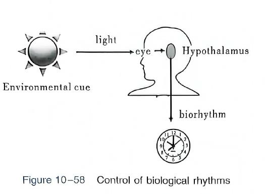
9.4.5 Summary
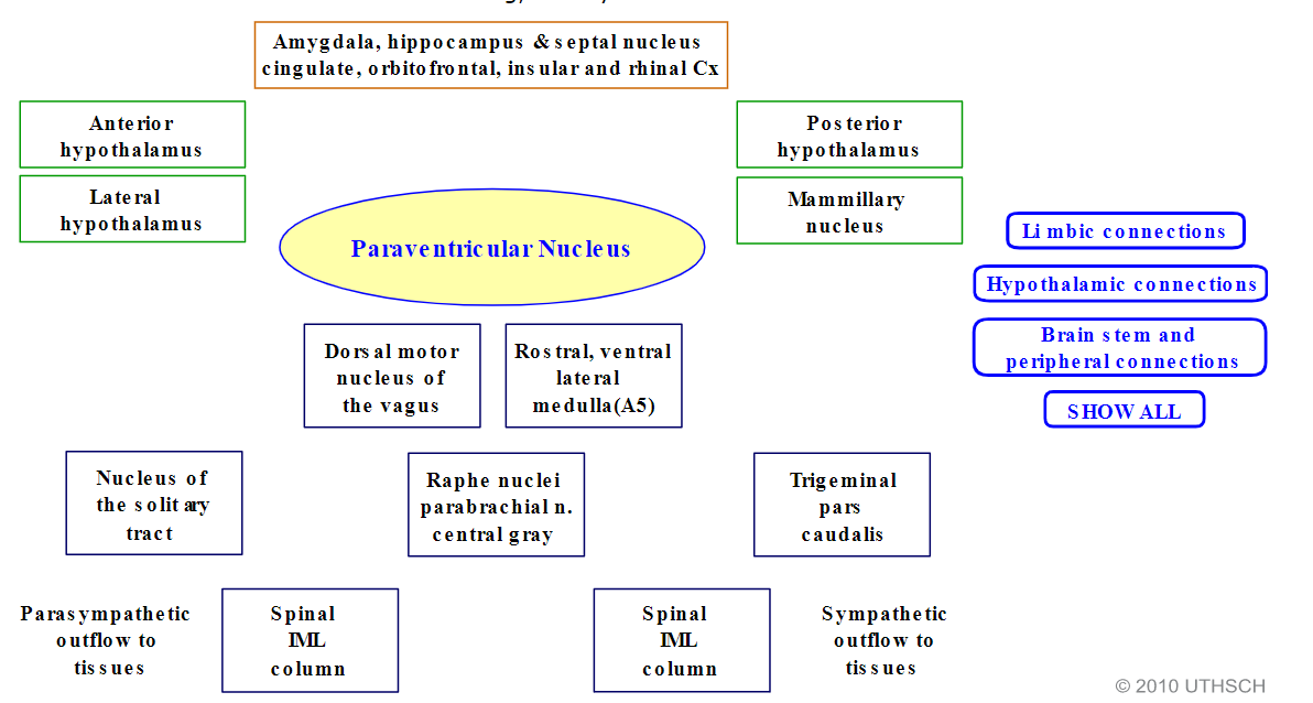
9.5 Electrical Activity of the Brain, Wakefulness and Sleep
9.5.1 Electrical Activity of the brain
Electrodes, which are wires attached to the head by a salty paste that conducts electricity, pick up electrical signals generated in the brain and transmit them to a machine that records them as the EEG. 电极是通过导电的咸糊状物连接到头部的电线,接收大脑中产生的电信号并将其传输到机器,将其记录为脑电图。
The wave’s amplitude, measured in microvolts
electrical activity of a similar type is occurring beneath the recording electrodes at any given time.
Large → neuron activity/synchronously firing
Small → less activated/ are firing asynchronously
The amplitude may range from 0.5 to 100 mV, which is about 1000 times smaller than the amplitude of an action potential.
The frequency of the wave → the frequency of its cycles from the maximal to the minimal amplitude and back. (Hz, or cycles per second) from 0.5 to 40 Hz or higher.
Four distinct frequency ranges that define different states of consciousness are characteristic of EEG patterns.
Lower EEG frequencies, less responsive states 脑电图频率越低,反应状态越低
Higher EEG frequencies, increased alertness 脑电图频率越高,警觉性越高。
Wave patterns vary not only as a function of state of consciousness but also according to where on the scalp they are recorded. 波浪模式不仅随意识状态而变化,而且根据记录在头皮上的位置而变化。
An EEG might also be helpful for diagnosing or treating:
- Brain tumors 脑肿瘤
- Brain damage from head injury 头部损伤造成的脑损伤
- Brain dysfunction that can have a variety of causes (encephalopathy) 多种原因导致的脑功能障碍(脑病)
- Sleep disorders 睡眠障碍
- Inflammation of the brain (herpes encephalitis) 大脑炎症(疱疹性脑炎)
- Stroke 中风
- Sleep disorders 睡眠障碍
- Creutzfeldt-Jakob disease 克雅氏病
Magnetic resonance imaging (MRI) is a medical imaging technique that uses a magnetic field and computer-generated radio waves to create detailed images of the organs and tissues in your body. 磁共振成像 (MRI) 是一种医学成像技术,它使用磁场和计算机生成的无线电波来创建体内器官和组织的详细图像。
To help diagnose:
- Aneurysms of cerebral vessels •Disorders of the eye and inner ear •Multiple sclerosis •Spinal cord disorders •Stroke •Tumors •Brain injury from trauma
- • 脑血管瘤 • 眼睛和内耳疾病 • 多发性硬化症 • 脊髓疾病 • 中风 • 肿瘤 • 外伤造成的脑损伤
A positron emission tomography (PET) scan is an imaging test that can help reveal the metabolic or biochemical function of your tissues and organs. 正电子发射断层扫描 (PET) 扫描是一种成像测试,可以帮助揭示组织和器官的代谢或生化功能。
The PET scan uses a radioactive drug (tracer) to show both normal and abnormal metabolic activity. PET 扫描使用放射性药物(示踪剂)来显示正常和异常的代谢活动。
A PET scan can often detect the abnormal metabolism of the tracer in diseases before the disease shows up on other imaging tests, such as computerized tomography (CT) and magnetic resonance imaging (MRI). PET 扫描通常可以在其他影像学检查(例如计算机断层扫描 (CT) 和磁共振成像 (MRI))显示疾病之前检测到疾病中示踪剂的异常代谢。
Types of EEG waves and related status
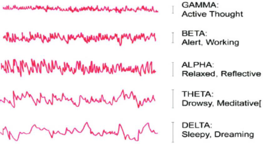
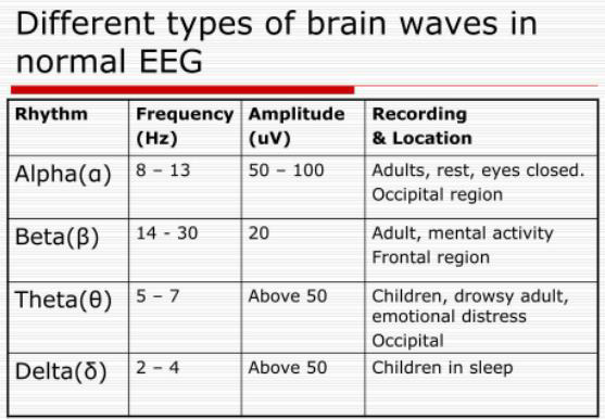
Alpha rhythm: The most prominent EEG wave pattern of an awake, relaxed adult whose eyes are closed is an oscillation of 8 to 12 Hz.
Beta rhythm: When people are attentive to an external stimulus or are thinking hard about something, the alpha rhythm is replaced by smaller-amplitude, higher-frequency (>12 Hz) oscillations.
Gamma rhythm: These are high-frequency oscillations (30–100 Hz) that spread across large regions of the cortex, which seem in some cases to emanate from the thalamus.
Delta rhythm (approximately 1-3 Hz): slowest rhythm normally recorded
Theta rhythm: oscillations (theta waves) with a frequency of 4 to 7 hertz.
Alpha 节律:清醒、放松、闭眼的成年人最显着的脑电图波形是 8 至 12 Hz 的振荡。 Beta节律:当人们注意外部刺激或认真思考某件事时,阿尔法节律是被更小幅度、更高频率(>12 Hz)的振荡所取代。 Gamma 节律:这些是高频振荡(30-100 Hz),分布在大脑皮层的大片区域,在某些情况下似乎是从丘脑发出的。 Delta 节律(大约 1-3 Hz):通常记录的最慢节律 Theta 节律:频率为 4 到 7 赫兹的振荡。
9.5.2 Wakefulness and Sleep
States of Consciousness: Awake / Drowsy / Asleep / Coma
Conscious Experiences: Thoughts / Perceptions / Reasoning / Motivation / Mood
Altered States of Consciousness
Consciousness
States of consciousness: levels of alertness such as being awake, drowsy, or asleep. 意识状态:警觉程度,例如清醒、昏昏欲睡或睡着。
(1) by behavior, covering the spectrum from maximum attentiveness to comatose, (2) by the pattern of brain activity that can be recorded electrically. (1)通过行为,涵盖从最大注意力到昏迷的范围,(2)通过可以电记录的大脑活动模式。
Conscious experiences : experiences a person is aware of—thoughts, feelings, perceptions, ideas, dreams, reasoning—during any of the states of consciousness. 意识体验:一个人在任何意识状态下所意识到的体验——思想、感觉、感知、想法、梦想、推理。
9.5.2.1 Wakefulness
waking state reflects the wide variety of activities you may be engaged in at any given moment 清醒状态反映了您在任何特定时刻可能从事的各种活动
EEG arousal, is associated with the act of paying attention to a stimulus rather than with the act of perception itself. 唤醒脑电图与注意刺激的行为相关,而不是与感知行为本身相关。
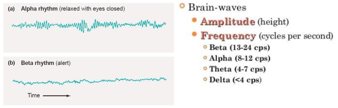
9.5.2.2 Sleep

Sleep states: NREM and REM
There are two phases of sleep, the names of which depend on whether or not the eyes move behind the closed eyelids:
- NREM (non–rapid eye movement)
- REM (rapid eye movement) sleep. The initial phase of sleep
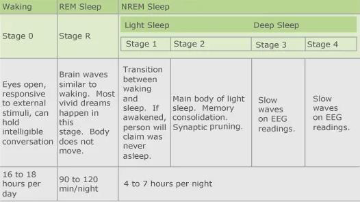
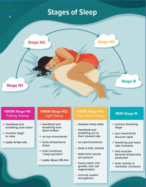
As you transition from drowsiness to stage N1 sleep, there is a considerable tension in the postural muscles, and brief muscle twitches called hypnic jerks sometimes occur. 当您从困倦过渡到 N1 阶段睡眠时,姿势肌肉会出现相当大的张力,有时会出现称为催眠抽搐的短暂肌肉抽搐。
The rate of breathing is frequently increased compared to the awake, relaxed state. In one form of a disease known as sleep apnea, however, stimulation of the respiratory muscles temporarily ceases, sometimes hundreds of times during a night. 与清醒、放松的状态相比,呼吸频率经常加快。然而,在一种称为睡眠呼吸暂停的疾病中,呼吸肌的刺激会暂时停止,有时一晚会停止数百次。
REM sleep is also called paradoxical sleep, because even though a person is asleep and difficult to arouse, his or her EEG pattern shows intense activity that is similar to that observed in the alert, awake state. 快速眼动睡眠也称为矛盾睡眠,因为即使一个人处于睡眠状态且难以醒来,他或她的脑电图模式也显示出与警觉、清醒状态下观察到的类似的强烈活动。
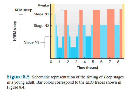
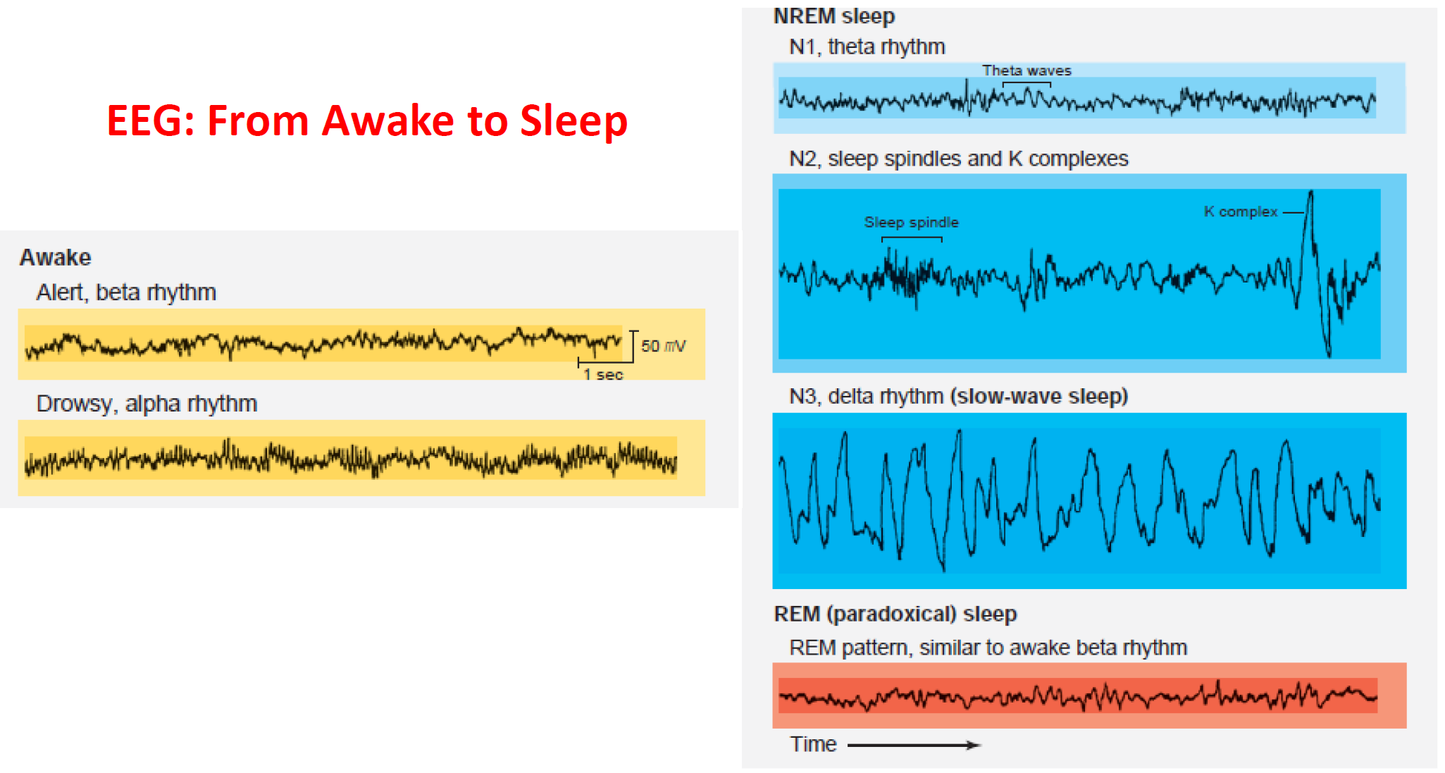
9.5.3 Mechanism of Waking and Sleeping
(Ascending) Reticular Activating System
Reticular Activating System (RAS): This system consists of clusters of neurons and neural pathways originating in the brainstem and hypothalamus, distinguished by both their anatomical distribution and the neurotransmitters they release. 网状激活系统(RAS):该系统由起源于脑干和下丘脑的神经元和神经通路簇组成,其特征在于它们的解剖分布和它们释放的神经递质。
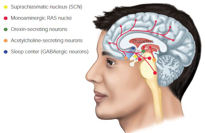
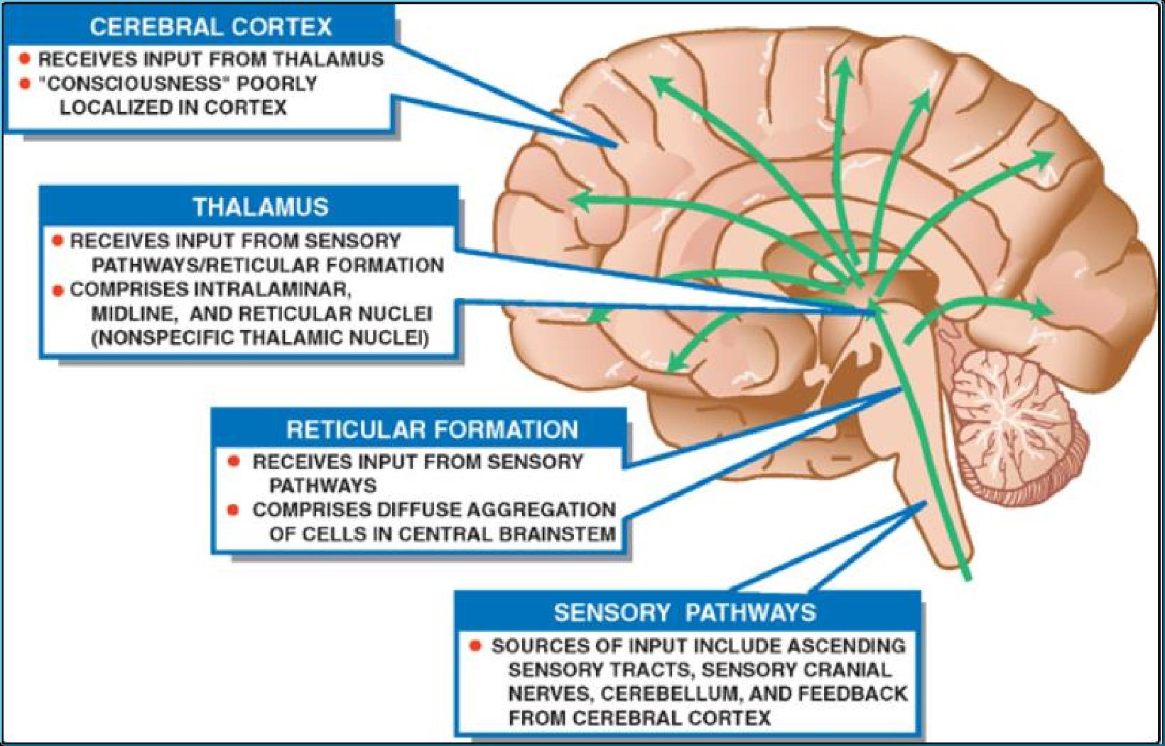
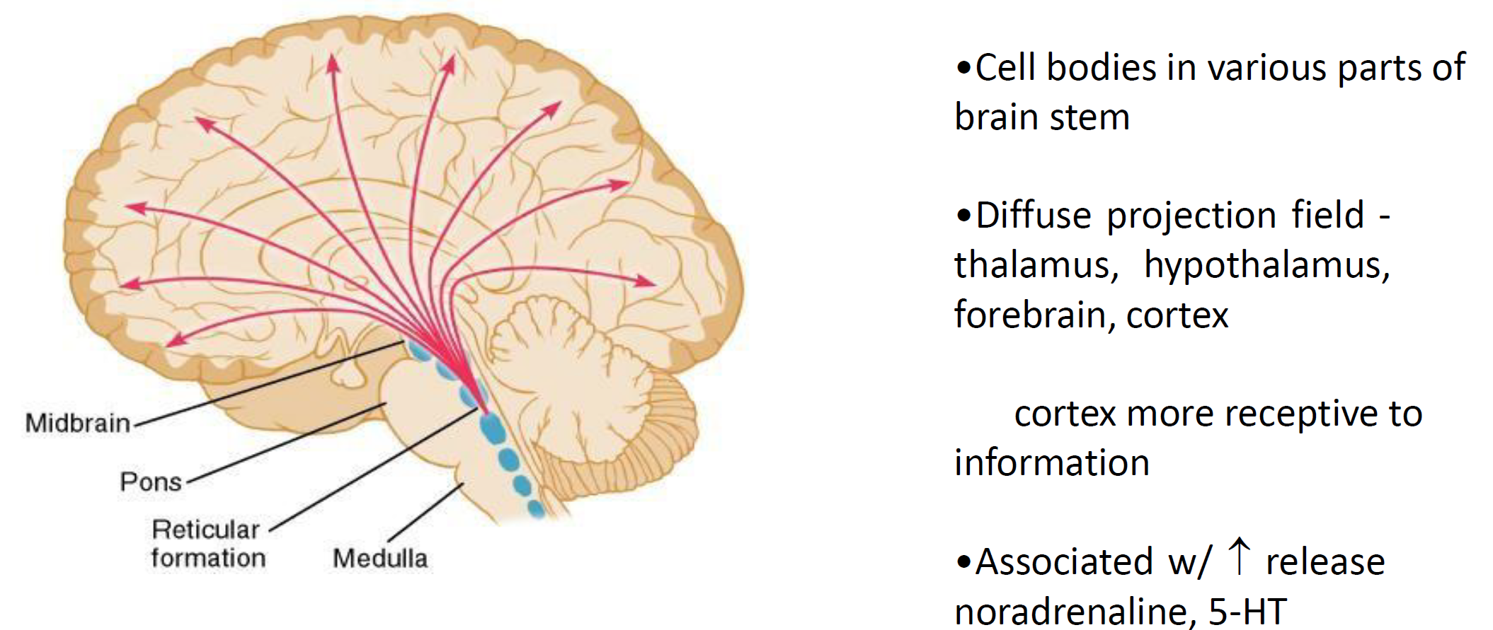
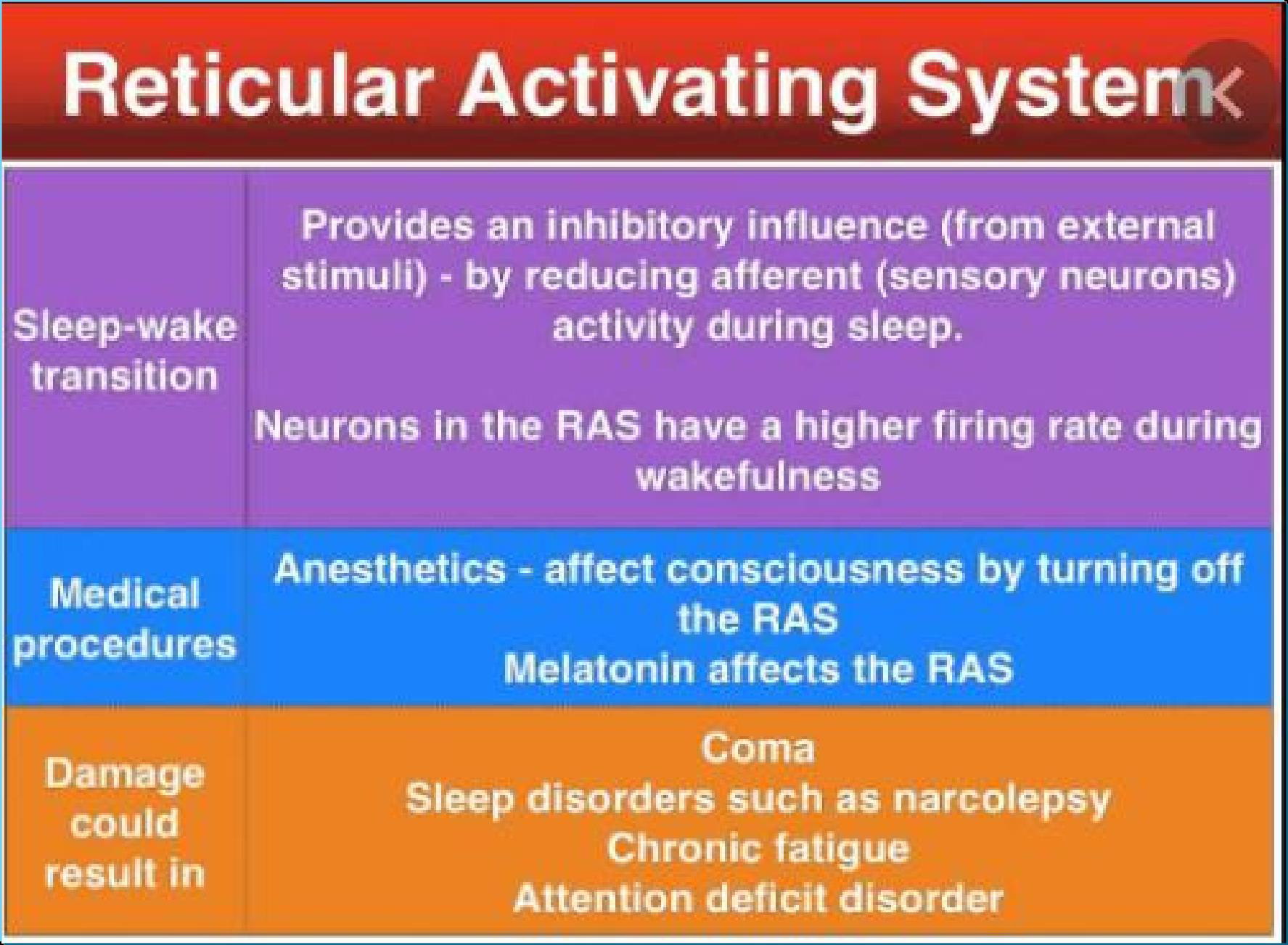
Here shows a model of factors involved in regulating the transition between waking and sleeping states. Transition to the wakeful state is favored by three main inputs to orexin-secreting cells:
- action potential firing from the suprachiasmatic nucleus (SCN)
- indicators of negative energy balance
- arousing emotional states signalled by the limbic system
- The SCN is the principle circadian pacemaker of the body
这里显示了一个涉及调节清醒和睡眠状态之间转换的因素模型。食欲素分泌细胞的三个主要输入有利于向清醒状态的转变:(1)从视交叉上核(SCN)发出的动作电位(2)负能量平衡的指标(3)边缘系统发出的唤起情绪状态的信号。 (4) SCN是人体昼夜节律的主要起搏器
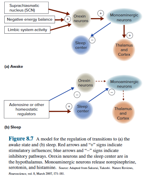
9.5.4 Conscious Experiences

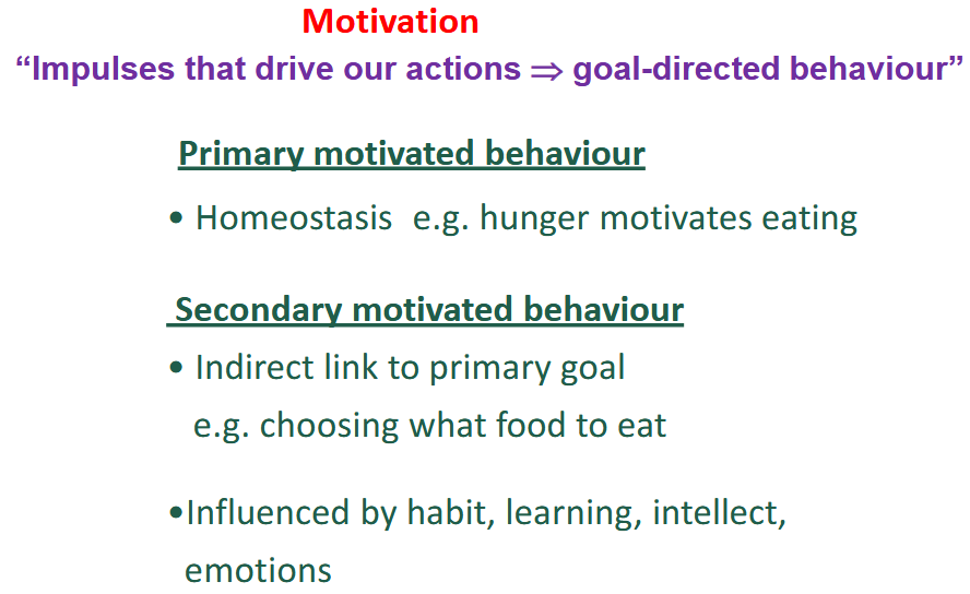
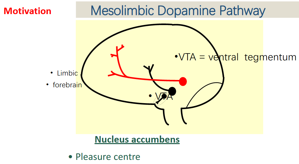
9.6 High Functions of the Brain
9.6.1 Learning
Learning is the acquisition and storage of information as a consequence of experience. It is measured by an increase in the likelihood of a particular behavioral response to a stimulus. 学习是作为经验的结果而获取和存储信息。它是通过对刺激做出特定行为反应的可能性的增加来衡量的。
- Rewards or punishments are crucial ingredients of learning, as are contact with and manipulation of the environment. 奖励或惩罚是学习的重要组成部分,与环境的接触和操纵也是如此。
Types:
- non-associated learning: 1) Habituation 2) Sensitization
- associated learning

9.6.1.1 Non-associative Learning
Non-associative learning is when you are not pairing a stimulus with a behavior. Non-associative learning can be either habituation or sensitization. (Simplest form of learning) 非联想学习是指你没有将刺激与行为配对。非联想学习可以是习惯化或敏化。
The strength of a reflex response can be weakened through Habituation or strengthened through Sensitization. 反射反应的强度可以通过习惯化减弱或通过敏化增强。
Habituation: When there is a reduction in response to a specific stimulus after repeated exposures to it this is known as habituation. 习惯化:当反复接触特定刺激后,对特定刺激的反应减弱,这称为习惯化。
Sensitization: Sensitization occurs when repeated exposure or a single exposure to a stimulus increases the intensity of the response. 敏化:当重复暴露或单次暴露于刺激物增加反应强度时,就会发生敏化。
Habituation requires repeated presentations whereas sensitization does not and habituation is related to the specific stimulus being exposed to whereas sensitization sensitizes to other stimuli. 习惯化需要重复呈现,而敏化则不需要,习惯化与所受到的特定刺激有关,而敏化则对其他刺激敏感。
9.6.1.2 Associative Learning
Associative learning refers to a variety of learning in which ideas and experiences are connected. 联想学习是指将想法和经验联系起来的各种学习。
The process of associative learning takes place through two types of conditioning. → Classical conditioning | Operant conditioning 联想学习的过程是通过两种类型的条件作用发生的。 经典条件反射、操作性条件反射
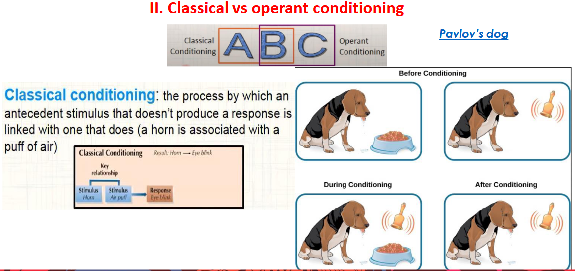
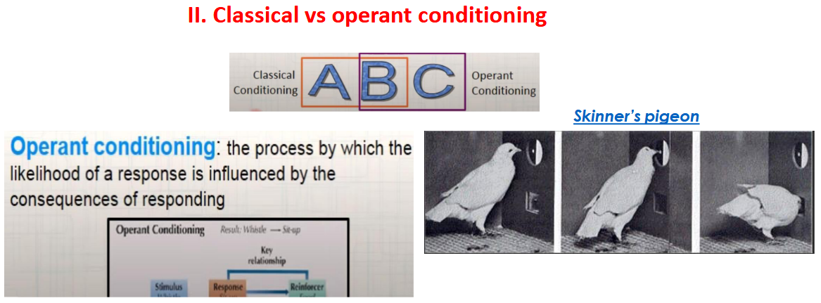
9.6.2 Memory
Memory is the relatively permanent storage form of learned information, although, as we will see, it is not a single, unitary phenomenon. Rather, the brain processes, stores, and retrieves information in different ways to suit different needs. 记忆是所学信息相对永久的存储形式,尽管正如我们将看到的,它不是单一的、单一的现象。相反,大脑以不同的方式处理、存储和检索信息以满足不同的需求。
Memory encoding defines the neural processes that change an experience into the memory of that experience—the physiological events that lead to memory formation. 记忆编码定义了将经历转变为对该经历的记忆的神经过程——导致记忆形成的生理事件。
9.6.2.1 Types of Memory
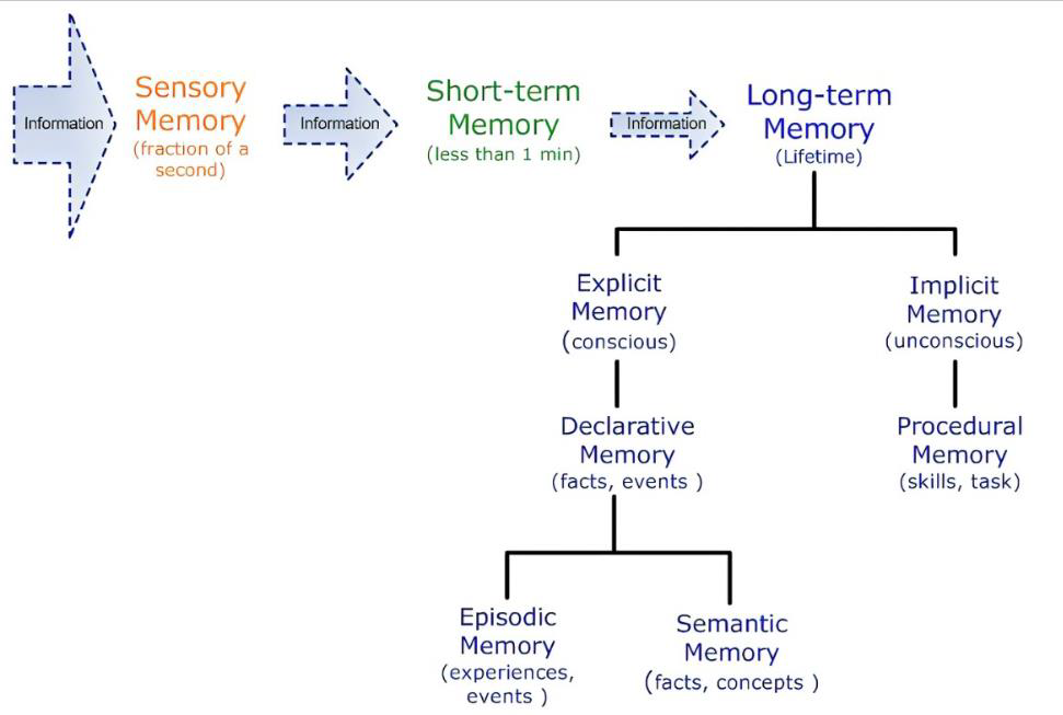
Short-term memory
Short-term memory is the information we are currently aware of or thinking about. It is the information that is held in our mind for a very short period of time before it is either dismissed or transferred to long-term memory. WORKING Memory can be regarded as a distinct segment of short-term memory. 短期记忆是我们当前意识到或正在思考的信息。它是在被忽略或转移到长期记忆之前在我们脑海中保留很短一段时间的信息。工作记忆可以被视为短期记忆的一个独特部分。
Working Memory
◦ Current, temporary information ◦ Lasts seconds-minutes ◦ Limited capacity Important factors ◦ Attention ◦ Language/visual associations ◦ Relevant data retrieved from long term memory
◦ 当前的临时信息 ◦ 持续数秒-分钟 ◦ 容量有限 重要因素 ◦ 注意力 ◦ 语言/视觉关联 ◦ 从长期记忆中检索到的相关数据
Long-Term Memory
◦ Information not currently in our attention ◦ Lasts hours, days, years ◦ Influenced by attention, motivation ◦ 2 types: procedural - how to do something | declarative - facts and events ◦ Unlimited capacity
◦ 当前不在我们注意力范围内的信息 ◦ 持续数小时、数天、数年 ◦ 受注意力、动机影响 ◦ 2 种类型:程序性 - 如何做某事 陈述性 - 事实和事件 ◦ 无限容量
Forgetting
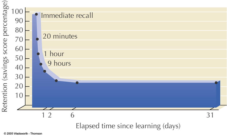
9.6.2.2 Neural basis of learning and memory
Hippocampus & cerebral cortex involved in memory
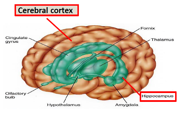
Short-term encoding and long-term memory storage occur in different brain areas for both declarative and procedural memories. 陈述性记忆和程序性记忆的短期编码和长期记忆存储发生在不同的大脑区域。
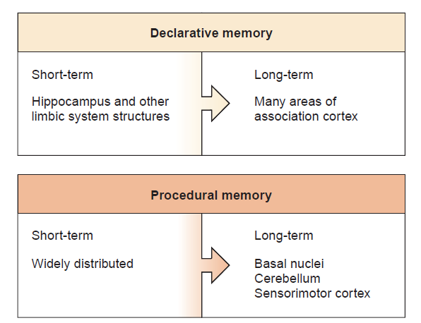
9.6.2.3 Disorder of Memory
Hippocampal Damage
◦ Retrograde amnesia – previous 1-2 years lost – older memories intact ◦ Anterograde amnesia – no new long-term memories
◦ 逆行性遗忘 – 之前 1-2 年的记忆丢失 – 较旧的记忆完好无损
◦ 顺行性遗忘 – 没有新的长期记忆
Alzheimer’s Disease
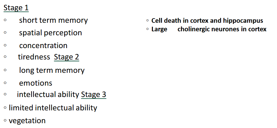
9.6.2.4 Long-Term Potentiation
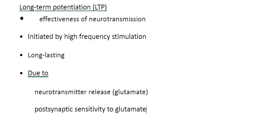
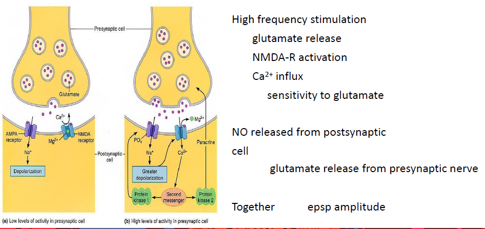
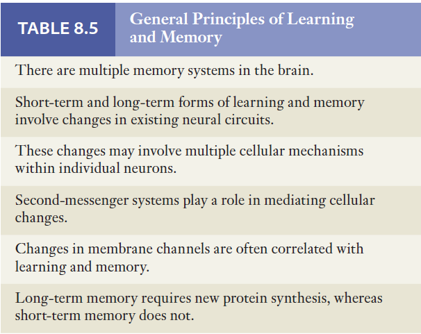
9.6.3 Cerebral Dominance and Language
Chapter 7 Energy Metabolism and body Temperature
7.1 Energy Metabolism
7.1.1 Basic Concept
Metabolism (新陈代谢) - Anabolism (合成代谢) - catabolism (异化作用)
Energy metabolism
Metabolic process by which the energy release ,transfers, utilizes, and heat loss stores in all cells, from bacterial to human.
ATP:
- Adenosine triphosphate, also known as ATP, is a molecule that carries energy within cells. 三磷酸腺苷,也称为 ATP,是一种在细胞内携带能量的分子。
- It is the main energy currency of the cell, and it is an end product of the processes of photophosphorylation (adding a phosphate group to a molecule using energy from light), cellular respiration, and fermentation. 它是细胞的主要能量货币,是光磷酸化(利用光能将磷酸基团添加到分子中)、细胞呼吸和发酵过程的最终产物。
- All living things use ATP.
- Also used in signal transduction pathways for cell communication and is incorporated into deoxyribonucleic acid (DNA) during DNA synthesis. 还用于细胞通讯的信号转导途径,并在 DNA 合成过程中掺入脱氧核糖核酸 (DNA)。
Energy expenditure:
External work - Internal work - Total energy expenditure
BMR (basal metabolic rate) 基础代谢率
is the rate of energy expenditure per unit time by endothermic animals at rest. It is reported in energy units per unit time ranging from watt (joule/second) to ml O~2~/min or joule per hour per kg body mass J/ (h·kg). 是吸热动物在休息时每单位时间的能量消耗率。以每单位时间的能量单位报告,范围从瓦特(焦耳/秒)到ml O~2~/分钟或焦耳每小时每公斤体重J/(h·kg)。
SDA (specific dynamic action) 特定动态动作
used to refer to the increased metabolic expenditure that occurs in postprandial animals.
Energy Balance 能量平衡
Relationship between “energy in” (food calories taken into the body through food and drink) and “energy out” (calories being used in the body for our daily energy requirements). This relationship, which is defined by the laws of thermodynamics, dictates whether weight is lost, gained, or remains the same. “能量输入”(通过食物和饮料摄入体内的食物热量)和“能量输出”(体内用于满足日常能量需求的卡路里)之间的关系。这种由热力学定律定义的关系决定了体重是减轻、增加还是保持不变。
BMI (body mass index)
a value derived from the mass (weight) and height of a person. The BMI is defined as the body mass divided by the square of the body height, and is expressed in units of kg/m^2^, resulting from mass in kilograms and height in meters. 从人的质量(重量)和身高得出的值。BMI被定义为体重除以身高的平方,并以kg/m^2^为单位表示,由以千克为单位的质量和以米为单位的身高得出。
7.1.2 Sources and expenditures of Energy
7.1.2.1 Sources of Energy
Three types of sources of Energy:
- Carbohydrates 碳水化合物
- C~6~H~12~O~6~+6O~2~→ 6H~2~O+6CO~2~+686 Kcal/mol
- 40% transfer to ATP
- Aerobic metabolic pathways - consume 38 ATP; Anaerobic metabolic pathways - consume 2 ATP 有氧代谢途径-消耗38 ATP;无氧代谢途径-消耗2个ATP
- Fats 脂肪
- Functions
- 18-carbon saturated fat → 146 ATP 18碳饱和脂肪→146个ATP
- glucose → fat, but fat cannot → glucose (That is the reason you cannot lose weight so rapidly)
- Proteins 蛋白质
- Involved in Energy consumption via glycolytic pathway or krebs cycle 通过糖酵解途径或克雷布斯循环参与能量消耗
The two functional states or periods to supply energy:
- Absorptive state, during which ingested nutrients enter the blood from the gastrointestinal tract. 吸收状态,在此期间摄入的营养物质从胃肠道进入血液。
- Postabsorptive state, during which the gastrointestinal tract is empty of nutrients and the body’s own stores must supply energy. 吸收后状态,在此期间胃肠道没有营养,身体自身的储存必须提供能量。
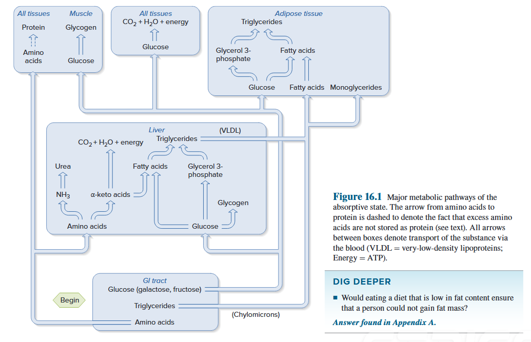
Summary of critical points in transition from the absorptive state to the postabsorptive state
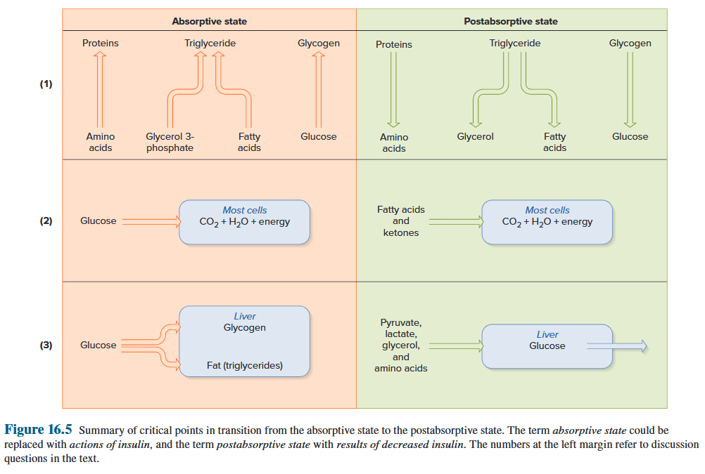
Key biochemical events response of target cells to insulin
- green arrow denotes a process stimulated by insulin. 绿色箭头表示胰岛素刺激的过程。
- dashed red arrow denotes inhibition by insulin 红色虚线箭头表示胰岛素的抑制
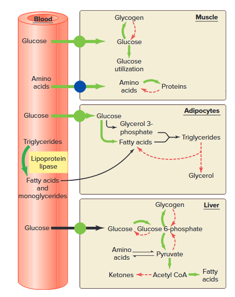
7.1.2.2 Expenditure of Energy
Types of expenditure of energy: muscle contraction, active transport, molecular synthesis 能量消耗类型:肌肉收缩→主动运输→分子合成
Energy can be neither created nor destroyed but can be converted from one form to another. Therefore, internal energy liberated (ΔE) during breakdown of an organic molecule can either appear as heat (H) or be used to perform work (W). 能量既不能被创造也不能被消灭,但可以从一种形式转换为另一种形式。 因此,有机分子分解过程中释放的内能 (ΔE) 可以表现为热量 (H) 或用于做功 (W)。
$$
\Delta E = 5H + 1W
$$
Biological work can be divided into two general categories:
- external work — the movement of external objects by contracting skeletal muscles; 外部功——通过收缩骨骼肌来运动外部物体
- internal work — all other forms of work, including skeletal muscle activity not used in moving external objects 内部功——所有其他形式的功,包括不用于移动外部物体的骨骼肌活动
Calorie = 4.184 J: One calorie is the amount of heat required to raise the temperature of one gram of water from 14.58 C to 15.58 C. 一卡路里是将一克水的温度从 14.58 C 升高到 15.58 C 所需的热量。
BMR (basal metabolic rate) is the rate of energy expenditure per unit time by endothermic animals at rest. It is reported in energy units per unit time ranging from watt (joule/second) to ml O~2~/min or joule per hour per kg body mass J/(h·kg). BMR(基础代谢率)是吸热动物在休息时单位时间内能量消耗的比率。以每单位时间的能量单位报告,范围从瓦特(焦耳/秒)到ml O~2~/分钟或焦耳每小时每公斤体重J/h·kg)。
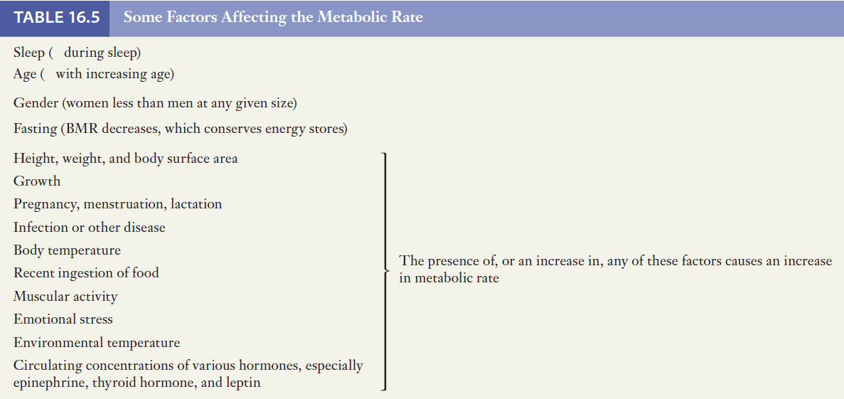
Major factors affecting metabolic rate
- Thyroid Hormones
- Epinephrine
- Diet-induced thermogenesis
- Muscle activity
- Emotional stress
- Environmental temperature
影响代谢率的主要因素 - 甲状腺激素 - 肾上腺素 - 饮食引起的产热 - 肌肉活动 - 情绪压力 - 环境温度
Thyroid Hormones
- Thyroid hormones (T3 and T4) are the single most important determinant of BMR regardless of body size, age, or gender. - T3 and T4 increase the oxygen consumption and heat production of most body tissues, a notable exception being the brain. 甲状腺激素(T3和T4)是 BMR 最重要的决定因素,无论体型、年龄或性别如何。 - T3 和 T4 会增加大多数身体组织的耗氧量和产热量,但大脑是一个值得注意的例外。
- Thyroid Hormones increase, metabolic rate increases - Calorigenic effect - Hyperthyroidism 甲状腺激素增加,代谢率增加 - 生热效应 - 甲状腺功能亢进
Epinephrine - Hormone - Calorigenic effect - Stimulation of glycogen and triglyceride catabolism - Epinephrine increases, metabolic rate increases 肾上腺素 - 激素 - 生热作用 - 刺激糖原和甘油三酯分解代谢 - 肾上腺素增加,代谢率增加
The calorigenic effect is a result of the increased metabolic rate and increased oxygen consumption by cells. When the metabolic rate increases, more heat is generated and body temperature rises. 生热效应是细胞代谢率增加和耗氧量增加的结果。当新陈代谢率增加时,会产生更多热量,体温升高。
Diet-induced thermogenesis
- The ingestion of food increases the metabolic rate by 10% to 20% for a few hours after eating.
- Protein > glucose and fat
- prolonged alterations in food intake also have significant effects on metabolic rate.
- 摄入食物可使新陈代谢率在饭后几个小时内增加 10% 至 20%。 - 蛋白质 > 葡萄糖和脂肪 - 食物摄入量的长期改变也会对代谢率产生显着影响。
Muscle activity
- The most important fact that can increase energy expenditure.
- depending on the degree of physical activity, total energy expenditure may vary for a healthy young adult from a value of approximately 1500 kcal/24 h (for a sedentary individual) to more than 7000 kcal/24 h (for someone who is extremely active).
- 可以增加能量消耗的最重要事实。 - 根据身体活动的程度,健康年轻人的总能量消耗可能会有所不同,从大约 1500 kcal/24 小时(对于久坐的人)到超过 7000 kcal/24 小时(对于极度活跃的人) )。
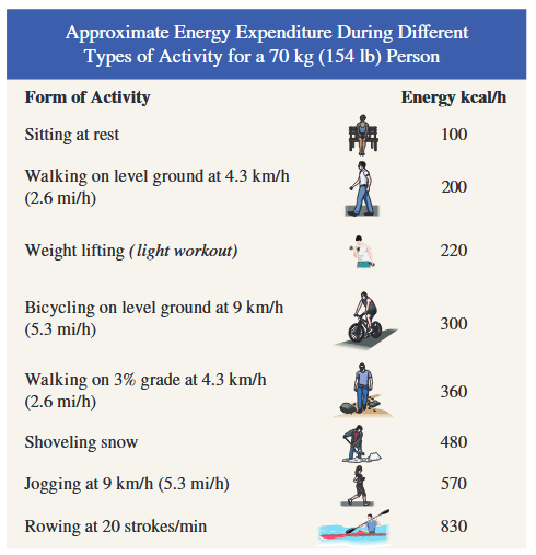
Emotional stress
- indirect, impact on CNS, to increase catecholamine (epinephrine and norepinephrine)
- Sympathetic stimulation increases, MR increases.
- 间接,对中枢神经系统的影响,增加儿茶酚胺(肾上腺素和去甲肾上腺素) - 交感神经刺激增加,MR增加。
Environmental temperature
- ET: 20~30 C, Stable
- ET: below 20, MR increase
- ET: increase, MR rises about 14% for each Celsius degree increases.
- ET:20~30℃,稳定 - ET:低于20,MR增加 - ET:增加,每增加摄氏度,MR增加约14%。
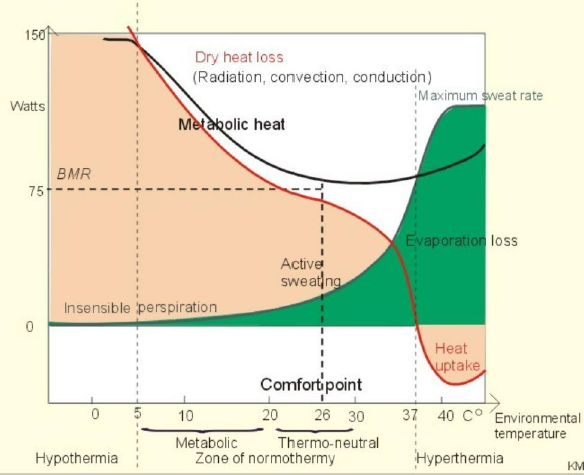
7.1.3 Energy Balance and its Control
Energy intake differs from the sum of internal heat produced and external work, changes in energy storage occur; the total-body energy content increases or decreases. 摄入的能量与内部产生的热量和外部做功的总和不同,能量储存发生变化;全身能量含量增加或减少。
7.1.3.1 Control of Food Intake
Leptin
- Synthesized by adipocytes
- Acts on the hypothalamus to cause a decrease in food intake, in part by inhibiting the release of neuropeptide Y.
- Increases BMR
- Functions in a negative feedback system to maintain a stable total-body energy content by signaling to the brain how much fat is stored.
- 由脂肪细胞合成 - 作用于下丘脑,部分通过抑制神经肽 Y 的释放来减少食物摄入量。 - 增加基础代谢率 - 在负反馈系统中发挥作用,通过向大脑发出信号来维持稳定的全身能量含量储存了多少脂肪。
Neuropeptide Y
- a hypothalamic neurotransmitter that stimulates appetite
- 刺激食欲的下丘脑神经递质
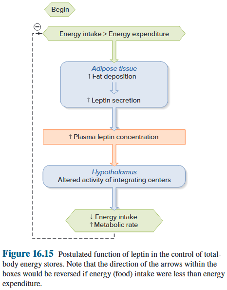
Satiety signals
- factors that decrease appetite cause the person to cease feeling hungry and set the time period before hunger returns.
- Insulin-dependent glucose utilization by certain areas of the hypothalamus
- Diet-induced thermogenesis
- 降低食欲的因素会导致人不再感到饥饿,并在饥饿再次出现之前设定一段时间。 - 下丘脑某些区域依赖胰岛素的葡萄糖利用 - 饮食诱导的生热作用
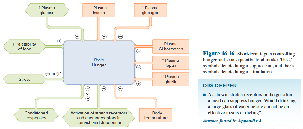
7.1.3.2 Overweight and Obesity
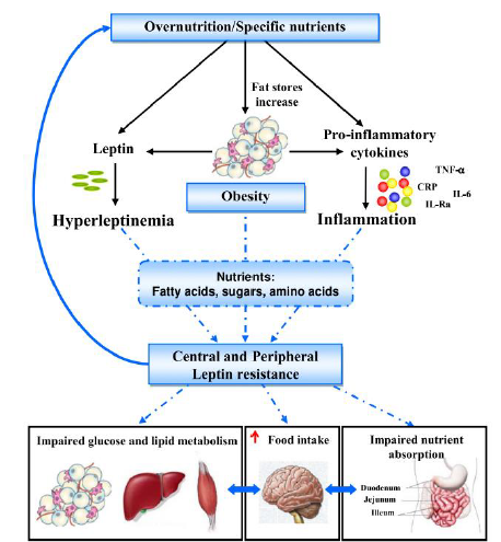
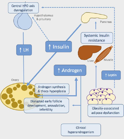
7.2 Body temperatures and its control
7.2.1 Basic concepts
Endotherms
Endotherms - generate their own internal body heat and do not rely on the energy of sunlight to warm the body. - humans maintain their body temperatures within very narrow limits despite wide fluctuations in ambient temperature and are, therefore, also known as homeotherms.
吸热动物——自身产生体内热量,不依赖阳光的能量来温暖身体。 - 尽管环境温度波动很大,人类仍将体温维持在非常窄的范围内,因此也被称为恒温动物。
- Radiation is the process by which the surfaces of all objects constantly emit heat in the form of electromagnetic waves.
- Conduction is the loss or gain of heat by transfer of thermal energy during collisions between adjacent molecules.
- Convection is the process whereby conductive heat loss or gain is aided by movement of the air or water next to the body.
- Evaporation of water from the skin and membranes lining the respiratory tract is the other major process causing loss of body heat.
辐射是所有物体表面不断以电磁波形式散发热量的过程。 传导是相邻分子碰撞期间通过热能转移而损失或获得热量。 对流是通过身体附近的空气或水的运动来帮助传导热量损失或增加的过程。 皮肤和呼吸道内膜的水分蒸发是导致体温损失的另一个主要过程。
7.2.2 Physiological Variances of Body Temperature
- Shell Temperature— the surface temperature —the temperature of the skin and tissues immediately at a constant value, and rises or falls with the temperature of the surrounding. 外壳温度——表面温度——皮肤和组织的温度立即处于恒定值,并随着周围温度的升高或降低而升高或降低。
- Core Temperature—the temperature of the deep tissues in the body—the core—remains almost exactly constant. 核心温度——身体深层组织——核心的温度——几乎保持恒定。
- Oral temperature averages about 0.58 C less than rectal, also known as core body temperature. 口腔温度平均比直肠温度低约 0.58 C,也称为核心体温。
- Internal temperature is not constant; although it does not vary much, it does change slightly in response to activity patterns and changes in external temperature. 内部温度不恒定;尽管变化不大,但它确实会根据活动模式和外部温度的变化而略有变化。
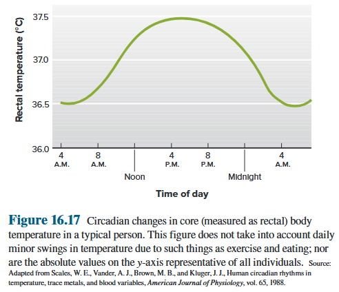

Factors affecting normal body temperature
- Circadian Rhythm 昼夜节律
- There is a circadian flunctuation, temperature being lowest during the early morning hours and highest in late afternoon or early morning hours and highest in late afternoon or early evening in a day. 昼夜节律波动,一天中清晨的温度较低,下午晚些时候或清晨的温度最高,下午晚些时候或傍晚的温度最高。
- The variation is not more than 1°C. This circadian rhythm is closely associated with the level of metabolic heat production. 变化不超过1°C。这种昼夜节律与代谢产热水平密切相关。
- Sex: The body temperature of a woman is slightly higher than a man and the difference is about 0.3 °C.
- Age: A new born child has a slight higher temperature than an adult, and the body temperature of a young child is more variable due to lack of control exercise by the nervous system and the higher metabolic rate. The old person have lower body temperature due to the decreased metabolic rate. 年龄:新生儿的体温比成人稍高,而幼儿由于缺乏神经系统的控制运动和较高的新陈代谢率,体温变化较大。老年人由于新陈代谢率下降,体温较低。
- Muscular Activity
- Others
7.2.3 Heat production and heat loss
- Main organ to produce heat
- at rest, liver and brain
- during exercise, the skeletal muscles
- Form of heat production
- shivering thermogenesis
- non-shivering thermogenesis
Because almost no external work is performed by shivering, virtually all the energy liberated by the metabolic machinery appears as internal heat and is known as shivering thermogenesis. 由于颤抖几乎不进行任何外部工作,因此几乎所有代谢机制释放的能量都表现为内热,被称为颤抖产热。
Chronic cold exposure induces an increase in metabolic rate (heat production) that is not due to increased muscle activity and is termed non-shivering thermogenesis. Its causes are an increased adrenal secretion of epinephrine and increased sympathetic activity to adipose tissue, with some contribution by thyroid hormone as well. 慢性寒冷暴露会导致代谢率(产热)增加,这不是由于肌肉活动增加所致,被称为非颤抖产热。其原因是肾上腺肾上腺素分泌增加和脂肪组织交感神经活动增加,甲状腺激素也有一定贡献。
Factors to regulate heat production
Metabolic rate of the body can change the rate of heat production.
- Hormones: thyroxin, growth hormone and testosterone. 甲状腺素、生长激素和睾酮。
- Nervous system: cold - decrease skin blood flow - reflex stimulation of sympathetic nerves to skin arterioles. 寒冷——皮肤血流量减少——交感神经对皮肤小动脉的反射性刺激。
Insensible perspiration is water loss of which a person is unware, in absence of sweating. Sensible perspiration. Sweating requires the active secretion of fluid by sweat glands and its extrusion into ducts that carry it to the skin surface. 不知不觉的出汗是指在没有出汗的情况下,人们没有意识到的水分流失。明显出汗。出汗需要汗腺主动分泌液体,并将其挤出到导管中,将液体输送到皮肤表面。
7.2.4 Regulations
Humans and other mammals are less affected by changes in the ambient temperature because they have the ability to maintain their body temperatures within a fairly narrow range through thermoregulation. 人类和其他哺乳动物受环境温度变化的影响较小,因为它们能够通过体温调节将体温维持在相当窄的范围内。
Animals with this ability are said to be homeothermic, whereas those lacking this ability are called poikilothermic. 具有这种能力的动物被称为恒温动物,而缺乏这种能力的动物被称为变温动物。
Temperature-Regulating Reflexes
- Peripheral thermoreceptors are in the skin
- Central thermoreceptors in deep body structures, including the hypothalamus, spinal cord, and abdominal organs.
- The skin thermoreceptors provide feedforward information, and also account for one’s ability to identify a hot or cold area of the skin. An area of the hypothalamus serves as the primary overall integrator of the reflexes, but other brain centers also exert some control over specific components of the reflexes.
外周温度感受器位于皮肤中。中枢温度感受器位于深层身体结构中,包括下丘脑、脊髓和腹部器官。皮肤温度感受器提供前馈信息,并且还解释了识别皮肤热或冷区域的能力。下丘脑的一个区域充当反射的主要整体整合器,但其他大脑中心也对反射的特定组成部分施加一些控制。
Relation of Controlling Signal to Thermal Integration
-
The hypothalamus serves as the primary overall integrator of the reflexes, but other brain centers also exert some control over specific components of the reflexes. 下丘脑是反射的主要整体整合器,但其他大脑中心也对反射的特定组成部分施加一些控制。
-
The resulting changes in body temperature are detected by thermoreceptors. These receptors initiate reflexes that change the output of various effectors so that heat production and/or loss are modified and body temperature is restored toward normal. 由此产生的体温变化由温度感受器检测到。这些受体启动反射,改变各种效应器的输出,从而改变热量的产生和/或损失,并使体温恢复正常。
Control of Skin Blood Vessels and Sweat Glands 皮肤血管和汗腺的控制
- Local cooling potentiates the constriction of blood vessels in response to nerve signals and vasoconstrictor substances.
- Local heating causes vasodilation through a direct action on the vessels, independent of nerve signals.
- Local thermal effects on sweat glands parallel those on blood vessels.
局部冷却增强血管对神经信号和血管收缩物质的反应的收缩。局部加热通过对血管的直接作用引起血管舒张,与神经信号无关。对汗腺的局部热效应与对血管的局部热效应相似。
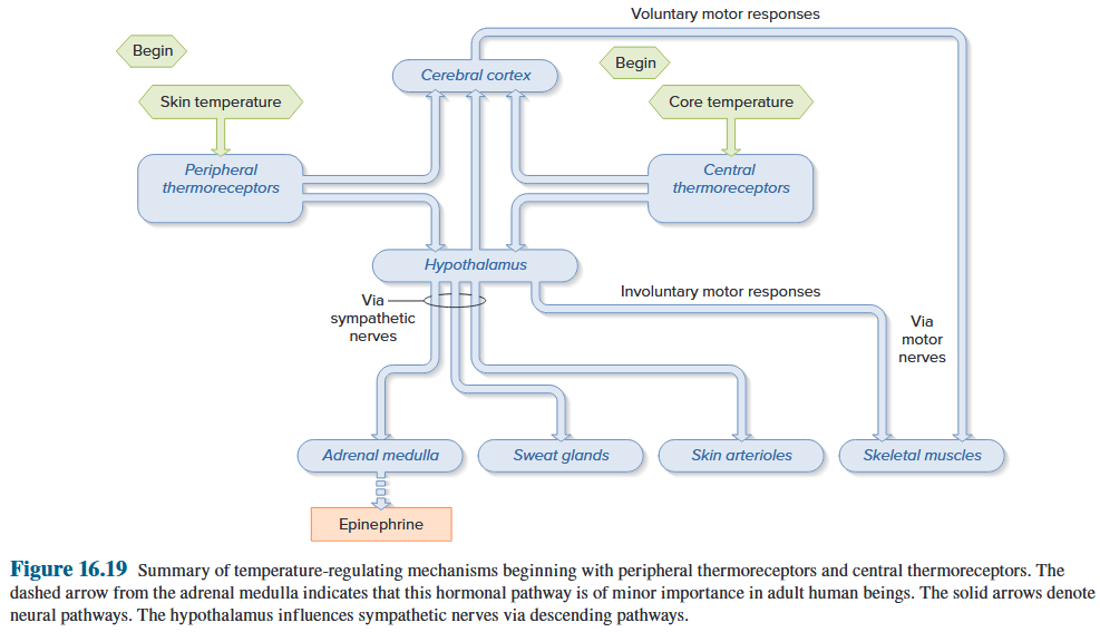
7.2.5 Clinical events (Fever and Hyperthermia)
Fever is an increase in body temperature due to a resetting of the “thermostat” in the hypothalamus. 发烧是由于下丘脑“恒温器”重置而导致的体温升高。
The most common cause of fever is infection, but physical trauma and tissue damage can also induce fever. 发烧最常见的原因是感染,但身体创伤和组织损伤也可诱发发烧。